Copyright © 2019 The Korean Society for Bone and Mineral Research
This is an Open Access article distributed under the terms of the Creative Commons Attribution Non-Commercial Li- cense (http://creativecommons.org/licenses/by-nc/4.0/) which permits unrestricted non-commercial use, distribu- tion, and reproduction in any medium, provided the original work is properly cited.
Effects of Di(2-ethylhexyl)phthalate on Bone Metabolism in Ovariectomized Mice
Jeong In Choi1, Hyun Hee Cho2
1Department of Obstetrics and Gynecology, Soonchunhyang University College of Medicine, Bucheon;
2 Department of Obstetrics and Gynecology, Eunpyeong St. Mary’s Hospital, College of Medicine, The Catholic University of Korea, Seoul, Korea
Background: The molecular pathways of how endocrine disruptors affect bone mineral density (BMD) and bone remodeling are still unclear. The purpose of this experimental study is to determine the effects of di(2-ethylhexyl)phthalate (DEHP) on bone metabo- lism in ovariectomized mice. Methods: Twenty-six-month-old female CD-1 mice were divided into 4 groups: control, low-dose DEHP, high-dose DEHP, and estrogen groups (n=5, each group). All mice were subjected to ovariectomy for the induction of artificial menopause and then exposed to corn oil, DEHP, and estrogen for 2 months. Micro-com- puted tomography (Micro-CT) of the bone and analysis of blood samples for bone mar- kers were performed to observe the changes in bone metabolism. Results: Osteocalcin level was decreased in the control, low-dose and high-dose DEHP group, the reduction width was greater in the high-dose DEHP group (-0.219 ng/mL) than control group (-0.077 ng/mL, P<0.05). C-terminal telopeptide of type I collagen level was increased in the control, low-dose and high-dose DEHP group, the increase range of low-dose DEHP group (0.329 ng/mL) showed greater than control group (0.093 ng/mL, P<0.05). Micro- CT analysis revealed that the BMD was significantly lower in the high-dose DEHP group (19.8×10⁻² g/cm3) than control group (27.2×10⁻² g/cm3, P<0.05). The structure model index was significantly higher in the high-dose DEHP group (2.737) than low-dose DEHP group (2.648) and estrogen group (2.63, P<0.05). It means the progression of osteopo- rosis in the high-dose DEHP group. Conclusions: These results confirm the negative ef- fects of DEHP on bone health in ovariectomized mice. Further continuous studies on ge- netic pathways and other endocrine disruptors will be necessary to validate these find- ings.
Key Words: Diethylhexyl phthalate · Osteoporosis · Ovariectomy · X-ray microtomography
INTRODUCTION
Osteoporosis is a disease with various causes; estrogen abnormality, thyroid hormone abnormality, calcium (Ca) metabolic disorder, steroid use, aging, and lack of exercise influence its occurrence. It is a disease where bone fractures de- velop easily due to the weakening of bone strength, as the overall bone metabo- lism declines. The balance between osteoclasts, which metabolize Ca by absorb- ing the bone, and the osteoblasts, which creates bone, also has a significant influ- ence on its occurrence.
Endocrine disruptors, which are known to respond to estrogen receptors due to Corresponding author
Hyun Hee Cho
Department of Obstetrics and Gynecology, Eunpyeong St. Mary’s Hospital, College of Medicine, The Catholic University of Korea, 1021 Tongil-ro, Eunpyeong-gu, Seoul 03312, Korea
Tel: +82-2-2030-4526 Fax: +82-2-595-1549 E-mail: drrabbit@catholic.ac.kr Received: June 11, 2019 Revised: August 2, 2019 Accepted: August 8, 2019
Original Article
pISSN 2287-6375 eISSN 2287-7029
their structural similarity with estrogen, initiate the struc- tural change of estrogen receptors, and act on several post- receptor responses associated with estrogen.[1] Due to such characteristics, the associations between diseases that develop in a variety of target organs affected by estro- gen are being discovered, of which the typical examples are breast cancer and other obstetric and gynecological diseases caused by estrogen imbalance. Among various previous studies on bone mineral density (BMD) and en- docrine disruptors, exposure to estrogen at the time of birth has been reported to have a significant effect on adult BMD, which is related not only to the duration of exposure but also to the concentration of estrogen.[2,3] In addition, there are studies reporting that exposure to environmental hormones during pregnancy and lactation affects BMD and bone remodeling.[4] In animal studies, phthalates have been observed to have an inhibitory effect on osteoblasts in mice, thereby affecting BMD, along with weak estrogen- ic and antiestrogenic activities, and anti-androgenic activi- ty.[5-8]
The molecular pathways of how endocrine disruptors af- fect BMD and bone remodeling are still unknown. Howev- er, in addition to their previously identified effects on es- trogen biosynthesis, estrogen metabolism and receptors, studies have also reported their effects on genes involved in the receptor sub-pathways or other genes that regulate osteocyte differentiation.[9] In our previous study, mice exposed to di(2-ethylhexyl)phthalate (DEHP) at prenatal and lactation period showed an abnormal amplification of genes related with bone remodeling in the adult stage.[10]
Considering that menopausal osteoporosis is caused by the decline in estrogen levels, the possibility of endocrine disruptors having similar functions as estrogen, i.e., affect- ing BMD after menopause should be fully considered. In a previous study by Min and Min,[11] which investigated urinary phthalate metabolites and BMD in elderly women using the US National Health Survey data, it was reported that the higher the concentration of phthalate metabo- lites, the lower the BMD and the higher the incidence of osteoporosis.
The purpose of this experimental study is to determine the effects of DEHP on bone health in ovariectomized mice.
Micro-computed tomography (Micro-CT) of the bone, and analysis of blood samples for bone markers were performed to observe the changes in bone metabolism.
METHODS
1. AnimalsFemale adult CD-1 (Crl:CD-1 [ICR] BR) mice were obtained from Orient Bio Inc. (Seongnam, Korea) and raised at the animal lab of the College of Medicine, Catholic University of Korea. In each cage, four mice were raised on an aspen bedding (Tapvei, Paekna, Estonia) under controlled light- ing (12 hr light/12 hr dark); appropriate conditions of tem- perature (22±5°C) and humidity (50%±10%) were main- tained. The mice were fed with Teklad Global 18% Protein Rodent Diet (Harlan Laboratories Inc., Madison, WI, USA), and sterilized tap water was supplied through polycarbon- ate bottles. Animal breeding and study protocols were per- formed in compliance with the Laboratory Animals Welfare Act, the Guide for the Care and Use of Laboratory Animals, and the Guidelines and Policies for Rodent Experiments, and were approved by the Institutional Animal Care and Use Committee (IACUC) of the School of Medicine, Catholic University of Korea (Approval no. CUMC-2014-0083-02). All animals were raised in humanitarian settings, and efforts were taken to minimize pain.
Twenty-six-month-old female CD-1 mice were divided into 4 groups: the control, low-dose DEHP treatment, high- dose DEHP treatment, and estrogen treatment groups; each group contained 5 mice. To prevent infection and reduce pain, gentamicin (5 mg/kg) and ketoprofen (5 mg/kg) were administered via subcutaneous injection once prior to sur- gery and for 3 days after surgery. The animals received in- halation anesthesia with isoflurane (1.5%) and were fixed to the anesthesia machine. An electric shaver was used to shave the surgical area, which was disinfected with alco- hol. An incision was made at the middle abdominal region, and the ovaries at both sides were checked and then re- moved.
2. DEHP treatment and blood sampling
Ovariectomized mice were exposed to the following concentrations of DEHP and estrogen for 2 months after surgery; the control group received corn oil (0.15 mL/wk) through subcutaneous injection, low-dose DEHP treat- ment group received 35 μg/kg/wk (5 μg/kg/d) DEHP dis- solved in 0.15 mL corn oil once a week via subcutaneous injection, high-dose DEHP treatment group received 350 μg/kg/wk (50 μg/kg/d) DEHP dissolved in 0.15 mL corn oil
once a week via subcutaneous injection, and estrogen treat- ment group received conjugated equine estrogen (Prema- rin 0.3 mg/kg/day) once a day via per oral route. In the con- trol and low-dose DEHP and high-dose DEHP treatment groups, venous blood was collected (0.3 mL) through the retro-orbital plexus once a month after ovariectomy to test for 2 types of bone markers (osteocalcin, C-terminal telo- peptide of type I collagen [CTX-1]), and serum Ca, phos- phorus (P), alkaline phosphatase (ALK-P), and magnesium (Mg).
A specific sandwich enzyme-linked immunosorbent as- say (ELISA) was used to quantify CTX-1 (MyBioSource, Inc., San Diego, CA, USA; MB703094) and Gla-Osteocalcin (Ta- kara Bio Inc., Otsu, Japan; MK127) in mouse serum. Aliquots of mouse serum samples were each tested in triplicate at several dilutions and compared to reference standards of CTX-1 and Gla-Osteocalcin. The concentration of CTX-1 and Gla-Osteocalcin was measured using ELISA kit accord- ing to the manufacture’s protocol.
Mouse Gla-Osteocalcin High Sensitive EIA Kit (TAKARA MK127); Prepare reagents and samples (100 μL each) in a separate 96 well plate in advance so that they can be add- ed to the Antibody Coated Microtiterplate quickly (within 5 min) using an 8-channel pipette or similar apparatus.
Perform this reaction at room temperature (20-30°C) for 1 hr; incubation at 37°C may compromise antigenicity (First reaction). Discard reaction mixtures, followed by 3 washes with Washing Buffer. Then add 100 μL of the POD-labeled Antibody Solution per well using an 8-channel pipette and allow to react for 1 hr at room temperature (20-30°C) (Sec- ond reaction). Discard reaction mixtures, followed by 4 wash- es with Washing Buffer. Then add 100 μL of Substrate Solu- tion (TMBZ) per well using an 8-channel pipette and allow to react at room temperature (20-30°C) for 10 to 15 min (Third reaction). Add 100 μL of Stop Solution to each well to stop the reaction in the same order as for Substrate So- lution (TMBZ). Then mix well. Use distilled water as a con- trol to make zero adjustment and measure absorbance at 450 nm.
Mouse cross linked CTX-1 ELISA Kit (mybiosource MB70- 3094); Prepare all reagents and samples as directed in the previous sections. Determine the number of wells to be used and put any remaining wells and the desiccant back into the pouch and seal the ziploc, store unused wells at 4°C. Set a Blank well without any solution. Add 100 μL of
Standard or Sample per well. Standard need test in dupli- cate. Add 100 μL of HRP-conjugate to each well (not to Blank well), then 100 μL Antibody to each well. Mix well and then incubate for 1 hr at 37°C. Aspirate each well and wash, repeating the process 2 times for a total of 3 washes.
Wash by filling each well with Wash Buffer (200 μL) using a squirt bottle, multi-channel pipette, manifold dispenser and let it stand for 10 sec, complete removal of liquid at each step is essential to good performance. After the last wash, remove any remaining Wash Buffer by aspirating or- decanting. Invert the plate and blot it against clean paper towels.
Add 50 μL of Substrate A and 50 μL of Substrate B to each well, mix well. Incubate for 15 min at 37°C. Keeping the plate away from drafts and other temperature fluctuations in the dark. Add 50 μL of Stop Solution to each well, gently tap the plate to ensure thorough mixing. Determine the optical density of each well within 10 min, using a micro- plate reader set to 450 nm.
3. Micro-CT analysis
All mice were sacrificed four months after surgery using carbon dioxide, the tibia samples were fixed with 70% eth- anol. BMD, microstructures (structural thickness, structural separation, structural linear density), structure model in- dex (SMI) of the tibia head were analyzed by Micro-CT (A SkyScan 1,176 instrument; Bruker microCT, Kontich, Bel- gium). Micro-CT analysis was carried out by SecondAnaly- sis corporation (Korea). CT Analysis and CT Volume pro- grams (Bruker microCT) were used to obtain images and analyze data. For analyze data, we used Excel (Microsoft office 2017), paired t-test (P<0.05). Statistical comparisons were performed using student’s t-test, and data were re- garded as being significant when P<0.05.
RESULTS
1. OsteocalcinOsteocalcin is a well-known bone formation marker. Lev- el of osteocalcin showed increase in estrogen group (3.021 ng/mL and 3.11 ng/mL, at 1-2 months), but showed de- crease in control group (3.355 ng/mL and 3.278 ng/mL), low-dose DEHP group (3.478 ng/mL and 3.39 ng/mL) and high-dose DEHP group (3.194 ng/mL and 2.975 ng/mL) (Fig. 1). The calculated subtraction value (2 months value
minus 1 month value) is statistically greater in the high- dose DEHP group (-0.219 ng/mL) than the control group (-0.077 ng/mL, P<0.05). These data means bone formation was promoted in the estrogen treated group, but in the other groups, bone formation was decreased, especially in high-dose DEHP group.
2. CTX-1
In bone physiology, the CTX-1 is a telopeptide that can be used as a bone resorption biomarker in the serum to measure the rate of bone turnover. Increased CTX-1 means higher bone resorption status means higher bone turnover status. Serum CTX assay shows greater utility for assessing efficacy of antiresorptive treatment. Serum CTX-1 levels showed a decrease in the estrogen treatment group (1.336 ng/mL and 1.134 ng/mL, at 1 and 2 months), while an in- crease in the control (1.384 ng/mL and 1.477 ng/mL), low- dose DEHP (1.079 ng/mL and 1.408 ng/mL), and high-dose DEHP groups (1.406 ng/mL and 1.533 ng/mL) (Fig. 2). These data means bone resorption was inhibited in estrogen treatment group, but not in the other groups. Additionally, the calculated subtraction value (2 months value minus 1 month value) of low-dose DEHP treatment groups (0.329 ng/mL) showed greater than the control group (0.093 ng/mL, P<0.05).
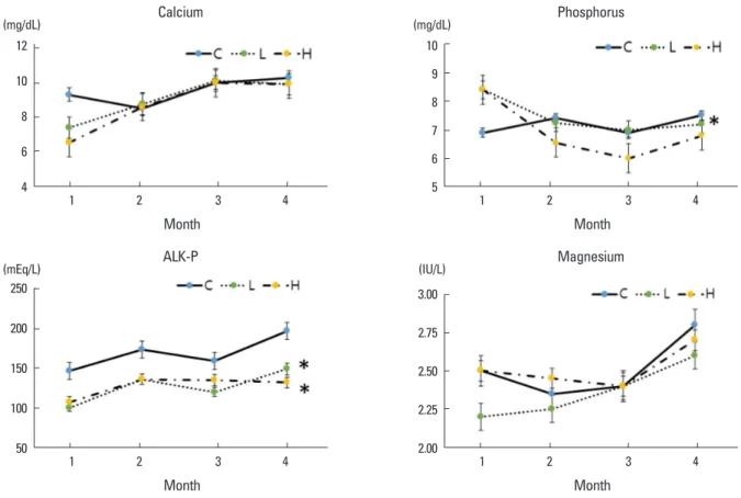
3. Biochemical assay; serum Ca, P, ALK-P and Mg Biochemical assays were performed using samples from the control, low-dose DEHP, and high-dose DEHP groups.
There was no significant difference in the serum Ca levels and serum Mg levels between control and treatment groups.
Serum P level in the high-dose DEHP group showed signif- icant decrease (8.4 mg/dL and 6.8 mg/dL, at 1 and 4 months) than control group (6.9 mg/dL and 7.5 mg/dL), and the se- rum ALK-P levels were decreased to a greater extent in the low-dose DEHP group (100 mEq/L and 149 mEq/L, at 1 and 4 months) and high-dose DEHP groups (107 mEq/L and 132 mEq/L) than in the control group (147 mEq/L and 197 mEq/L, P<0.05) (Fig. 3).
4. Bone marrow density measurement
After 4 months from ovariectomy, the BMD was significant- ly lower in the high-dose DEHP group (19.8×10⁻² g/cm3) and significantly higher in the estrogen treatment group (30.9×10⁻² g/cm3) than control group (27.2×10⁻² g/cm3, P<0.05) (Fig. 4).
5. Microstructural analysis
In the microstructural analysis using Micro-CT, structural thickness, structural separation, and structural linear den- sity of tibia head were calculated. The structural linear den- Fig. 1. Comparison of serum bone formation marker (osteocalcin) con-
centrations at month 1 and 2 after di(2-ethylhexyl)phthalate (DEHP) treatment in ovariectomized mice. A gradual increase was shown in the estrogen treatment group (E), but a decrease was shown in the control group (C), low-dose DEHP group (L), and high-dose DEHP group (H). The calculated subtraction value is statistically greater in the H (*) than the C. *P<0.05 vs. C.
4.0
3.5
3.0
2.5
Osteocalcin (ng/mL)
1 2
Month
Fig. 2. Change in serum bone resorption marker (C-terminal telopep- tide of type 1 collagen [CTX-1]) concentrations at month 1 and 2 after di(2-ethylhexyl)phthalate (DEHP) treatment. CTX-1 level showed a decrease in the estrogen treatment group (E), while an increase the other groups. The calculated subtraction value of CTX-1 is statistical- ly greater in the low-dose DEHP group (L) (*) than the control group (C). H, high-dose di(2-ethylhexyl)phthalate treatment group. *P<0.05 vs. C.
1.8
1.6
1.4
1.2
1.0
CTX-1 (ng/mL)
1 2
Month
sity was significantly lower in the high-dose DEHP group (0.16±0.01/mm) than in the estrogen treatment (0.23±
0.03/mm) and low-dose DEHP (0.24±0.01/mm) groups (P<0.05). No other significant structural differences were
observed among the four groups (Table 1).
After the microstructural analysis using Micro-CT, tra- becular area was also identified with yellow color (Fig. 5).
Trabecular area is more prominent in estrogen treatment group. In high-dose DEHP group, trabecular area is smaller than the others (estrogen treatment, control, and low-dose DEHP groups).
Table 1. Microstructural analysis of the tibia head of ovariectomized mice
St. thickness
(mm) St. separation
(mm) St. linear density (1/mm)
Estrogen 0.09±0.01 0.67±0.009 0.23±0.03
Low-dose DEHP 0.08±0.008 0.79±0.008 0.24±0.01 High-dose DEHP 0.08±0.007 0.74±0.01 0.16±0.01a)
Control 0.05±0.01 0.87±0.01 0.19±0.02
The data is presented as mean±standard deviation.
a)P<0.05.
St, structural; DEHP, di(2-ethylhexyl)phthalate.
Fig. 3. Change in serum calcium (Ca), phosphorus (P), alkaline phosphatase (ALK-P), and magnesium (Mg). There was no significant difference in the serum Ca levels and Mg levels among the three groups. Although, there was a significant decrease in the serum P level in the high-dose di(2- ethylhexyl)phthalate (DEHP) group, and the serum ALK-P levels were decreased to a greater extent in the low-dose DEHP groups (L) and high- dose DEHP groups (H) than in the control group (C). *P<0.05 vs. C.
12 10 8 6 4
(mg/dL) Calcium
1 2 3 4
Month
250
200
150
100
50
(mEq/L) ALK-P
1 2 3 4
Month
10 9 8 7 6 5
(mg/dL) Phosphorus
1 2 3 4
Month
3.00
2.75
2.50
2.25
2.00
(IU/L) Magnesium
1 2 3 4
Month
Fig. 4. Comparison of bone marrow density (BMD) in the tibia of ovariectomized mice. The BMD was significantly lower in the high- dose di(2-ethylhexyl)phthalate (DEHP) group (H) (*) and significantly higher in the estrogen-treated group (E) than control group (C). L, low- dose d di(2-ethylhexyl)phthalate treatment group. *P<0.05 vs. C.
35 30 25 20 15 10 5
-23Bone marrow density (10 g/cm) 0
E DEHP L DEHP H C
*
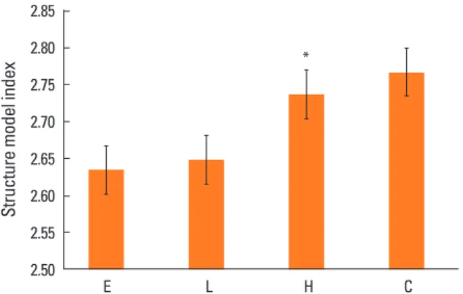
6. Structural model index
SMI suggests that the shape of the bone is dominant, ei- ther bar-like (score 3) or plate-like (score 0), and higher SMI means predominate bar shape, which means that the bone strength is weakened. SMI was significantly higher in the high-dose DEHP group (2.737) than in the estrogen treatment (2.63) and low-dose DEHP group (2.648, P<0.05).
No significant differences were observed in the high-dose DEHP group and control group (2.767) (Fig. 6).
DISCUSSION
Osteoporosis is a major disease that increases mortality and the prevalence of the elderly in an aging society. In addition to age and menopause, there are various factors that increase the incidence of osteoporosis, including amount of exercise, dietary Ca, vitamin D, other underlying diseas- es, etc. The incidence of pelvic fracture, which has the high- est mortality, increases by 13-fold in the age group of 60 to 80 years. Osteoporosis is the most important predictor of pelvic fracture in both men and women. Therefore, efforts to reduce the incidence of osteoporosis along with preven-
tion of falls and appropriate treatment according to BMD are needed. In addition, attention should also be paid to a variety of endocrine disruptors that can affect bone me- tabolism.
DEHP is a widely known endocrine disruptor that is used to soften plastic. There are 2 pathways for DEHP exposure:
the oral and parenteral routes. In this paper, we used sub- cutaneous injection for the sustained constant blood level of DEHP and slower rise of body burden. Absorption through airborne pollutants, perfumes, cosmetics, personal hygiene products, etc., constitute DEHP exposure via the parenteral route.[12] Although DEHP contents in cosmetics and per- sonal hygiene products are regulated, there are reports that it is still detected in many countries.[13] Various equip- ment made of plastics that are used in the medical indus- try are the cause of parenteral phthalate exposure; Luo et al.[14] have reported phthalate detection in medical prod- ucts made of polyvinyl chloride. In addition, it has been re- ported that serum DEHP and DEHP metabolite concentra- tions in urine among intensive care unit patients increase by 100- to 1,000-fold.[15]
Osseous tissue is synthesized by osteoblasts and absorbed by osteoclasts. The synthesis and resorption of osseous tis- sue play an important role in maintaining its strength. The functions of osteoblasts and osteoclasts are affected by various hormones, among which testosterone, estrogen, and cortisol are hormones that influence the synthesis and resorption of osseous tissue. Since the cells in osseous tis- sues respond to estrogen, endocrine disruptors that mimic Fig. 5. Tibia head microstructure analysis using micro-computed to-
mography scan. Trabecular area (yellow) is more prominent in estro- gen treatment group (E). In high-dose di(2-ethylhexyl)phthalate (DEHP) group (H), trabecular area is smaller than the others (E, control group [C], low-dose DEHP treatment group [L]).
Fig. 6. Structure model index (SMI) analysis in the tibia of ovariecto- mized mice. SMI was significantly higher in the high-dose di(2-ethyl- hexyl)phthalate (DEHP) group (H) (*) than in the estrogen treatment (E) and low-dose DEHP groups (L). No significant differences were ob- served in the H and control group (C). *P<0.05 vs. C.
2.85 2.80 2.75 2.70 2.65 2.60 2.55 2.50
Structure model index
E L H C
*
estrogen were believed to influence the functions of os- teoblasts and osteoclasts; many previous studies regarding this aspect have been published.
Animal studies have shown that the oral exposure of en- docrine disruptors such as phthalates to pregnant mice causes malformation and deformation in the bones of pre- natal mice,[3] and affects the actin fiber of the osteoblasts, thereby transforming their shape.[16] Phthalates like ben- zyl butyl phthalate or di-n-butyl phthalate enter and accu- mulate within mice osteoblasts.[5] The structure of micro- filaments in the cell is disrupted, osteoblast DNA is dam- aged, synthesis of apoptosis promoter proteins like P53 is promoted, and synthesis of lipid metabolism and blood is affected.[17] The differentiation of calvarial osteoblasts of mice exposed to DEHP is also affected, which is known to be due to the effects of DEHP on collagen synthesis and ALK-P expression.[18] Metabolites of phthalate like mono (2-ethylhexyl) phthalate (MEHP) or monobenzyl phthalate (MBzP) have been identified as peroxisome proliferator ac- tivated receptor γ (PPAR-γ) agonists. Increase in the PPAR-γ level further leads to a decrease in the BMD, which is known to show effects especially in postmenopausal women.[19,20]
Selective estrogen receptor modulator (SERM) and phy- toestrogen are substances that affect estrogen action in the body, such as hormone disruptants. SERM drugs act on the estrogen receptor. It act as partial estrogen receptors agonists for maintaining bone density bone for applica- tions in osteoporosis treatment, and same time act as es- trogen receptor antagonists in breast tissues. Phytoestro- gens are chemicals synthesized from plants, and show low estrogenic activity or anti estrogenic activity.[21] They binds to estrogen receptor and occupies it to prevent es- trogen from binding to the receptor. Unlike SERM or phy- toestrogen, the mechanism of action of DEHP is thought not to be through the estrogen receptor. In hepatic tissues, DEPH modulates some genetic pathways like PPAR-α sig- naling pathways and Janus kinase/signal transducers and activators of transcription pathway [22] and in ovarian tis- sues DEHP dysregulated proapoptotic factors and antiapop- totic factors and altered levels of proteins in phosphatidyli- nositol 3 kinase (PIsK) signaling pathways.[23,24] In a re- cently reported study by Chiu et al.[25], they suggested that DEHP and MEHP exposure may inhibit osteoblasto- genesis and promote adipogenesis of bone marrow stro- mal cells in a mouse model. The downregulation of Wnt/
β-catenin signaling and the upregulation of PPAR-γ path- way may contribute to the inhibitory effects of DEHP or MEHP on osteoblast differentiation and thus triggering bone loss.[25]
In human study, some authors reported about phthalate and bone health. Min and Min [11] claimed in a study with 398 women older than 50 years of age that urinary con- centration of mono-n-butyl phthalate, mono-(3-carboxy- prophyl) phthalate, MBzP correlates with low BMD, which increases the risk of osteoporosis in postmenopausal wom- en. DeFlorio-Barker and Turyk [26] have demonstrated that there is a negative correlation between the total low-mo- lecular weight phthalate metabolite contents and BMD in postmenopausal women. The relationship between phthal- ate metabolites and BMD is affected by body fat percent- age and age; postmenopausal women younger than 65 years of age with low body fat percentage showed a nega- tive correlation between BMD and phthalate metabolites, while women older than 65 years of age with a high body fat percentage showed a positive correlation between BMD and phthalate metabolites. The average phthalate expo- sure is 0.003 to 0.03 mg/kg/day (7.7-77 μM),[27] and the concentration of low dose DEHP in this paper is 30 µg/kg/
day, which is relevant to the clinical situation. The dosage of high dose is over 10 times of mean exposure level of hu- man as previously reported.[28]
The results of this study showed that in mice that were exposed to DEHP, bone formation marker levels significant- ly decreased, while the bone resorption marker levels sig- nificantly increased; these results differed clearly from those observed for the estrogen treatment group. In biochemical assessment, serum P level was significantly low in high dose DEHP group and serum ALK-P levels were significantly low in low dose and high dose DEHP group than control. In postmenopausal osteoporosis women, serum ALK-P is in- creased because of high bone turnover and serum Ca and serum P levels are decreased.[29] In other words, the effect of DEHP that act on bones is not simply due to their estro- gen or anti-estrogen like function. Further studies about biochemical changes in DEHP exposed mice are needed. In addition, BMD was significantly reduced in mice treated with a high dose of DEHP, and the results of Micro-CT showed that the SMI in this group increased significantly, compared to that for other groups. SMI is the distribution of rods and plates obtained from the structural analysis of trabecular
bone, which is represented as a ratio [30]; it increases with the progression of osteoporosis.[26] The increase of the SMI value indicates the onset of osteoporosis.[31,32] For a structure with both plates and rods of equal thickness the value lies between 0 and 3, epending on the volume ratio of rods and plates. In this paper, high dose DEPH group showed decreased BMD and higher SMI, which means weak- ened bone strength.
This study was performed under well controlled same environment to control and treatment group. In the think of many factors which could influence to bone metabo- lism, well controlled animal study is highly valuable. Small sample size is the limitation of this paper. In blood analysis data, we took the baseline samples, but the amount of blood was not sufficient for analysis. So we analyzed the data after 1 month. This should be taken into consideration when interpreting the results. As estrogen is a powerful protector of bone, there is likely to be a difference in the effect of DEHP on bone in situations with and without es- trogen. This study was carried out in the absence of estro- gen because absence of estrogen could better explain the effects of DEHP. In the further, the study with estrogen should proceed more.
The DEHP treatment groups showed lesser bone forma- tion and greater bone resorption than the control group.
In addition, Low BMD and increase of the SMI value were observed in the high-dose DEHP group, it means the pro- gression of osteoporosis. In summary, based on the results of this study, the negative effects of DEHP on bone metab- olism in ovariectomized mice were confirmed. So we could suggest that DEHP may have the possibility of adverse ef- fect on bone metabolism in postmenopausal women. As a result of this study, it is difficult to say with certainty how DEHP affects the bones. However, it is certain that DEHP affects bone metabolism, and avoiding DEHP in meno- pausal women is likely to have a positive effect on bone metabolism as it gives a negative effect on high dose. Fur- ther studies on genetic pathways and other endocrine dis- ruptors will be necessary for a deeper understanding of these effects and treatment of bone disorders.
DECLARATIONS
Ethics approval and consent to participate All studies involving animals were approved by the IACUC
at the Catholic University of Korea (CUMC-2014-0083-02).
Conflict of interest
No potential conflict of interest relevant to this article was reported.
ORCID
Jeong In Choi https://orcid.org/0000-0001-8023-084X
REFERENCES
1. Roy JR, Chakraborty S, Chakraborty TR. Estrogen-like en- docrine disrupting chemicals affecting puberty in humans- -a review. Med Sci Monit 2009;15:Ra137-45.
2. Migliaccio S, Newbold RR, Bullock BC, et al. Alterations of maternal estrogen levels during gestation affect the skel- eton of female offspring. Endocrinology 1996;137:2118- 25.
3. Migliaccio S, Newbold RR, Teti A, et al. Transient estrogen exposure of female mice during early development per- manently affects osteoclastogenesis in adulthood. Bone 2000;27:47-52.
4. Hermsen SA, Larsson S, Arima A, et al. In utero and lacta- tional exposure to 2,3,7,8-tetrachlorodibenzo-p-dioxin (TCDD) affects bone tissue in rhesus monkeys. Toxicology 2008;253:147-52.
5. Sabbieti MG, Agas D, Santoni G, et al. Involvement of p53 in phthalate effects on mouse and rat osteoblasts. J Cell Biochem 2009;107:316-27.
6. Agas D, Sabbieti MG, Marchetti L. Endocrine disruptors and bone metabolism. Arch Toxicol 2013;87:735-51.
7. Harris CA, Henttu P, Parker MG, et al. The estrogenic activity of phthalate esters in vitro. Environ Health Perspect 1997;
105:802-11.
8. Okubo T, Suzuki T, Yokoyama Y, et al. Estimation of estro- genic and anti-estrogenic activities of some phthalate di- esters and monoesters by MCF-7 cell proliferation assay in vitro. Biol Pharm Bull 2003;26:1219-24.
9. Hurst CH, Waxman DJ. Activation of PPARalpha and PPAR- gamma by environmental phthalate monoesters. Toxicol Sci 2003;74:297-308.
10. Cheon KY, Kil KH, Choi JI, et al. Parenteral exposure to DEHP and its effect on the microstructure of bone and Wnt sig- naling pathway in F2 female mice. Biochip J 2016;10:233- 40.
11. Min KB, Min JY. Urinary phthalate metabolites and the risk of low bone mineral density and osteoporosis in older wom- en. J Clin Endocrinol Metab 2014;99:E1997-2003.
12. Koo HJ, Lee BM. Estimated exposure to phthalates in cos- metics and risk assessment. J Toxicol Environ Health A 2004;
67:1901-14.
13. Guo Y, Wang L, Kannan K. Phthalates and parabens in per- sonal care products from China: concentrations and hu- man exposure. Arch Environ Contam Toxicol 2014;66:113-9.
14. Luo H, Sun G, Shi Y, et al. Evaluation of the Di(2-ethylhexyl) phthalate released from polyvinyl chloride medical devic- es that contact blood. Springerplus 2014;3:58.
15. Huygh J, Clotman K, Malarvannan G, et al. Considerable exposure to the endocrine disrupting chemicals phthal- ates and bisphenol-A in intensive care unit (ICU) patients.
Environ Int 2015;81:64-72.
16. Gordon SR. Microfilament disruption in a noncycling or- ganized tissue, the corneal endothelium, initiates mitosis.
Exp Cell Res 2002;272:127-34.
17. Agarwal DK, Maronpot RR, Lamb JCt, et al. Adverse effects of butyl benzyl phthalate on the reproductive and hema- topoietic systems of male rats. Toxicology 1985;35:189-206.
18. Bhat FA, Ramajayam G, Parameswari S, et al. Di 2-ethyl hexyl phthalate affects differentiation and matrix mineral- ization of rat calvarial osteoblasts--in vitro. Toxicol In Vitro 2013;27:250-6.
19. Akune T, Ohba S, Kamekura S, et al. PPARgamma insuffi- ciency enhances osteogenesis through osteoblast forma- tion from bone marrow progenitors. J Clin Invest 2004;
113:846-55.
20. Grey A, Bolland M, Gamble G, et al. The peroxisome prolif- erator-activated receptor-gamma agonist rosiglitazone decreases bone formation and bone mineral density in healthy postmenopausal women: a randomized, controlled trial. J Clin Endocrinol Metab 2007;92:1305-10.
21. Lecomte S, Demay F, Ferrière F, et al. Phytochemicals tar- geting estrogen receptors: Beneficial rather than adverse effects? Int J Mol Sci 2017;18.
22. Rusyn I, Peters JM, Cunningham ML. Modes of action and
species-specific effects of di-(2-ethylhexyl)phthalate in the liver. Crit Rev Toxicol 2006;36:459-79.
23. Hannon PR, Brannick KE, Wang W, et al. Di(2-ethylhexyl) phthalate inhibits antral follicle growth, induces atresia, and inhibits steroid hormone production in cultured mouse antral follicles. Toxicol Appl Pharmacol 2015;284:42-53.
24. Hannon PR, Peretz J, Flaws JA. Daily exposure to Di(2-eth- ylhexyl) phthalate alters estrous cyclicity and accelerates primordial follicle recruitment potentially via dysregula- tion of the phosphatidylinositol 3-kinase signaling path- way in adult mice. Biol Reprod 2014;90:136.
25. Chiu CY, Sun SC, Chiang CK, et al. Plasticizer di(2-ethylhex- yl)phthalate interferes with osteoblastogenesis and adi- pogenesis in a mouse model. J Orthop Res 2018;36:1124- 34.
26. DeFlorio-Barker SA, Turyk ME. Associations between bone mineral density and urinary phthalate metabolites among post-menopausal women: a cross-sectional study of NHANES data 2005-2010. Int J Environ Health Res 2016;26:326-45.
27. Agency for Toxic Substances and Disease Registry. Toxico- logical profile for di(2-Ethylhexyl)phthalate (DEHP). 2002 [cited by 2017 Sep 1]. Available from: https://www.atsdr.
cdc.gov/ToxProfiles/tp.asp?id=684&tid=65
28. Cho HH, Kim GW, Ryu JC. The effects of Di-2-ethylhexyl phthalates (DEHP) on the cell cycle of the endometrial cancer cell lines (ECC-1). Toxicol Environ Health Sci 2014;
6:217-23.
29. Khadka B, Tiwari ML, Gautam R, et al. Correlates of bioche- mical markers of bone turnover among post-menopausal women. JNMA J Nepal Med Assoc 2018;56:754-8.
30. Hildebrand T, Rüegsegger P. Quantification of bone micro- architecture with the structure model index. Comput Me- thods Biomech Biomed Engin 1997;1:15-23.
31. Grote HJ, Amling M, Vogel M, et al. Intervertebral variation in trabecular microarchitecture throughout the normal spine in relation to age. Bone 1995;16:301-8.
32. Ding M, Hvid I. Quantification of age-related changes in the structure model type and trabecular thickness of hu- man tibial cancellous bone. Bone 2000;26:291-5.
![Fig. 2. Change in serum bone resorption marker (C-terminal telopep- telopep-tide of type 1 collagen [CTX-1]) concentrations at month 1 and 2 after di(2-ethylhexyl)phthalate (DEHP) treatment](https://thumb-ap.123doks.com/thumbv2/123dokinfo/5249900.133824/4.892.464.805.111.361/resorption-terminal-telopep-collagen-concentrations-ethylhexyl-phthalate-treatment.webp)