Background and Purpose We investigated the frequency and clinical features of restless legs syndrome (RLS) in patients with Parkinson’s disease (PD).
Methods This study included 74 PD patients. RLS was diagnosed in face-to-face assessments of all of the subjects based on diagnostic criteria of the International Restless Legs Syndrome Study Group revised in 2003. We analyzed the clinical features of PD patients with and with- out RLS and compared the data to idiopathic RLS.
Results The frequency of RLS in the cohort was 21.6% (n=16). Two (12.5%) of the patients with RLS were not treated with dopaminergic drugs, while 14 (24.1%) of the 58 patients with- out RLS received treatment with dopaminergic drugs. Anxiety, depression, and quality of life (QoL) were significantly worst in patients with RLS. PD patients with RLS had significantly worse sleep quality (p=0.003) and worse scores on the cardiovascular subscale of the Scales for Outcomes in Parkinson’s Disease for Autonomic Symptoms (p=0.031) compared to those with- out RLS. In the group of PD patients with RLS, RLS preceding PD onset was related to a lower Hoehn and Yahr stage.
Conclusions We found that the frequency of RLS in the present patients with PD was higher than that in our previous study of a general population of RLS subjects. Compared to the PD pa- tients without RLS, the present PD patients with RLS suffered from worse sleep quality and QoL, depression, anxiety, and autonomic disturbances, especially those with cardiovascular problems.
Key Words Parkinson disease, restless legs syndrome, prevalence, dysautonomia, sleep.
Restless Legs Syndrome in Parkinson’s Disease Patients:
Clinical Features Including Motor and Nonmotor Symptoms
INTRODUCTION
Restless legs syndrome (RLS) is a common neurological disorder that is characterized by an urge to move the legs.1 Various previous studies have found that the prevalence of RLS in the general population is typically 5–15%,2 and can reportedly be as high as 45%.3 In a large epidemiological study in South Korea, 3.9% of the population fulfilled the criteria for RLS.4 The worldwide prevalence of RLS in Parkinson’s disease (PD) patients was found to be 14–16%, but this was lower among East Asians, at 0.98–12%.5,6 However, a study in Korea reported on in 2013 found that the prevalence of RLS in drug-naïve PD patients was 16.5%.7 These previous findings indicate that the prevalence of RLS in PD patients is relatively high, and there might be a link between these two diseases since they both respond well to dopaminergic drugs. There have been various studies of the effects of RLS on PD, including motor symptoms,7 poor sleep quality, and quality of life (QoL),8 and the associations of RLS with autonomic functions.9,10
In this study we aimed to determine the frequency of RLS in PD, and identify various Sooyeoun Youa,b
Soo Myeong Jeona So Young Doa Yong Won Choa
a Department of Neurology,
Keimyung University School of Medicine, Daegu, Korea
b Ewha Womans University, School of Medicine, Seoul, Korea
pISSN 1738-6586 / eISSN 2005-5013 / J Clin Neurol 2019;15(3):321-327 / https://doi.org/10.3988/jcn.2019.15.3.321
Received August 20, 2018 Revised January 10, 2019 Accepted January 14, 2019 Correspondence Yong Won Cho, MD, PhD Department of Neurology, Keimyung University School of Medicine,
1035 Dalgubeol-daero, Dalseo-gu, Daegu 42601, Korea
Tel +82-53-258-7832 Fax +82-53-258-4380 E-mail neurocho@gmail.com
cc This is an Open Access article distributed under the terms of the Creative Commons Attribution Non-Com- mercial License (https://creativecommons.org/licenses/by-nc/4.0) which permits unrestricted non-commercial use, distribution, and reproduction in any medium, provided the original work is properly cited.
JCN
Open Access ORIGINAL ARTICLERestless Legs Syndrome in Parkinson’s Disease Patients
JCN
clinical characteristics including motor and nonmotor symp- toms in PD patients with and without RLS. PD with RLS was compared to idiopathic RLS (iRLS). We also investigated the differences between RLS in PD patients and iRLS, and the response of RLS in PD patients to treatment with a dopamine agonist.
METHODS
This study was approved by the Institutional Review Board of Dongsan Medical Center (IRB No. 2014-04-051), and writ- ten informed consent was obtained from all patients enrolled in the study.
All of the patients with PD in this study were clinically diagnosed using criteria of the Brain Bank of the Parkin- son’s Disease Society of the United Kingdom and enrolled in a movement disorder clinic at a regional tertiary hospital from 2013 to 2017. Patients with Parkinson-plus syndrome, vascular parkinsonism, and secondary parkinsonism were excluded, as were patients with diseases that could cause other secondary RLS, such as iron-deficiency anemia, pregnancy, chronic renal disease, peripheral neuropathy, myelopathy, and medication-induced RLS. RLS was diagnosed through face-to-face interviews and examinations by a neurologist (YWC) who is an expert in RLS in order to exclude any mim- icking diseases based on diagnostic criteria of the International Restless Legs Syndrome (IRLS) Study Group revised in 2003.1
When enrolling the patients after RLS diagnosis, motor symptoms of PD were assessed using the Unified Parkin- son’s Disease Rating Scale Part III (UPDRS-III), and the se- verity of PD was evaluated using the Hoehn and Yahr (H&Y) stage. Also, the levodopa equivalent dose (LED) was calculated in each treated PD patient. Nonmotor symptoms were evalu- ated using a battery of rating scales, including the Beck Anxiety Index (BAI), Beck Depression Index (BDI), Insomnia Sever- ity Index (ISI), Parkinson’s Disease Sleep Scale (PDSS), Pitts- burgh Sleep Quality Index (PSQI), Scales for Outcomes in Parkinson’s Disease for Autonomic Symptoms (SCOPA-AUT) for autonomic dysfunction, and Mini Mental State Exami- nation (MMSE) for assessing cognitive impairment. Blood tests for hemoglobin, serum ferritin, iron, and total iron-bind- ing capacity (TIBC) were performed simultaneously. In addi- tion, the IRLS for RLS severity and an RLS-related QoL ques- tionnaire were completed prior to starting RLS treatment.
The PD group was divided into those with RLS [PD-RLS (+)] and those without RLS [PD-RLS(–)]. When patients were enrolled into the PD-RLS(+) group in this study, they were asked about the onset timing between motor symptoms of PD and RLS, in terms of whether their RLS symptoms or their first symptoms of PD appeared first. Depending on the
onset time of RLS, we divided PD-RLS(+) patients into two groups: those with RLS preceding PD onset and those with RLS after PD onset.
In addition, to compare PD-RLS(+) and iRLS we randomly selected age- and sex-matched iRLS patients from our hos- pital sleep-disorder database.
We observed how the RLS-related symptoms of patients diagnosed with RLS changed when adding adjustable dosag- es of a dopamine agonist (pramipexole) based on the clinical response at 2 weeks after the RLS diagnosis. The question- naires on RLS severity, RLS-related QoL, depression, anxiety, sleep quality, and autonomic symptoms were reapplied after 2 weeks of administering pramipexole at 0.125 mg/day, and the scores were compared with those before treatment.
Statistical analyses were performed with SPSS Statistics (version 24.0, IBM Corp., Armonk, NY, USA). Probability values of p<0.05 were considered significant. Demographic variables, motor symptoms, nonmotor symptoms, levels of iron and related proteins in serum, and the results of sleep questionnaires were compared between PD-RLS(+) and PD- RLS(-). The independent t-test, chi-square test, and Fisher’s exact test were used to compare the characteristics of patients in the PD-RLS(+) and PD-RLS(–) groups. The same methods were used to compare the PD-RLS(+) and iRLS groups.
Pearson correlation analyses were performed between RLS severity and the clinical data of PD-RLS(+). Comparisons be- tween before and after administering a dopamine agonist were performed in the PD-RLS(+) group using the paired t- test. The PD-RLS(+) group comprised 16 patients who con- formed to a normal distribution according to a Kolmogorov- Smirnov test. After confirming the results of the tests, a parametric analysis was performed using the t-test.
RESULTS
The study enrolled 74 PD patients. The subjects were aged 65.5±9.1 years (mean±SD) and 30 (40.5%) of them were male. The age at PD onset was 63.0±9.7 years and the PD du- ration was 2.8±3.9 years. The UPDRS-III score was 24.0±12.2 and the H&Y stage was 2.1±0.7. Sixteen (21.6%) of the 74 sub- jects were diagnosed with RLS. The severity of RLS symp- toms using the IRLS was 22.7±10.7.
Sixteen (21.6%) of the 74 PD patients were drug-naïve, with the remainder treated by dopaminergic drugs such as levodopa/aromatic L-amino acid decarboxylase inhibitor, monoamine oxidase-B inhibitor, and amantadine. Two (12.5%) of the drug-naïve PD patients were diagnosed with RLS, and 14 (24.1%) of the 58 patients who were already being treat- ed were diagnosed with RLS (p=0.496).
You S et al.
JCN
Comparison of PD-RLS(+) and PD-RLS(-) groups None of the demographic variables differed significantly be- tween PD subjects with and without RLS (Table 1). The only significant intergroup difference was in the LED dose be- tween naïve and treated patients (0 and 406.8±291.1 mg, re- spectively; p<0.001), as expected. The durations of PD and RLS were 3.0±2.6 and 2.0±2.6 years, respectively, in patients with RLS, and their IRLS score was 22.7±10.7.
There were no statistically significant differences between PD-RLS(+) and PD-RLS(-) in terms of motor symptoms, dis- ease duration, H&Y stage, UPDRS-III score, or LED (Table 2), or in serum iron, ferritin, or TIBC. Comparisons of nonmotor symptoms in indicated that the BAI, BDI, ISI, PSQI, and SCOPA-AUT score (cardiovascular subscore) were signifi- cantly higher in PD-RLS(+) than in PD-RLS(-). There was a particularly large difference in BDI between the PD-RLS(+) and PD-RLS(-) groups (28.3±9.9 vs. 18.0±9.5), indicating that depression was more severe in the latter. The PDSS score was significantly lower in PD-RLS(+) than in PD-RLS(-) (Table 2). However, the MMSE score did not differ signifi- cantly between PD-RLS(+) and PD-RLS(-) (Table 2).
Three (18.8%) of the 16 PD-RLS(+) patients experienced RLS symptoms prior to the onset of PD. Most of the demo- graphic variables and clinical data did not differ significantly with the onset timing, with only the H&Y stage being signifi- cantly lower in RLS preceding PD onset than in RLS after PD onset (1.3±0.6 vs. 2.4±0.8, p=0.042).
Comparison of PD-RLS(+) and iRLS groups
To compare PD-RLS(+) and iRLS, we reviewed data from 32 age- and sex-matched iRLS patients. Table 3 compares the demographic variables and clinical data between these two groups. The results from laboratory tests and for RLS severity, sleep quality, and anxiety did not differ significantly between
the two groups. However, compared to PD-RLS(+), the iRLS group showed a significantly younger onset age, longer dis- ease duration, and less-severe depression (p<0.001) (Table 3).
Correlation between RLS severity and clinical data in PD-RLS(+)
Correlation analyses revealed statistical significance only for the BDI and PDSS score with RLS severity. No association was found between RLS severity and parkinsonism based on the H&Y stage and scores for parkinsonism, which were significantly higher in patients with RLS. Significant correla-
Table 1. Demographic variables of PD patients with RLS [PD-RLS (+)] and without RLS [PD-RLS(-)]
PD-RLS(+) (n=16)
PD-RLS(–)
(n=58) p
Age, years 66.5±11.3 65.3±8.5 0.637
Age at PD onset, years 63.50±11.48 62.86±9.30 0.845 Sex
Male 4 (25.0) 26 (44.8) 0.153
Female 12 (75.0) 32 (55.2) -
Duration of PD, years 3.0±2.6 2.8±4.2 0.812
Duration of RLS, years 2.0±2.6 - -
IRLS score 22.7±10.7 - -
Drug-naïve patients 2 (12.5) 14 (24.1) 0.496 Data are mean±SD or n (%) values.
IRLS: international restless legs syndrome, PD: Parkinson’s disease, RLS: restless legs syndrome
Table 2. Motor and nonmotor symptoms of PD-RLS(+) and PD-RLS(-) patients
PD-RLS(+) PD-RLS(–) p Laboratory tests
Hemoglobin, g/dL 13.0±1.7 13.6±1.4 0.120 Serum ferritin, ng/mL 58.2±60.3 83.1±74.5 0.223 Serum iron, mg/dL 75.9±37.1 79.3±31.6 0.718
TIBC, mg/dL 329.0±47.2 331.8±37.1 0.803
Motor symptoms
H&Y stage 2.2±0.8 2.0±0.7 0.448
UPDRS-III score 25.8±13.9 23.5±11.8 0.513
LED, mg 365.6±238.9 375.6±324.7 0.912
Nonmotor symptoms
MMSE score 24.6±3.3 25.4±3.8 0.420
MMSE score <26 10 (62.5) 234 (39.7) 0.104
BAI 17.4±11.7 10.1±7.4 0.003
BDI 28.3±9.9 18.0±9.5 <0.001
PDSS score 86.6±21.8 116.9±24.1 <0.001
ISI 12.1±5.9 7.4±6.6 0.013
PSQI 12.4±4.4 7.4±4.5 <0.001
ESS score 6.5±5.9 4.6±4.0 0.230
SSS score 3.9±1.5 2.6±1.3 0.002
RBDSQ score 5.6±2.6 3.2±2.2 0.001
SCOPA-AUT, total score 19.9±8.2 15.4±9.3 0.082 Gastrointestinal subscore 5.4±3.3 4.6±3.5 0.371
Urinary subscore 8.5±4.1 6.3±4.6 0.092
Cardiovascular subscore 3.1±2.9 1.6±2.0 0.023
Sexual subscore 1.1±2.4 1.6±2.2 0.506
Pupillomotor subscore 0.8±0.9 0.8±1.2 0.951 Thermoregulatory subscore 1.0±1.3 0.5±0.8 0.194 Data are mean±SD or n (%) values.
BAI: Beck Anxiety Index, BDI: Beck Depression Index, ESS: Epworth Sleepiness Scale, H&Y: Hoehn and Yahr, ISI: Insomnia Severity Index, LED: levodopa equivalent dose, MMSE: Mini Mental State Examination, PD: Parkinson’s disease, PDSS: Parkinson’s Disease Sleep Scale, PSQI:
Pittsburgh Sleep Quality Index, RBDSQ: Rapid Eye Movement Sleep Behavior Disorder Screening Questionnaire, RLS: restless legs syn- drome, SCOPA-AUT: Scales for Outcomes in Parkinson’s Disease for Au- tonomic Dysfunction, SSS: Stanford Sleepiness Scale, TIBC: total iron- binding capacity, UPDRS-III: Unified Parkinson’s Disease Rating Scale Part III.
Restless Legs Syndrome in Parkinson’s Disease Patients
JCN
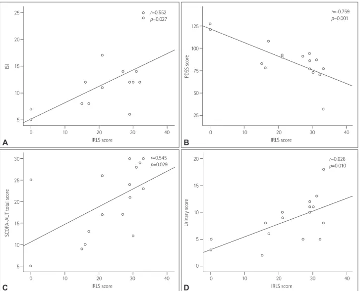
tions were found between the IRLS score and the PDSS score (r=-0.759, p=0.001), ISI (r=-0.552, p=0.027), SCOPA-AUT total score (r=0.545, p=0.029), and SCOPA-AUT urinary sub- score (r=0.626, p=0.010) (Fig. 1). There were no correlations between IRLS scores and demographic variables, motor symp- toms, and laboratory data.
Changes after dopamine-agonist treatment in PD-RLS(+)
In 16 PD-RLS(+) patients, the RLS severity decreased signifi- cantly after the administration of the dopamine agonist prami- pexole at 0.125–0.5 mg/day. In addition, RLS-related QoL, depression, anxiety, and sleep quality all improved, with signifi- cant improvements observed on the SCOPA-AUT scale (total score and urinary subscore) (Table 4). Motor symptoms did not change significantly between before and after dopamine- agonist treatment.
DISCUSSION
We observed that the frequency of RLS is higher in PD pa- tients than in the general population. In addition, we found that PD-RLS(+) patients had worse nonmotor symptoms such as depression, anxiety, sleep quality, and autonomic dysfunc-
tion than did PD-RLS(-) patients. The severity of RLS was mainly related to sleep quality and autonomic dysfunction. It was also observed that the H&Y stage was lower in RLS pre- ceding PD than in PD preceding RLS. PD-RLS(+) patients were older and had a shorter duration of RLS than did those with iRLS. In addition, RLS symptoms improved after dopa- mine-agonist administration in the PD-RLS(+) group, al- though this result was from a small sample (n=16).
The frequency of RLS in PD patients was markedly higher than in our previously published study involving the general Korean population (21.6% vs. 3.9%),4 and also higher than in a previous study of Koreans with PD (21.6% vs. 16.5%).7 The latter difference may have been due to the previous study targeting untreated PD patients while this study included both treated and untreated patients. A previous study in Western countries found that the prevalence of RLS was 21% in treated PD patients,11 while in this study it was 12.5% in the untreated group and 24.1% in the treated group. However, there was no difference in age, PD onset age, or duration of PD symp- toms between the present treated and untreated groups. These observations suggest that the use of dopaminergic drugs is associated with the development of RLS symptoms. Lee et al.12 reported that a longer duration of dopaminergic treat- ment was related to the presence of RLS in PD patients. Also, RLS severity was found to improve with decreasing dopami- nergic treatment after subthalamic nucleus deep-brain stim- ulation (STN-DBS).13 These factors suggest that overstimula- tion of dopamine receptors can cause RLS in PD patients.14
Comparisons of demographic variables revealed no signif- icant differences in age, onset age of PD, sex, or disease dura- tion between PD-RLS(+) and PD-RLS(-). These findings suggest that demographic variables are not related to RLS in PD patients. However, previous studies of PD-RLS(+) have found a younger age of PD onset and the predominance of males in Malaysia,15 an older age at the onset of PD in India,16 and a prolonged disease duration in China.10 These findings could have been due to selection bias of study samples, the smallness of the included samples, or race-specific genetic characteristics affecting the demographic variables of PD- RLS(+) patients. The H&Y stage and UPDRS-III score did not differ significantly between our PD-RLS(+) and PD-RLS(-) groups. Previous studies found RLS to be associated with PD motor symptoms, suggesting that both motor symptoms and RLS are associated with the dopaminergic system;7,10,17 that is, the decrease in dopamine production with PD progression could lead to RLS aggravation.18
A possible reason for an absence of significant differences in motor symptoms in the present patients is that they had already been treated with dopaminergic drugs and were at H&Y stage 3 or lower, and thus their motor symptoms were Table 3. Demographic variables and clinical data of PD-RLS(+) and
iRLS patients
PD-RLS(+) (n=16)
iRLS
(n=32) p
Demographic variables
Age, years 66.5±11.3 64.5±5.2 0.508
Sex
Male 4 (25.0) 8 (25.0) 1.000
Female 12 (75.0) 24 (75.0)
Age at RLS onset, years 64.8±12.2 49.7±12.0 <0.001 Duration of RLS, years 2.0±2.6 13.5±11.3 <0.001 Laboratory tests
Hemoglobin, g/dL 13.0±1.7 14.1±6.1 0.456 Serum ferritin, ng/mL 58.2±60.3 74.4±58.8 0.377 Serum iron, mg/dL 75.9±37.1 83.5±26.9 0.422 TIBC, mg/dL 329.0±47.0 321.5±34.5 0.533 Clinical data
ISI 12.1±5.91 15.8±6.9 0.067
PSQI 2.4±4.4 12.5±5.1 0.967
IRLS score 22.7±10.7 26.6±7.2 0.138
RLS-related QoL score 58.8±25.0 55.8±27.4 0.729
BDI 28.3±9.9 14.0±8.0 <0.001
Data are mean±SD or n (%) values.
BDI: Beck Depression Index, ISI: Insomnia Severity Index, IRLS: interna- tional restless legs syndrome, iRLS: idiopathic restless legs syndrome, PD:
Parkinson’s disease, PSQI: Pittsburgh Sleep Quality Index, QoL: quality of life, RLS: restless legs syndrome, TIBC: total iron-binding capacity.
You S et al.
JCN
Fig. 1. Correlation analysis between RLS severity and PD nonmotor symptom severity. RLS severity was positively correlated with ISI (A) and nega- tively correlated with PDSS score (B). The SCOPA-AUT total score (C) and urinary subscore (D) were positively correlated with RLS severity. IRLS: in- ternational restless legs syndrome, ISI: Insomnia Severity Index, PDSS: Parkinson’s Disease Sleep Scale, RLS: restless legs syndrome, SCOPA-AUT:
Scales for Outcomes in Parkinson’s Disease for Autonomic Symptoms.
not severe. While an H&Y stage of 3 or lower was not an in- clusion criterion for this study, most of the subjects were ear- ly-stage PD patients.
The scores on the scales related to nonmotor symptoms were worse in PD-RLS(+) than in PD-RLS(-), showing more depression and anxiety, lower sleep quality, and more auto- nomic dysfunction, which has also been reported previous- ly.9,16,19-20 Depression and anxiety are believed to result from decreases in dopamine and 5-hydroxytryptamine in the central nervous system contributing to poor sleep quality in PD-RLS(+). A previous study found that the levels of these two neurotransmitters in CSF were lower in PD-RLS(+) than in PD-RLS(-).10 Autonomic dysfunction in PD-RLS(+) may be a consequence of a lower cardiovascular subscore, which is believed to be associated with the A11 dopaminergic dien-
cephalospinal pathway that innervates preganglionic sympa- thetic neurons and the dorsal horn in the spinal cord.21 Cog- nitive dysfunction in nonmotor symptoms was not observed in either group, suggesting that cognitive dysfunction is more closely related to acetylcholine than dopamine, and is mainly observed in the late phases of PD.10 Therefore, cognitive dys- function was unlikely to be observed in the present study since the included patients were all in the early or middle stage of PD.
The serum iron, ferritin, and TIBC results did not differ between the PD-RLS(+) and PD-RLS(-) groups, which was similar to previous findings,7,22,23 suggesting that iron metab- olism is not affect by RLS in PD patients. However, a previ- ous study found that PD-RLS(+) patients had reduced lev- els of iron and ferritin and an increased transferrin level in
25
20
15
10
5
125
100
75
50
25
20
15
10
5
0 30
25
20
15
10
5
ISI PDSS scoreUrinary score
SCOPA-AUT total score
0 10 20 30 40 0 10 20 30 40
0 10 20 30 40 0 10 20 30 40
IRLS score IRLS score
IRLS score IRLS score
r=-0.759 p=0.001
r=0.626 p=0.010 r=0.545
p=0.029 r=0.552 p=0.027
A
C
B
D
Restless Legs Syndrome in Parkinson’s Disease Patients
JCN
CSF compared to PD-RLS(-) patients.10 This may mean that measurements of iron and iron-related proteins in the pe- ripheral system do to accurately reflect iron metabolism in the brain of PD-RLS(+) patients.
Previous studies comparing the characteristics of PD- RLS(+) and iRLS patients have shown that the onset age is younger in iRLS than in PD-RLS(+), RLS is more severe in iRLS, and that the two groups exhibit opposite seasonal pat- terns.17,24,25 In addition, a family history of RLS is more com- mon and the ferritin level is higher in iRLS than in PD-RLS (+).24 In our study the RLS onset age was younger and the RLS duration was longer in iRLS. However, serum ferritin levels and RLS severity were not significantly different between the two groups. These results are consistent with those of pre- vious studies. Depression was more severe in PD-RLS(+) than in iRLS, which suggests that depression results from PD-re- lated motor disability or other comorbidities, as well as RLS per se.26 Information on family histories and the seasonal patterns of RLS was not collected in the present study.
In the PD-RLS(+) group, depression, anxiety, QoL, sleep quality, and severity of RLS symptoms all improved after administering the dopamine agonist. Although the number of patients in the PD-RLS(+) group was small and there was no blinding for dopamine-agonist treatment and no control arm, it is possible that RLS in PD patients responds to dopa-
mine agonists regardless of the conventional dopamine treat- ment. Of course, it is difficult to clearly determine whether this result was due to the dopamine agonist itself or the effect of increasing the LED. There is still no established treatment for RLS in PD patients. One previous study administered le- vodopa, dopamine agonists (ropinirole, pramipexole, and per- golide), opioid, and clonazepam to patients with RLS after STN-DBS.27 Another study showed that RLS that had devel- oped during PD treatment improved after STN-DBS.28 There is a recent case report of safinamide being effective for RLS treatment in PD patients.29 However, more research is need- ed into how to treat RLS in PD.
Three (18.8%) of our 16 PD-RLS(+) patients had RLS be- fore the onset of PD, which is a higher prevalence than in pre- vious studies.10 Recent studies suggest that RLS is a nonmo- tor symptom that precedes PD motor symptoms.30,31 Other studies have suggested that RLS slows the onset and progres- sion of PD.32,33 This explains why the H&Y stage was signifi- cantly lower in our group with RLS preceding PD onset than in the group where RLS occurred after the PD onset.
The limitations of this study include the inclusion of a rel- atively small number of patients from a single tertiary hos- pital, its cross-sectional design, and the heterogeneity of the patients due to dopaminergic treatment. We enrolled patients from an outpatient clinic of a tertiary hospital, and so patients with advanced motion limitations could not be enrolled due to attendance difficulties. It is possible that this enrollment procedure caused selection bias. In addition, the ability to de- tect effects of dopamine-agonist treatment in the PD-RLS(+) group would have been reduced by the smallness of the sample, the short treatment period, the lack of blinding, and the ab- sence of a control arm.
In conclusion, in this study we found that the prevalence of RLS was higher in treated patients with PD than in our previously published study that involved a general Korean population, and that RLS had a negative effect on nonmotor symptoms. Depression was more severe in the PD-RLS(+) patients than in those with iRLS.
Conflicts of Interest
The authors have no potential conflicts of interest to disclose.
Acknowledgements
This research was supported by the Basic Science Research Program through the National Research Foundation of Korea (NRF) funded by the Ministry of Education (grant no. NRF-2017R1C1B5017796 and NRF- 2017R1D1A3B03031021) and the Korean Government (MSIP) (grant no.
2014R1A5A2010008).
REFERENCES
1. Allen RP, Picchietti DL, Garcia-Borreguero D, Ondo WG, Walters AS, Winkelman JW, et al. Restless legs syndrome/Willis-Ekbom disease Table 4. Comparison between pretreatment and posttreatment in
patients with RLS
Pretreatment Posttreatment p
IRLS score 23.6±9.2 12.6±6.6 <0.001
RLS-related QoL score 56.4±23.8 75.0±15.9 0.008
ISI 12.6±6.2 8.1±2.6 0.001
SSS score 4.1±1.6 2.9±1.2 0.004
ESS score 6.1±6.1 5.6±4.4 0.601
PSQI 12.1±4.1 7.9±1.2 <0.001
PDSS score 86.4±22.5 111.8±19.5 0.001
BDI 28.3±9.9 16.6±8.0 <0.001
BAI 17.2±11.9 9.6±8.1 <0.001
SCOPA-AUT, total score 18.8±8.5 15.3±6.8 0.031 Gastrointestinal subscore 5.1±3.5 4.1±3.7 0.181
Urinary subscore 8.4±4.1 6.6±3.5 0.048
Cardiovascular subscore 2.9±3.0 2.6±2.9 0.371
Sexual subscore 0.8±2.0 0.6±1.6 0.835
Pupillomotor subscore 0.8±1.2 0.8±1.1 1.000 Thermoregulatory subscore 0.9±0.9 0.9±1.6 0.751 Data are mean±SD values.
BAI: Beck Anxiety Index, BDI: Beck Depression Index, ESS: Epworth Sleepiness Scale, IRLS: international restless legs syndrome, ISI: Insom- nia Severity Index, PDSS: Parkinson’s Disease Sleep Scale, PSQI: Pitts- burgh Sleep Quality Index, QoL: quality of life, RLS: restless legs syn- drome, SCOPA-AUT: Scales for Outcomes in Parkinson’s Disease for Autonomic Dysfunction, SSS: Stanford Sleepiness Scale.
You S et al.
JCN
diagnostic criteria: updated International Restless Legs Syndrome Study Group (IRLSSG) consensus criteria--history, rationale, descrip- tion, and significance. Sleep Med 2014;15:860-873.
2. Yeh P, Walters AS, Tsuang JW. Restless legs syndrome: a comprehen- sive overview on its epidemiology, risk factors, and treatment. Sleep Breath 2012;16:987-1007.
3. Högl B, Kiechl S, Willeit J, Saletu M, Frauscher B, Seppi K, et al. Rest- less legs syndrome: a community-based study of prevalence, severity, and risk factors. Neurology 2005;64:1920-1924.
4. Cho YW, Shin WC, Yun CH, Hong SB, Kim JH, Allen RP, et al. Epide- miology of restless legs syndrome in Korean adults. Sleep 2008;31:
219-223.
5. Yang X, Liu B, Shen H, Li S, Zhao Q, An R, et al. Prevalence of restless legs syndrome in Parkinson’s disease: a systematic review and meta- analysis of observational studies. Sleep Med 2018;43:40-46.
6. Trenkwalder C, Allen R, Högl B, Paulus W, Winkelmann J. Restless legs syndrome associated with major diseases: a systematic review and new concept. Neurology 2016;86:1336-1343.
7. Shin HY, Youn J, Yoon WT, Kim JS, Cho JW. Restless legs syndrome in Korean patients with drug-naïve Parkinson’s disease: a nation-wide study. Parkinsonism Relat Disord 2013;19:355-358.
8. Pont-Sunyer C, Hotter A, Gaig C, Seppi K, Compta Y, Katzenschlager R, et al. The onset of nonmotor symptoms in Parkinson’s disease (the ONSET PD study). Mov Disord 2015;30:229-237.
9. Oh YS, Kim JS, Park IS, Song IU, Son YM, Park JW, et al. Association between nocturnal/supine hypertension and restless legs syndrome in patients with Parkinson’s disease. J Neurol Sci 2014;344:186-189.
10. Piao YS, Lian TH, Hu Y, Zuo LJ, Guo P, Yu SY, et al. Restless legs syn- drome in Parkinson disease: clinical characteristics, abnormal iron metabolism and altered neurotransmitters. Sci Rep 2017;7:10547.
11. Rana AQ, Siddiqui I, Mosabbir A, Athar A, Syed O, Jesudasan M, et al. Association of pain, Parkinson’s disease, and restless legs syndrome.
J Neurol Sci 2013;327:32-34.
12. Lee JE, Shin HW, Kim KS, Sohn YH. Factors contributing to the de- velopment of restless legs syndrome in patients with Parkinson dis- ease. Mov Disord 2009;24:579-582.
13. Chahine LM, Ahmed A, Sun Z. Effects of STN DBS for Parkinson’s disease on restless legs syndrome and other sleep-related measures.
Parkinsonism Relat Disord 2011;17:208-211.
14. Rijsman RM, Schoolderman LF, Rundervoort RS, Louter M. Restless legs syndrome in Parkinson’s disease. Parkinsonism Relat Disord 2014;
20 Suppl 1:S5-S9.
15. Azmin S, Khairul Anuar AM, Nafisah WY, Tan HJ, Raymond AA, Hanita O, et al. Restless legs syndrome and its associated risk factors in Parkinson’s disease. Parkinsons Dis 2013;2013:535613.
16. Krishnan PR, Bhatia M, Behari M. Restless legs syndrome in Parkin- son’s disease: a case-controlled study. Mov Disord 2003;18:181-185.
17. hu XY, Liu Y, Zhang XJ, Yang WH, Feng Y, Ondo WG, et al. Clinical characteristics of leg restlessness in Parkinson’s disease compared with
idiopathic restless legs syndrome. J Neurol Sci 2015;357:109-114.
18. Möller JC, Unger M, Stiasny-Kolster K, Oertel WH. Restless legs syn- drome (RLS) and Parkinson’s disease (PD)-related disorders or differ- ent entities? J Neurol Sci 2010;289:135-137.
19. Fereshtehnejad SM, Shafieesabet M, Shahidi GA, Delbari A, Lökk J.
Restless legs syndrome in patients with Parkinson’s disease: a compara- tive study on prevalence, clinical characteristics, quality of life and nu- tritional status. Acta Neurol Scand 2015;131:211-218.
20. Shneyder N, Adler CH, Hentz JG, Shill H, Caviness JN, Sabbagh MN, et al. Autonomic complaints in patients with restless legs syndrome.
Sleep Med 2013;14:1413-1416.
21. Walters AS, Rye DB. Review of the relationship of restless legs syn- drome and periodic limb movements in sleep to hypertension, heart disease, and stroke. Sleep 2009;32:589-597.
22. Gjerstad MD, Tysnes OB, Larsen JP. Increased risk of leg motor rest- lessness but not RLS in early Parkinson disease. Neurology 2011;77:
1941-1946.
23. Nomura T, Inoue Y, Miyake M, Yasui K, Nakashima K. Prevalence and clinical characteristics of restless legs syndrome in Japanese patients with Parkinson’s disease. Mov Disord 2006;21:380-384.
24. Ondo WG, Vuong KD, Jankovic J. Exploring the relationship between Parkinson disease and restless legs syndrome. Arch Neurol 2002;59:
421-424.
25. Suzuki K, Miyamoto M, Miyamoto T, Hirata K. Restless legs syn- drome and leg motor restlessness in Parkinson’s disease. Parkinsons Dis 2015;2015:490938.
26. Marsh L. Depression and Parkinson’s disease: current knowledge. Curr Neurol Neurosci Rep 2013;13:409.
27. Kedia S, Moro E, Tagliati M, Lang AE, Kumar R. Emergence of rest- less legs syndrome during subthalamic stimulation for Parkinson dis- ease. Neurology 2004;63:2410-2412.
28. Klepitskaya O, Liu Y, Sharma S, Sillau SH, Tsai J, Walters AS. Deep brain stimulation improves restless legs syndrome in patients with Par- kinson disease. Neurology 2018;91:e1013-e1021.
29. Liguori C, Mercuri NB, Stefani A, Pierantozzi M. Effective treatment of restless legs syndrome by safinamide in Parkinson’s disease patients.
Sleep Med 2018;41:113-114.
30. Wong JC, Li Y, Schwarzschild MA, Ascherio A, Gao X. Restless legs syndrome: an early clinical feature of Parkinson disease in men. Sleep 2014;37:369-372.
31. Szatmari S Jr, Bereczki D, Fornadi K, Kalantar-Zadeh K, Kovesdy CP, Molnar MZ. Association of restless legs syndrome with incident Par- kinson’s disease. Sleep 2017;40:zsw065.
32. Dragan EM, Chen Z, Ondo WG. Does idiopathic restless legs syn- drome delay onset and reduce severity of Parkinson’s disease: a pilot study. Int J Neurosci 2015;125:526-530.
33. Moccia M, Erro R, Picillo M, Santangelo G, Spina E, Allocca R, et al.
A four-year longitudinal study on restless legs syndrome in Parkinson disease. Sleep 2016;39:405-412.
![Table 1. Demographic variables of PD patients with RLS [PD-RLS (+)] and without RLS [PD-RLS(-)] PD-RLS(+) (n=16) PD-RLS(–) (n=58) p Age, years 66.5±11.3 65.3±8.5 0.637](https://thumb-ap.123doks.com/thumbv2/123dokinfo/5263465.139824/3.892.465.806.381.918/table-demographic-variables-patients-rls-rls-age-years.webp)