ISSN 2234-3806 • eISSN 2234-3814
254 www.annlabmed.org http://dx.doi.org/10.3343/alm.2015.35.2.254 Ann Lab Med 2015;35:254-256
http://dx.doi.org/10.3343/alm.2015.35.2.254
Letter to the Editor
Diagnostic Hematology
Pediatric B-cell Lymphoma, Unclassifiable, With
Intermediate Features Between Those of Diffuse Large B-cell Lymphoma and Burkitt Lymphoma: A Report of Two Cases
Shanxiang Zhang, M.D.1, David Wilson, M.D.2, and Magdalena Czader, M.D.1
Department of Pathology and Laboratory Medicine1, Indiana University; AmeriPath2, Indianapolis, IN, USA
B-cell lymphoma, unclassifiable, with intermediate features be- tween those of diffuse large B-cell lymphoma (DLBCL) and Burkitt lymphoma (BL; intermediate BL/DLBCL) commonly ex- hibits a morphology similar to that of typical BL and an atypical immunophenotype such as diffuse and moderate-to-strong BCL2 positivity and/or a Ki67 proliferation index <90%. Alterna- tively, the tumor cells exhibit greater variations in nuclear size and contouring than would be considered acceptable for BL [1, 2]. Intermediate BL/DLBCL is most commonly observed in adults [3-5]. Among pediatric populations, to date, the English literature only includes reports of one case of intermediate BL/DLBCL in a 2-yr-old Korean boy and eight cases in Chinese children [6, 7].
Here, we report two additional cases that represent the first re- port from a western population.
Case 1
A 15-yr-old girl with sudden-onset abdominal pain and emesis was found to have a small bowel obstruction and intussuscep- tion during a computed tomography (CT) study. An urgent ex- ploratory laparotomy revealed a mass comprising mildly to mod- erately pleomorphic lymphoid cells that were mostly medium-
sized with rare large cells and slightly irregular nuclear contours;
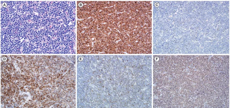
these cells primarily contained 2-3 inconspicuous nucleoli or, occasionally, single prominent nucleoli. Mitotic figures were fre- quent. Focally, tingible body macrophages imparted a “starry sky” appearance. The atypical lymphoid cells were positive for CD20, CD10, BCL-6 (focal, weak), MUM-1, and BCL-2 and neg- ative for terminal deoxynucleotidyl transferase (TdT). The prolif- erative index, as indicated by Ki-67 staining, was high (>90%;
Fig. 1). An Epstein-Barr virus (EBV) study with late membrane protein 1 (LMP1) immunohistochemical stain (IHC) and in situ hybridization for EBV-encoded RNAs (EBER), flow cytometric analysis, and karyotyping were not performed. FISH analysis re- vealed MYC rearrangement but no BCL2 or BCL6 rearrangement or copy number changes. Three of the 16 resected lymph nodes were also positive for intermediate BL/DLBCL. The patient under- went chemotherapy with cyclophosphamide, vincristine, predni- sone, doxorubicin, and methotrexate. She remained in complete remission 11 months after completing chemotherapy.
Case 2
A 4-yr-old boy with a 2-week history of abdominal distension and
Received: January 8, 2014
Revision received: February 15, 2014 Accepted: November 25, 2014 Corresponding author: Shanxiang Zhang Department of Pathology and Laboratory Medicine, Indiana University, 350 West 11th Street, Room 5042, Indianapolis, IN 46202, USA
Tel: +1-317-491-6175, Fax: +1-317-491-6114, E-mail: [email protected]
© The Korean Society for Laboratory Medicine.
This is an Open Access article distributed under the terms of the Creative Commons Attribution Non-Commercial License (http://creativecommons.org/licenses/by-nc/3.0) which permits unrestricted non-commercial use, distribution, and reproduction in any medium, provided the original work is properly cited.
Zhang S, et al.
Intermediate pediatric B-cell lymphoma
http://dx.doi.org/10.3343/alm.2015.35.2.254 www.annlabmed.org 255
diarrhea was found to have diffuse bowel wall thickening on CT.
A transrectal biopsy revealed a diffuse infiltrate of predominantly medium-sized with rare large atypical lymphoid cells; these cells had slightly irregular nuclear contours, vesicular chromatin, and mostly inconspicuous nucleoli. Apoptotic bodies and mitotic fig- ures were frequently observed. The atypical lymphoid cells were positive for CD20, PAX-5, CD10, BCL-6, and BCL-2 and exhib- ited a high proliferative index, as indicated by Ki-67 staining (>
95%). MUM-1 staining was negative. The EBV study was nega- tive according to LMP1 IHC and EBER in situ hybridization. Con- current flow cytometric analysis revealed a monoclonal B-cell
population with CD19, CD20, CD22, CD10, and surface kappa light-chain expression. The monoclonal B cells were negative for CD5, CD23, CD34, and TdT. A cytogenetic analysis (karyotyping) was not performed. FISH analysis with a MYC dual-color break apart probe revealed MYC rearrangement. There was no evi- dence of BCL2/IGH fusion, BCL6 rearrangement, or copy num- ber changes in BCL2 or BCL6. The bone marrow and central nervous system were not involved (stage III). This patient re- ceived the same chemotherapy regimen as patient 1 and re- mained in remission for 12 months, as per the last follow-up.
We conducted a systematic review of the pediatric high-grade Fig. 1. Morphologic (A, Hematoxylin and eosin staining, ×50) and immunophenotypic (B-F, ×20) features of B-cell lymphoma, unclassifi- able, with intermediate features between DLBCL and Burkitt lymphoma. (B) CD20, (C) BCL-6, (D) BCL-2, (E) MUM-1, and (F) Ki-67.
A
D
B
E
C
F
Table 1. Comparison of the current cases with previously reported intermediate BL/DLBCL cases in English literature Case No. Sex
(N)
Age (yr)
Tissue / Location
(Case No.) Immunophenotype* FISH for gene rearrangement*
EBER* Karyotype c-MYC BCL2 BCL6 Follow-up Ahn et al. [6] 1 M 2 Femur neck (1) CD20, CD10, BCL6, MUM1 (not
mentioned), BCL2, Ki-67 (-90%)
0/1 Complex 1/1 0/1 0/1 died at 5 months Lu et al. [7] 8 M (6),
F (2) 4-13 Abdomen/
intestine (7), gingiva (1)
CD20 (8/8), CD10 (6/8), BCL6 (5/8), MUM1 (5/8), BCL2 (4/8), Ki-67
(60-95%)
6/6 ND 4/8 0/8 0/8† NA
Current study 2 M (1),
F (1) 4, 15 Intestine (2) CD20 (2/2), CD10 (2/2), BCL6 (1 strong, 1 weak), MUM1 (1/2),
BCL2 (2/2), Ki-67 (90%, 95%)
1/1 ND 2/2 0/2 0/2 remission at
11, 12 months
*positive case number/tested case number; †2 out of 8 cases showed 3 copies of BCL6.
Abbreviations: DLBCL, diffuse large B-cell lymphoma; BL, Burkitt lymphoma; M, male; F, female; ND, not done; NA, not available; EBER, in situ hybridization for Epstein-Barr virus-encoded RNAs.
Zhang S, et al.
Intermediate pediatric B-cell lymphoma
256 www.annlabmed.org http://dx.doi.org/10.3343/alm.2015.35.2.254 mature B-cell lymphoma cases in our archives from 1988 to
2012 and identified 2 cases of intermediate BL/DLBCL. Both cases exhibited histologic morphology compatible with BL but with moderate-to-strong BCL-2 expression. Weak BCL-2 expres- sion might be observed in BL [2]. In our experience, BCL-2 staining has yielded consistently negative or weakly positive (few cells) results in cases of BL. In addition, Case 1 exhibited focal and weak BCL6 expression and moderate-to-strong MUM-1 ex- pression, which would be unusual for a diagnosis of BL [2]. The focal and weak BCL6 staining in Case 2 was not likely caused by poor tissue preservation and/or the stain itself, as the staining of samples from the same tissue block for other nuclear mark- ers (MUM-1 and Ki-67) was successful. To the best of our knowl- edge, these two cases represent the first report of pediatric in- termediate BL/DLBCL in a western population. A comparison of the current cases with previously reported pediatric intermediate BL/DLBCL cases in Asian populations is summarized in Table 1.
The current treatments for pediatric BL, DLBCL, and interme- diate BL/DLBCL are similar [8]. However, treatment for patients aged 15-20 yr has been controversial because adolescent DLBCL patients fare better when treated with more aggressive regimens, compared with those who receive a cyclophospha- mide, doxorubicin, vincristine, prednisone (CHOP)-like regimen [9, 10]. Both of our patients were treated with a BL chemother- apeutic regimen. It will be interesting to observe the responses of our patients, particularly the 15-yr-old female patient, during long-term follow-up. With the development of individualized tar- geted therapies, the recognition of pediatric intermediate BL/
DLBCL might become more clinically relevant.
Authors’ Disclosures of Potential Conflicts of Interest
No potential conflicts of interest relevant to this article were re- ported.
REFERENCES
1. Carbone A, Gloghini A, Aiello A, Testi A, Cabras A. B-cell lymphomas with features intermediate between distinct pathologic entities. From pathogenesis to pathology. Hum Pathol 2010;41:621-31.
2. Kluin PM, Harris NL, Stein H, Leoncini L, Raphael M, Campo E, et al.
B-cell lymphoma, unclassifiable, with features intermediate between diffuse large B-cell lymphoma and Burkitt lymphoma. In: Swerdlow SH, Harris NL, Jaffe ES, Pileri SA, Stein H, Thiele J, Vardiman JW, eds. WHO classification of tumours of haematopoietic and lymphoid tissues. Lyon:
IARC Press, 2008:265-6.
3. Liang X, Greffe B, Cook B, Giller R, Graham DK, McGranahan AN, et al.
Gray zone lymphomas in pediatric patients. Pediatr Dev Pathol 2011;
14:57-63.
4. Lin P, Dickason TJ, Fayad LE, Lennon PA, Hu P, Garcia M, et al. Prog- nostic value of MYC rearrangement in cases of B-cell lymphoma, un- classifiable, with features intermediate between diffuse large B-cell lym- phoma and Burkitt lymphoma. Cancer 2012;118:1566-73.
5. Salaverria I and Siebert R. The gray zone between Burkitt’s lymphoma and diffuse large B-cell lymphoma from a genetics perspective. J Clin Oncol 2011;29:1835-43.
6. Ahn JY, Seo YH, Park PW, Kim KH, Park MJ, Jeong JH, et al. A case of B-cell lymphoma, unclassifiable, with features intermediate between diffuse large B-cell lymphoma and Burkitt lymphoma in a Korean child.
Ann Lab Med 2012;32:162-6.
7. Lu B, Zhou C, Yang W, Huang H, Gao Z, He Y, et al. Morphological, im- munophenotypic and molecular characterization of mature aggressive B-cell lymphomas in Chinese pediatric patients. Leuk Lymphoma 2011;
52:2356-64.
8. Reiter A, Schrappe M, Parwaresch R, Henze G, Müller-Weihrich S, Sau- ter S, et al. Non-Hodgkin’s lymphomas of childhood and adolescence:
results of a treatment stratified for biologic subtypes and stage--a report of the Berlin-Frankfurt-Münster Group. J Clin Oncol 1995;13:359-72.
9. Cairo MS, Gerrard M, Sposto R, Auperin A, Pinkerton CR, Michon J, et al. Results of a randomized international study of high-risk central ner- vous system B non-Hodgkin lymphoma and B acute lymphoblastic leu- kemia in children and adolescents. Blood 2007;109:2736-43.
10. Patte C, Auperin A, Gerrad M, Michon J, Pinkerton R, Sposto R, et al.
Results of the randomized international FAB/LMB96 trial for intermedi- ate risk B-cell non-Hodgkin lymphoma in children and adolescents: it is possible to reduce treatment for the early responding patients. Blood 2007;109:2773-80.