Received August 25, 2015, Revised November 18, 2015, Accepted for publication December 14, 2015
Corresponding author: You Won Choi, Department of Dermatology, Ewha Womans University School of Medicine, 1071 Anyangcheon-ro, Yang- cheon-gu, Seoul 07985, Korea. Tel: 82-2-2650-5159, Fax: 82-2-2652-6925, E-mail: uwon313@ewha.ac.kr
This is an Open Access article distributed under the terms of the Creative Commons Attribution Non-Commercial License (http://creativecommons.
org/licenses/by-nc/4.0) which permits unrestricted non-commercial use, distribution, and reproduction in any medium, provided the original work is properly cited.
Copyright © The Korean Dermatological Association and The Korean Society for Investigative Dermatology
Ann Dermatol Vol. 28, No. 5, 2016 http://dx.doi.org/10.5021/ad.2016.28.5.548
ORIGINAL ARTICLE
Expression of Phosphatase and Tensin Homologue, phospho-Akt, and p53 in Acral Benign and Malignant Melanocytic Neoplasms (Benign Nevi, Dysplastic Nevi, and Acral Melanomas)
So Min Lyu, Ju Yeon Wu, Ji Yeon Byun, Hae Young Choi, Sang Hee Park1, You Won Choi
Departments of Dermatology and 1Pathology, Ewha Womans University School of Medicine, Seoul, Korea
Background: The role of the phosphatidylinositol-3 kinase signaling pathway in the development of acral melanoma has recently gained evidence. Phosphatase and tensin homo- logue (PTEN), one of the key molecules in the pathway, acts as a tumor suppressor through either an Akt-dependent or Akt-independent pathway. Akt accelerates degradation of p53. Objective: We assessed the expression of PTEN, phos- pho-Akt (p-Akt), and p53 by immunohistochemistry in be- nign acral nevi, acral dysplastic nevi, and acral melanomas in the radial growth phase and with a vertical growth component. Methods: Ten specimens in each group were included. Paraffin-embedded specimens were immunostained with antibodies for PTEN, p-Akt, and p53. We scored both the staining intensity and the proportion of positive cells. The final score was calculated by multiplying the intensity score by the proportion score. Results: All specimens of benign ac- ral nevi except one showed some degree of PTEN-negative cells. The numbers of p-Akt and p53-positive cells were high- er in acral dysplastic nevi and melanoma than in benign nevi.
P-Akt scores were 1.7, 1.8, 2.6, and 4.4, and p53 scores were 2.0, 2.1, 3.8, and 4.1 in each group. PTEN and p-Akt scores
in advanced acral melanoma were higher than in the other neoplasms. Conclusion: The expression of PTEN was de- creased and the expression of p-Akt was increased in acral melanoma, especially in advanced cases. The PTEN-in- duced pathway appears to affect the late stage of melano- magenesis. Altered expression of p-Akt is thought to be due to secondary changes following the loss of PTEN. (Ann Dermatol 28(5) 548∼554, 2016)
-Keywords-
Acral, Akt, Melanocytic, p53, PTEN
INTRODUCTION
Both environmental and genetic factors contribute to the pathogenesis of malignant melanoma (MM). A few of the representative genes mutated in MM include B-raf proto- oncogene, serine/threonine kinase (BRAF), neuroblastoma ras viral (V-ras) oncogene homolog (NRAS), CDK4, and p16, which are part of the mitogen-activated protein kin- ases (MAPK) pathway known to regulate cellular pro- liferation. There is increasing evidence for a role of the phosphoinositide 3-kinase pathway in the pathogenesis of MM, and it appears that different subtypes of MM show distinct patterns of genetic mutations1. Alterations in BRAF and NRAS are usually found in non-chronically sun-dam- aged skin melanomas, which frequently occur in Western countries, while melanomas associated with alterations in KIT, cyclin D1, and CDK4 are most frequently found in Eastern countries2. However, it remains unclear whether there exists a relationship between the involved genes and why MM occurs more frequently in Asians.
Phosphatase and tensin homologue (PTEN) is a tumor sup- pressor gene located on chromosome 10q23.3 that enc- odes the protein PTEN, which dephosphorylates lipids and proteins, therefore inhibiting the PI3K pathway. PTEN regulates cell growth and survival through Akt-dependent and -independent pathways; formation of tumor cells is as- sociated with alterations of PTEN acting in the Akt-in- dependent pathway, although Akt may be involved in- directly as well3. Akt is a protein kinase B that functions as a signaling molecule and receives a phosphate group from PIP3; it phosphorylates proteins such as Bad, caspase-9, and mdm2, and also accelerates degradation of p53, a tu- mor suppressor4. PTEN/PI3K/Akt also promotes p53 trans- lation and protein stability, suggesting that additional mechanisms may be involved in the Akt-mediated regu- lation of p53 in tumors5.
We have analyzed the degree of expression of PTEN, Akt, and p53 in different types of acral melanocytic lesions in- cluding benign acral nevi, acral dysplastic nevi, acral mel- anoma in the radial growth phase and acral melanoma with a vertical growth component, and our results suggest a possible role of the abovementioned proteins in the for- mation of acral melanoma.
MATERIALS AND METHODS
Materials
A total of 40 specimens were obtained from 40 patients who were clinicopathologically diagnosed with different types of acral melanocytic lesions from 2005 to 2013 at Ewha Womans University Mokdong Hospital (Seoul, Korea). This study was approved by the Ethics Committee of Ewha Womans University Mokdong Hospital (IRB no.
2014-08-016-003). The 40 specimens included 10 of each of following disorders: benign acral nevi, acral dysplastic nevi, acral melanoma in the radial growth phase, and ac- ral melanoma with vertical growth.
The disorders are defined as follows: Benign acral nevi consist of nests of nevoid to small epithelioid melanocytes predominating near the dermal-epidermal junction. These nevi have no severe uniform atypia or mitotic activity or continuous proliferation of single cells between the nests.
Dysplastic nevi have elongated rete ridges, randomly dis- tributed junctional nests, a clearly visible shoulder, and fi- broplasia in the dermis. They also contatin nests of le- sional cells that tend to be oriented parallel to the surface and to bridge the adjacent elongated rete. Nevus cells are present, and a minority have slightly irregular nuclei.
Acral lentiginous melanoma in the radial growth phase consists of irregular epidermal hyperplasia and scattered, basally located, atypical melanocytes. In the vertical
growth phase, in addition to the radial growth phase tu- mor, there are multiple nests in the dermis.
Methods
1) Immunohistochemical staining
Three 3∼4 μm deparaffinized and hydrated, paraffin em- bedded tissue sections were prepared per specimen.
Sections were treated with 3% H2O2 for 10 minutes and then washed with tap water and phosphate buffer solution (PBS) for 5 minutes each to prevent endogenous perox- idase activity. Heat-induced antigen retrieval was per- formed by treating the sections with 100oC, pH 6 citrate buffer solution for 20 minutes. Sections were then in- cubated for 30 minutes with primary antibodies against PTEN (Santa Cruz Biotechnology, Santa Cruz, CA, USA;
1:400), phosphor-Akt (p-Akt, the active form; Cell Signaling Technology, Danvers, MA, USA; 1:50), and p53 (CM-1; Novocastra Claremont Place, Newcastle upon Tyne, UK; 1:500) (all polyclonal from rabbit). Sections were then washed with PBS for five minutes, incubated in the secondary antibody (mouse anti-rabbit IgG; Dako, Carpinteria, CA, USA; 1:100) for 30 minutes, washed with PBS for five minutes, and treated with streptavidin-perox- idase conjugate for 30 minutes, followed by PBS for an- other 10 minutes. Sections were stained by treating with 3,3’-diaminobenzidine tetrahydrochloride for 10 minutes followed by counterstaining with Mayer’s hematoxylin.
Finally, they were mounted with water-based mounting medium and observed with an optical microscope.
2) Assessment of immunohistochemical staining Two dermatologists observed 100 randomly chosen im- munohistochemically stained nevus/tumor slides under an optical microscope (×400). Three parameters were con- sidered: (1) number of PTEN, p-Akt, and p53 stain-positive cells in each group of 10, (2) stain intensity of the pos- itively stained cells, (3) proportion of positively stained cells. Loss of PTEN expression was also considered.
Staining intensity was scored by agreement of the two der- matologists, and scores of the proportion of positively stained samples and PTEN loss were averaged.
(1) PTEN: Staining for PTEN was observed only in the nu- cleus because although PTEN proteins in both the nucleus and cytoplasm can be stained, those in the cytoplasm are hard to distinguish from melanin. The staining intensity was somewhat constant, so it was not included in the cal- culation of total score. Out of 100 nevi/tumor cells/slide, the proportion of negatively stained cells in each group was calculated. One point was given if the total PTEN-loss
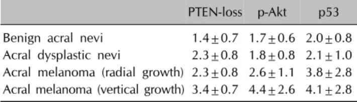
Table 1. Final staining scores of PTEN-loss, p-Akt, and p53 in benign acral nevi, acral dysplastic nevi, and acral melanomas
PTEN-loss p-Akt p53 Benign acral nevi 1.4±0.7 1.7±0.6 2.0±0.8 Acral dysplastic nevi 2.3±0.8 1.8±0.8 2.1±1.0 Acral melanoma (radial growth) 2.3±0.8 2.6±1.1 3.8±2.8 Acral melanoma (vertical growth) 3.4±0.7 4.4±2.6 4.1±2.8 Values are presented as mean±standard deviation. PTEN:
phosphatase and tensin homologue, p-Akt: phospho-Akt. Final score=proportion score×intensity score.
was less than 10%, 2 points for a reduction of 10%∼
25%, 3 points for 25%∼50%, and 4 points for 50% or more.
(2) p-Akt: p-Akt resides in the cytoplasm; therefore, to distinguish it from melanin pigments, stained nevi/tumor cells in the nest/slide, where less melanin is found, were chosen if possible. Stain intensity and the proportion of positively stained cells were measured. The scoring system used for stain intensity was 0 for no stain, 1 for lightly stained, and 2 for strongly stained, and for scoring of the proportion of positively stained cells, 0 for no stain, 1 point for 10% of the cells, 2 points for 10%∼25%, 3 for 25%∼50%, and 4 points for more than 50%. Total Akt score was calculated by multiplying the scores for stain intensity and the proportion of positively stained cells.
(3) p53: As p53 resides in the nucleus, the proportion and intensity of cells stained brown in the nucleus was measured. The intensity scoring system was 0 points for no staining, 1 point for lightly stained, and 2 points for strongly stained. Points assigned for the proportion of positively stained cells were 0 for less than 1%, 1 for 1%∼
5%, 2 for 5%∼15%, 3 for 15%∼35%, and 4 for over 35%. The total p53 score was calculated by multiplying the scores for the proportion and the intensity of stain.
3) Statistical analysis
The average, variance, and standard deviations of total scores for PTEN-loss, p-Akt, and p53 of the four groups were calculated, and ANOVA and Tukey’s test were per- formed to evaluate statistical significance with significance level of p<0.05.
RESULTS
Patient information
The average age of the 40 patients was 49.3 years, with a sex ratio of 1:3, with more female patients. The average ages 36.5 years in the benign acral nevi group, 45.7 for acral dysplastic nevi, 56.4 for acral melanoma in the radi- al growth phase, and 58.6 for acral melanoma with verti- cal growth. Benign acral nevi were found on the sole (n=8), toe (n=1), and finger (n=1), while acral dysplastic nevi were localized on the sole (n=6), palm (n=2), toe (n=1), and finger (n=1). Acral melanoma in the radial growth phase was found on the sole (n=5), toe (n=2), top of the foot (n=1), palm (n=1), and finger (n=1), and acral melanoma with vertical growth on the sole (n=4), toe (n=2), heel (n=1), finger (n=2), and near the fingernail (n=1).
PTEN, p-Akt, and p53 staining
1) PTEN
Positive staining for PTEN was 9 benign acral nevi (90%) and all 10 of the specimens (100%) in the other three groups.
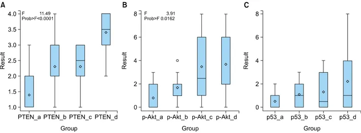
A PTEN-loss score of 1.4±0.7 was calculated for benign acral nevi, 2.3±0.8 for acral dysplastic nevi, 2.3±0.8 for acral melanoma in the radial growth phase, and 3.4±0.7 for acral melanoma with vertical growth (Table 1). PTEN-loss significantly differed between samples of ac- ral melanoma with vertical growth component and the other neoplasm (Fig. 1A) (p<0.0001).
Nevus/tumor cells lacking PTEN in their nuclei showed random arrangements, while some acral melanomas with vertical growth tended to be clustered (Fig. 2N).
2) p-Akt
Three benign acral nevi specimens were p-Akt-positive (30%), slightly higher than the proportions in other neo- plasms: 6 acral dysplastic nevi (60%), 5 acral melanomas in the radial growth phase (50%), and 5 acral melanomas with vertical growth (50%).
Evaluating p-Akt-positive tissues separately, the average stain intensity score for each group was 1.3, 1.0, 1.2, and 1.8, with acral melanoma with vertical growth scoring the highest. The average score for the proportion of positively stained cells for the melanoma was higher than that of the nevi, with the neoplasm types scoring 1.3, 1.8, 2.6, and 2.6. The total p-Akt score for benign acral nevi was 1.7±0.6, for acral dysplastic nevi 1.8±0.8, for acral mela- noma in the radial growth phase 2.6±1.1, and for acral melanoma with vertical growth 4.4±2.6 (Table 1, Fig.
1B). The difference in p-Akt scores between the acral mel- anoma with vertical growth and benign acral nevi was sig- nificant (p=0.0162).
Cells with positive staining for cytoplasmic p-Akt were ir- regularly scattered with pattern of nests formed by pos-
Fig. 1. Comparison of the final staining scores between acral melanocytic lesions. (A) Phosphatase and tensin homologue (PTEN) (PTEN loss score), (B) phospho-Akt (p-Akt), (C) p53. a: benign acral nevi group, b: acral dysplastic nevi group, c: acral melanoma group in radial growth phase, d: acral melanoma group with a vertical growth. p<0.05.
Fig. 2. Representative figures of immunohistochemical staining for phosphatase and tensin homologue (PTEN) (B, F, J, N: ×400), phospho-Akt (p-Akt) (C, G, K, O, ×400), and p53 protein (D, H, L, P, ×400) in benign acral nevus (A∼D), acral dysplastic nevus (E∼H), acral melanoma in radial growth phase (I∼L), acral melanoma with vertical growth (M∼P). Patchy loss of nuclear PTEN is remarkable in advanced acral melanoma specimen (J, arrows: nuclear PTEN-lost cells). A nest of cytoplasmic p-Akt-positive tumor cells are noted in advanced acral melanoma. Both staining intensity and proportion of the p53-positive cells are higher in acral melanoma than two benign nevi groups. A, E, I, and M were stained with H&E (×100). Arrows: F, nuclear PTEN-lost cells; H, p53-positive cells; K, p-Akt-positive cells.
itively stained cells in acral melanomas with vertical growth (Fig. 2O).
3) p53
Tissue counts for p53-positivity were 4 for benign acral nevi (40%), 8 for acral dysplastic nevi (80%), 9 for acral melanoma in the radial growth phase (90%), and 9 for ac- ral melanoma with vertical growth (90%).
Similar to p-Akt staining, the other three neoplasms had a higher proportion of positively stained cells than benign acral nevi. The average score for stain intensity for each group was 1.2, 1.1, 1.6, and 1.6 with the two melanoma groups more strongly stained. The calculated proportion score for positively stained cells was 1.8, 1.9, 2.2, and 2.4 for each group; the score for acral melanoma with vertical growth was highest. The total p53 score for benign acral nevi was 2.0±0.8, acral dysplastic nevi 2.1±1.0, acral melanoma in the radial growth phase 3.8±2.8, and acral melanoma with vertical growth 4.1±2.8 (Table 1, Fig.
1C). The average p53 scores for benign acral nevi and ac- ral dysplastic nevi were similar, while those of the two melanomas were higher, especially acral melanoma with vertical growth. However, analysis through Tukey’s test showed no significant difference between the total scores of the four groups (p=0.2182).
Cells with a nucleus positive for p53 mostly showed ran- dom arrangements, while some clusters were observed in a few melanoma tissues (Fig. 2P).
DISCUSSION
The number of patients with melnoma in Korea has been increasing. According to the Ministry of Health, Welfare and Family Affairs, 276 patients were diagnosed with mel- anoma in 2002, 351 in 2006 and 428 in 2010, showing an increase of 70∼80 patients for every 4 year6. Acral len- tiginous melanoma is the most commonly occurring type of melanoma in Eastern countries, accounting for about 60% of total melanoma patients in Korea, while it occurs in only about 2%∼8% of patients in Western countries7. Although the incidence rate as well as social interest of melanoma are increasing in Korea, the pathophysiology of the most commonly occurring type, acral lentiginous mel- anoma, is still not well known compared to other subtypes.
The most investigated gene in the study of MM patho- physiology is BRAF, a serine/threonine kinase in the MAPK pathway, and around 60%∼70% of total melano- ma patients carry the BRAF mutation8. The second most commonly mutated gene, found in about 15%∼20% of melanoma patients, is NRAS, which is associated with
both the MAPK and PI3K pathways9. However, the muta- tion rate is dependent on MM subtype, specifically on whether or not chronic sun-damage is present. Thus Bastian et al.10 have suggested a new classification system of melanoma, defining acral melanoma as melanoma that occurs in the acral regions regardless of prior classifications.
Subsequently, Zebary et al.11 have found that of all genetic mutations known for acral melanoma, BRAF accounts for around 14.8%, NRAS 13.3%, and KIT 13.4%, while ge- netic mutations in cyclin D1 and CDK4 are more frequent. These authors pointed out the importance of the PI3K signaling pathway.
PTEN is the most commonly researched protein in the PI3K/Akt pathway that is associated with melanoma. As mentioned earlier, PTEN is associated with both Akt-de- pendent and Akt-independent pathways in tumorigenesis3. First, PTEN blocks the pathway leading to PI3K, phospha- tidylinositol (3,4,5)-trisphosphate (PIP3), and Akt, through which it inhibits cellular apoptosis and expression of cy- clin D and, through mdm2, suppresses the degradation of p53 to stop tumor formation3. Also, PTEN Akt-indepen- dently interacts with p53 and cyclin D, and stabilizes p53, whose half-life is short in the nucleus, and promotes tran- scription of p5312,13. Furthermore, p53 binds the PTEN promoter region and activates translation of PTEN, form- ing a positive feedback loop14. PTEN-dependent tumori- genesis is thought to be associated with the Akt-in- dependent pathway, but the exact players in tumori- genesis are still unclear. However, it appears that Akt does more than just accumulate as a result of inhibited PTEN expression3.
In most tumors, a PTEN mutation usually appears as a loss of allele or point mutation, and is more frequently found in progressed or metastatic tumors15. Shull et al.16 reported that in 454 metastasized melanomas, around 22% con- tained inactive PTEN mutation; they therefore concluded that it is the predominant mutation. However, in another study investigating 88 primary acral lentiginous melanoma patients, only one patient (4%) was found to have a PTEN mutation11. According to our immunohistochemical stain- ing, except for one benign acral nevi sample, all others (39 samples) expressed PTEN. Moreover, a positive corre- lation between malignancy and the proportion of PTEN negative cells was observed, with the total PTEN-loss score for each group at 1.4, 2.3, 2.3, and 3.4. In partic- ular, the proportion of PTEN-negative cells was higher in the more advanced form of acral melanoma. In a previous study by Whiteman et al.17, they observed 84 out of 94 primary melanoma tissue samples lacked PTEN protein and 8 had normal expression. In an independent study by Tsao et al.18, 16 of 35 benign nevi (46%), 1 of 4 dysplastic
nevi (25%), and 26 of 30 melanomas (87%) showed PTEN loss in the nucleus. Additionally, melanoma types with greater PTEN loss have been reported to be associated with higher mitotic rate and tumor cell migration rate as well as worse prognosis17. In summary, PTEN mutation is thought to have an impact on melanoma progression and migration. In parallel with this hypothesis, our results show that acral melanomas with vertical growth had great- er PTEN loss compared to the other skin neoplasms, showing the possibility that this hypothesis may apply for acral melanoma.
Akt is a protein kinase B that functions as a signaling mol- ecule that receives a phosphate group from PIP3 and phosphorylates proteins such as BAD, caspase-9, and mdm2 and also accelerates degradation of p53, a tumor suppressor4. PTEN/PI3K/Akt also promotes p53 translation and protein stability, suggesting that additional mecha- nisms may be involved in Akt-mediated regulation of p53 in tumors5. Some reports have found Akt gene mutationS in MM samples, but the mutation rate was very low19. Therefore, altered expression of Akt protein in melanoma appears to be due to upstream mutations in PTEN or other molecules in the Akt pathway. In our experiment, overall p-Akt staining was low. The total p-Akt score for each group was 1.7, 1.8, 2.6, and 4.4, and as with the PTEN re- duction score, acral melanoma with vertical growth scor- ed the highest for both stain intensity and the proportion of positive cells. However, scores were only based on the positively p-Akt stained samples, so the sample number was too small for a significant ANOVA or Tukey’s test to be performed. An analysis of p-Akt stains by Dai et al.20, in nevus/tumor cells from different body parts found that 17% of benign nevi, 43% of dysplastic nevi, 49% of pri- mary melanoma, and 77% of metastasized melanoma were positively stained, and in parallel with our results, stain intensity was highest for metastasized melanoma.
They also suggested that higher p-Akt expression was asso- ciated with stronger local invasion and more advanced progression accompanied by worse patient prognosis. In contrast, Shanesmith et al.21 have reported the opposite re- sult by comparing benign nevi, including dysplastic nevi and Spitz nevi, against MM. They found p-Akt in 75% of benign nevi and 43% of the MM group, with weaker stain intensity in the MM group. We have obtained results sim- ilar to those of the former studies, suggesting that p-Akt ex- pression changes after the growth of melanoma but further investigations with larger sample sizes will be necessary.
The mutation rate of p53 in acral melanoma is very low compared to other types of melanoma, but nuclear p53 expression is known to be more frequent and intense in melanoma than other tissues22. The half-life of wild type
p53 is very short, only a few minutes, while the half-life of mutated p53 is much longer, up to several hours, which enables detection of mutated p53 through immunohisto- chemical staining23. Increased expression of mutated p53 in melanoma is more likely to be due to secondary changes resulting from abnormal signaling upstream of p53 rather than mutation of p53 the gene itself. Our data show that expression of p53 increased with malignancy, with total p53 scores for each group calculated to be 2.0, 2.1, 3.9, and 4.4, with the two melanoma groups scoring higher than the nevus groups. However, unlike PTEN and p-Akt staining, the degree of expression of p53 was also high in acral melanoma in the radial growth phase, sug- gesting that expression of p53 may also be associated with other signaling pathways besides PTEN and Akt. Mc- Gregor et al.24 were the first to report that p53 expression in metastasized melanoma is higher than that of primary melanoma, and subsequently Kanoko et al.25 have quanti- fied this and reported no p53 positivity in benign and dys- plastic nevi, while about 27%∼29% of primary melano- mas and 57%∼64% of metastasized melanomas were positive for p53. Therefore, change in expression of p53 protein may be a subsequent event in the development of melanoma.
In summary, we have performed immunohistochemical staining of benign and malignant acral melanocytic lesions against proteins involved in PI3K such as PTEN, p-Akt, and p53. PTEN expression was reduced, while p-Akt was increased in acral melanomas with vertical growth. Further, melanomas with mutated p53 showed greater stain in- tensity and a higher proportion of positively stained cells;
however, these differences were not statistically significant.
Therefore, we suggest that abnormal PTEN promotes tu- mor formation in the later phase of melanoma development.
Altered expression of p-Akt is thought to be secondary fol- lowing the loss of PTEN. This study is the first comparing the degree of expression of PTEN-related proteins of acral melanocytic lesions, and further investigations on muta- tions and expression of other proteins in different path- ways will shed light on the pathophysiology of acral mela- nocytic tumors. However, our study had certain limitations.
We had a small number of samples. In addition, con- comitant genomic analysis to corroborate immunohisto- chemical findings should be performed in future.
Moreover, with accumulating studies on the effectiveness of PI3K pathway blockers in melanoma resistant to BRAF-targeted therapy, drugs that target PI3K/Akt may be good candidates for future melanoma treatment26,27.
REFERENCES
1. Curtin JA, Fridlyand J, Kageshita T, Patel HN, Busam KJ, Kutzner H, et al. Distinct sets of genetic alterations in mela- noma. N Engl J Med 2005;353:2135-2147.
2. Broekaert SM, Roy R, Okamoto I, van den Oord J, Bauer J, Garbe C, et al. Genetic and morphologic features for mela- noma classification. Pigment Cell Melanoma Res 2010;
23:763-770.
3. Blanco-Aparicio C, Renner O, Leal JF, Carnero A. PTEN, more than the AKT pathway. Carcinogenesis 2007;28:
1379-1386.
4. Saraiva VS, Caissie AL, Segal L, Edelstein C, Burnier MN Jr.
Immunohistochemical expression of phospho-Akt in uveal melanoma. Melanoma Res 2005;15:245-250.
5. Abraham AG, O'Neill E. PI3K/Akt-mediated regulation of p53 in cancer. Biochem Soc Trans 2014;42:798-803.
6. Ministry of Health and Welfare. Annual report of cancer statistics in Korea. Sejong: Ministry of Health and Welfare, 2002, 2006, 2010.
7. Kim SY, Kim JS, Park K, Oh CW, Shin JH, Kang HY, et al.
Epidermal and adnexal nevi and tumors. In: Textbook of dermatology. 6th ed. Seoul: Daehan Medical Publishing Co., 2014:804.
8. Davies H, Bignell GR, Cox C, Stephens P, Edkins S, Clegg S, et al. Mutations of the BRAF gene in human cancer.
Nature 2002;417:949-954.
9. Goydos JS, Mann B, Kim HJ, Gabriel EM, Alsina J, Germino FJ, et al. Detection of B-RAF and N-RAS mutations in human melanoma. J Am Coll Surg 2005;200:362-370.
10. Bastian BC, Kashani-Sabet M, Hamm H, Godfrey T, Moore DH 2nd, Bröcker EB, et al. Gene amplifications charac- terize acral melanoma and permit the detection of occult tumor cells in the surrounding skin. Cancer Res 2000;60:
1968-1973.
11. Zebary A, Omholt K, Vassilaki I, Höiom V, Lindén D, Viberg L, et al. KIT, NRAS, BRAF and PTEN mutations in a sample of Swedish patients with acral lentiginous mela- noma. J Dermatol Sci 2013;72:284-289.
12. Sansal I, Sellers WR. The biology and clinical relevance of the PTEN tumor suppressor pathway. J Clin Oncol 2004;
22:2954-2963.
13. Freeman DJ, Li AG, Wei G, Li HH, Kertesz N, Lesche R, et al. PTEN tumor suppressor regulates p53 protein levels and activity through phosphatase-dependent and -independent mechanisms. Cancer Cell 2003;3:117-130.
14. Trotman LC, Pandolfi PP. PTEN and p53: who will get the upper hand? Cancer Cell 2003;3:97-99.
15. Reifenberger J, Wolter M, Boström J, Büschges R, Schulte
KW, Megahed M, et al. Allelic losses on chromosome arm 10q and mutation of the PTEN (MMAC1) tumour suppressor gene in primary and metastatic malignant melanomas.
Virchows Arch 2000;436:487-493.
16. Shull AY, Latham-Schwark A, Ramasamy P, Leskoske K, Oroian D, Birtwistle MR, et al. Novel somatic mutations to PI3K pathway genes in metastatic melanoma. PLoS One 2012;7:e43369.
17. Whiteman DC, Zhou XP, Cummings MC, Pavey S, Hay- ward NK, Eng C. Nuclear PTEN expression and clinico- pathologic features in a population-based series of primary cutaneous melanoma. Int J Cancer 2002;99:63-67.
18. Tsao H, Mihm MC Jr, Sheehan C. PTEN expression in normal skin, acquired melanocytic nevi, and cutaneous melanoma. J Am Acad Dermatol 2003;49:865-872.
19. Davies MA, Stemke-Hale K, Tellez C, Calderone TL, Deng W, Prieto VG, et al. A novel AKT3 mutation in melanoma tumours and cell lines. Br J Cancer 2008;99:1265-1268.
20. Dai DL, Martinka M, Li G. Prognostic significance of activated Akt expression in melanoma: a clinicopathologic study of 292 cases. J Clin Oncol 2005;23:1473-1482.
21. Shanesmith RP, Smart C, Cassarino DS. Tissue microarray analysis of ezrin, KBA.62, CD166, nestin, and p-Akt in melanoma versus banal and atypical nevi, and nonme- lanocytic lesions. Am J Dermatopathol 2011;33:663-668.
22. Ragnarsson-Olding BK, Karsberg S, Platz A, Ringborg UK.
Mutations in the TP53 gene in human malignant mela- nomas derived from sun-exposed skin and unexposed mucosal membranes. Melanoma Res 2002;12:453-463.
23. Talve L, Kainu J, Collan Y, Ekfors T. Immunohistochemical expression of p53 protein, mitotic index and nuclear morphometry in primary malignant melanoma of the skin.
Pathol Res Pract 1996;192:825-833.
24. McGregor JM, Yu CC, Dublin EA, Barnes DM, Levison DA, MacDonald DM. p53 immunoreactivity in human malignant melanoma and dysplastic naevi. Br J Dermatol 1993;128:
606-611.
25. Kanoko M, Ueda M, Nagano T, Ichihashi M. Expression of p53 protein in melanoma progression. J Dermatol Sci 1996;12:97-103.
26. Niessner H, Forschner A, Klumpp B, Honegger JB, Witte M, Bornemann A, et al. Targeting hyperactivation of the AKT survival pathway to overcome therapy resistance of mela- noma brain metastases. Cancer Med 2013;2:76-85.
27. Mao M, Tian F, Mariadason JM, Tsao CC, Lemos R Jr, Dayyani F, et al. Resistance to BRAF inhibition in BRAF- mutant colon cancer can be overcome with PI3K inhibition or demethylating agents. Clin Cancer Res 2013;19:657-667.