ISSN 2234-3806 • eISSN 2234-3814
Ann Lab Med 2012;32:298-303
http://dx.doi.org/10.3343/alm.2012.32.4.298
Lung Infection Caused by Mycobacterium riyadhense Confused with Mycobacterium tuberculosis:
The First Case in Korea
Jung-In Choi, M.D.1, Ji-Hun Lim, M.D.1, Sung-Ryul Kim, M.D.1, Seon Ho Lee, M.D.1, Jae-Sun Park, M.D.1, Kwang Won Seo, M.D.2, Jae Bum Jeon, M.D.2, and Joseph Jeong, M.D.1
Departments of Laboratory Medicine1 and Internal Medicine2, Ulsan University Hospital, University of Ulsan College of Medicine, Ulsan, Korea
A slowly growing, non-chromogenic mycobacterial strain was isolated from sputum and bronchial lavage fluid samples of a patient presenting with productive cough, blood-tinged sputum, low-grade fever, and weakness. A positive acid-fast bacilli sputum smear result prompted the initiation of an anti-tuberculosis regimen. Multiplex real-time PCR showed a negative result for Mycobacterium tuberculosis complex and a positive result for nontu- berculous mycobacteria. The DNA chip test confirmed this organism as a member of the genus Mycobacterium, but could not specify the species. Interestingly, the mycolic acid patterns obtained by HPLC nearly overlapped with those of M. simulans. The sequences of the Mycobacterium 16S rRNA gene and 16S-23S internal transcribed spacer region were unique and were found to have 100% similarity with those of M. riyadhense. After a review of the literature, we report this case as the first Korean case of M. riyadhense lung infection.
Key Words: Nontuberculous mycobacteria, Mycobacterium, Mycobacterium riyadhense, Mycobacterium simulans
Received: November 24, 2011 Revision received: February 10, 2012 Accepted: May 17, 2012
Corresponding author: Joseph Jeong Department of Laboratory Medicine, Ulsan University Hospital, University of Ulsan College of Medicine, 290-3 Jeonha 1-dong, Dong-gu, Ulsan 682-714, Korea
Tel: +82-52-250-7273 Fax: +82-52-250-8269 E-mail: [email protected]
© The Korean Society for Laboratory Medicine.
This is an Open Access article distributed under the terms of the Creative Commons Attribution Non-Commercial License (http://creativecom- mons.org/licenses/by-nc/3.0) which permits unrestricted non-commercial use, distribution, and reproduction in any medium, provided the original work is properly cited.
INTRODUCTION
Currently, there are more than 125 known species of nontuber- culous mycobacteria (NTM) [1, 2]. NTM are generally free-living organisms that are ubiquitous in the environment [3, 4], and are often found as contaminating organisms in laboratory or medi- cal equipment [3, 5]. This is true especially in Korea, which is a country with a relatively high prevalence of tuberculosis (TB) [6].
NTM infection results in a disease that is not severe; however, disseminated disease may be life threatening in immunocom- promised patients [3]. In recent years, NTM infections have been diagnosed in immunocompetent individuals without predispos- ing conditions [7, 8]. Therefore, the identification of mycobacte- ria that are responsible for a specific disease and the differentia- tion between environmental and pathogenic species are impor- tant diagnostic issues in the treatment of patients [3].
Herein, we report a case of NTM lung infection without pre- disposing conditions, in which an individual had been inade- quately treated, thus resulting in gradual progression to chronic pulmonary disease before the consultation at our institute. In this case, the patient’s condition improved only once the etiol- ogy of her disease was finally deciphered at our hospital. Of note, the recently characterized species, Mycobacterium riyadhense, was responsible for the tuberculosis-like clinical symptoms that provided our laboratory data [9, 10]. According to the literature, the following is the first case report of M. riyadhense lung infec- tion in Korea.
CASE REPORT
A 38-yr-old woman was admitted to the pulmonology department of the Ulsan University Hospital for productive cough, blood-ting ed
ISSN 2234-3806 • eISSN 2234-3814
sputum, low-grade fever, and weakness. Three months prior to her admission, she had been diagnosed with bronchiectasis at the secondary referral center but her symptoms persisted after she completed treatment there. One month before consulting our hospital, she had experienced a mild fever, weakness, and anorexia. Additionally, she was diagnosed with pneumonia. How- ever, her condition had progressed to a more constant cough and weight loss despite previous treatment, thus she was even- tually hospitalized at our institute. Upon hospitalization, a chest radiograph revealed poorly defined ground-glass opacities, which were consistent with the diagnosis of pneumonia, and a com- puted tomography (CT) scan showed bronchiectasis with multi- ple cavitary nodules.
Specimens obtained from sputum and bronchial lavage fluid revealed the presence of acid-fast bacilli, based on auramine- rhodamine-stained fluorescence microscopy. Acid-fastness was verified by Ziehl-Neelsen stained smears from colonies grown on Mycobacteria Growth Indicator Tube (MGIT; Becton Dickin- son, Sparks, MD, USA) liquid medium.
Multiplex real-time PCR performed with the AdvanSureTB/
NTM real-time PCR Kit (LG Lifescience, Seoul, Korea) showed a negative result for M. tuberculosis complex (MTBC) and a posi- tive result for NTM.
Clinical and radiologic signs and symptoms of pulmonary in- fection including cough, fever, weight loss, and multifocal bron- chiectasis with multiple small nodules and positive culture re- sults from a single bronchial lavage fulfilled the American Tho- racic Society diagnostic criteria of NTM lung disease [3]. Thus the patient was presumed to have NTM lung disease and treat- ment was started employing the standard regimen with isoniazid (INH), rifampicin (RIF), pyrazinamide (PZA), and ethambutol (EMB).
After a week of treatment, which was well tolerated, the gen- eral condition of the patient improved and the sputum smears became mycobacteria-negative. Therefore, the patient was dis- charged and advised to continue the same therapy until the fi- nal diagnosis was confirmed. Cultures grown in MGIT medium produced acid-fast bacilli in 7-9 days. In 3% Ogawa solid egg- based medium (Asan Pharmaceutical, Seoul, Korea), small, non- pigmented, smooth colonies grew in approximately 14 days at 37°C. Conventional techniques were used to test for growth and biochemical characteristics [11, 12]. The patient-derived strain UUH-10070721646 was positive for nitrate reductase, catalase, and urease, was tolerant to INH, but negative for thermotolerant catalase (Table 1). However, these phenotypic features were not sufficient to differentiate strain UUH-10070721646 from other
related Mycobacteria strains.
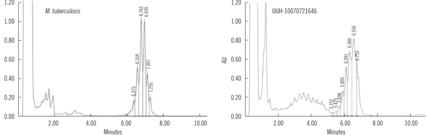
The mycolic acid analyses were also performed using HPLC, as described previously [13]. HPLC patterns were compared with patterns from standard mycobacterial species, which were obtained from 28 ATCC standard mycobacterial species and 5 Korean Type Culture Collection (KTCC) standard mycobacterial species. The mycolic acid pattern of strain UUH-10070721646 was characterized by a single, late cluster of peaks, which was clearly distinct from TB but nearly overlapping with those of M.
simulans (Fig. 1).
To identify the organism at the species level, a commercial DNA chip assay (CombiChip Mycobacteria Genotyping DNA Chip; Gene In Inc., Busan, Korea) was performed, which imple- ments the hybridization method by using an oligonucleotide chip containing internal transcribed spacer (ITS) sequence between the 16S rRNA and 23S rRNA of Mycobacterium, thereby identi- fying 20 species of mycobacteria (Panmycobacteria, MTBC, M.
avium-intracellular complex, M. fortuitum, M. chelonae, M. ab- scessus, M. kansasii, M. gordonae, M. scrofulaceum, M. szul- gai, M. vaccae, M. xenopi, M. terrae, M. flavescens, M. smeg- matis, M. malmoense, M. simiae, M. marinum-ulcerance, M.
gastri, and M. leprae). In this case, hybridization with the genus- specific probe and the failure to hybridize with species-specific probes indicated the presence of a Mycobacterium strain that did not belong to any species that was identifiable by the system.
For complete analysis, sequencing of the 16S rRNA gene and the 16S-23S ITS region was performed with a MJ Research PTC- 225 Peltier Thermal Cycler using Applied Biosystems (ABI) PRISM BigDye Terminator Cycle Sequencing Kits and ABI 3730xl se- Table 1. Biochemical identification results from strain UUH-10070 721646 and related Mycobacterium species
Test Strain UUH-
10070721646 Mycobacteri-
um riyadhense Mycobacte- rium szulgai
Nitrate reduction + + +
68°C catalase - - +
Catalase>45 mm + + +
Urease + + +
Pigmentation Absent Absent Photochro-
mogen
Colony morphology Smooth Rough Smooth/
rough
Growth at 25°C + + +
Pirazinamidase + + -
Tolerance to isoniazid (500 μg/mL) + + +/-*
*+/-, variable.
quencer (Applied Biosystems, Foster City, CA, USA) by using the standard protocol [14]. The primer pair used for amplifica- tion consisted of 27F (5′-AGA GTT TGA TC [A/C] TGG CTC AG- 3′) and 1492R (5′-G [C/T] T ACC TTG TTA CGA CTT-3′). This pri- mer pair amplifies a 1,500 bp fragment of the 16S rRNA gene between positions 8 and 1509 of the Escherichia coli 16S rRNA gene. We compared the obtained sequences with the GenBank and European Molecular Biology Laboratory (EMBL; National Center for Biotechnology Information, http://www.ncbi.nlm.nih.
gov) gene sequence databases. The sequencing results are list ed in Table 2. The sequences of the 16S rRNA gene and 16S-23S ITS regions of strain UUH-10070721646 were unique and closely related to the recently described species, M. riyadhense [9]. In the complete sequence of the 16S rRNA of the current isolate, the similarity to the latter species was 100%, with 1 mismatch in 1,438 bp. In the hypervariable region of hsp65 [15], there were 3 mismatches (in 423 bp; similarity 99%). In the ITS, the pres- ence of 7 mismatches in 278 bp was responsible for a similarity of 97%. In the 744 bp stretch of rpoB, M. riyadhense presented the closest similarity (95%, with 37 mismatches). In the same regions, the similarities with M. tuberculosis were clearly lower (98% in the 16S rRNA, 86% in hsp65, 88% in the ITS, and 91%
in rpoB).
The 16S rRNA gene sequence was compared with those of reference strains of the most closely related mycobacterial spe- cies present in major international nucleotide sequence data- bases (GenBank, EMBL, DNA Data Bank of Japan [DDBJ]) us- ing Clustal W software version 2 (http://www.ebi.ac.uk/tools/clust- alw2) [16]. The resulting topology and tree that were inferred by neighbor-joining and visualized using the Molecular Evolutionary Genetics Analysis (MEGA) software package were evaluated by bootstrap analyses based on 1,000 resamplings (Fig. 2).
Although the patient seemed to show both clinical and radio- logical improvement after the first regimen of INH, RIF, PZA, and EMB, INH was discontinued after 8 months of treatment due to the results of an in vitro drug susceptibility test. The drug sus- ceptibility test was performed according to the absolute concen- tration method (validated for MTB strains only) in Löwenstein- Jensen medium (Green Cross Reference Laboratory, Yongin, Korea), using the first-line and second-line drugs, and the mini- mal inhibitory concentrations were determined using the micro- dilution method recommended by the Clinical and Laboratory Standards Institute (CLSI) [17]. The results are interpreted fol- lowing the CLSI guideline for other slowly growing NTM and new ly
AU
1.20 1.00 0.80 0.60 0.40 0.20
0.00 2.00 4.00 6.00 8.00 10.00
Minutes
6.762 6.939
6.375 6.554 7.0977.255
M. tuberculosis
AU
1.20 1.00 0.80 0.60 0.40 0.20
0.00 2.00 4.00 6.00 8.00 10.00
Minutes
5.252 5.475 5.6985.895 6.091 6.300 6.7336.556
UUH-10070721646
Fig. 1. HPLC pattern of strain UUH-10070721646 (right) compared with that of Mycobacterium tuberculosis (left); the HPLC pattern of UUH-10070721646 is characterized by a single, late cluster of peaks.
Table 2. Sequence comparison between UUH-10070721646 and its closely related Mycobacterium species
Gene/region Most similar sequences
16S rRNA gene (full) Mycobacterium malmoense (99%), Mycobacterium szulgai (99%), Mycobacterium bohemicum (98%) 16S-23S ITS (273 bp) M. szulgai (91%), Mycobacterium kansasii (91%), Mycobacterium marinum (91%)
23S rRNA gene (full) M. kansasii (97%), Mycobacterium avium (97%), Mycobacterium ulcerans agy99 (97%)
rpoB (472 bp) M. avium 104 (93%), Mycobacterium paratuberculosis k10 (93%), Mycobacterium tuberculosis H37RvT (91%) hsp65 (421 bp) Mycobacterium genavense DSM 44424T (95%), M. bohemicum CIP 105811T (95%), M. malmoense CIP 105775T (95%)
described species, which are generally tested as for M. kansasii [17].
Strain UUH-10070721646 was found to be susceptible to RIF, EMB, kanamycin, rifabutin, amikacin, ethionamide, cycloserine, capreomycin, clarithromycin, and moxifloxacin, intermediately susceptible to ciprofloxacin, and was resistant to INH, strepto- mycin, ofloxacin, para-amino-salicylic acid, and levofloxacin by using the 2 methods above (Table 3).
The patient has been receiving clinical follow-up assessments for 13 months without recurrence of disease.
DISCUSSION
The incidence of pulmonary infection caused by NTM is increas- ing; however, it is not commonly described in Korean clinical settings. This may be explained by clinicians overlooking the possibility of an infection due to NTM, as Korea is still an en- demic area for TB. Many pulmonary NTM patients are inade- quately and unnecessarily treated for pulmonary TB. Further- more, some patients are even misdiagnosed with multidrug re- sistant TB and treated with the secondary anti-TB regimen, as the clinical presentation of NTM is often difficult to differentiate from that of MTBC [9, 10, 18].
M. riyadhense can infect a patient without predisposing fac- tors, resulting in the tuberculosis-like clinical symptoms that provided laboratory data from our patient. In this case, the pa- tient’s condition improved only once the etiology was finally un- covered. This is the first Korean report of a mycobacterial strain that was phenotypically and diagnostically confused with TB (but
clearly distinct from it) and responsible for severe disease.
There are 4 case reports of M. riyadhense before UUH-1007 0721646 (Table 4) [9, 10, 19], the major features shared by UUH- 10070721646 and these cases resulted in the confusion with MTBC. Commercial probes are frequently used for rapid identi- fication of mycobacterial species [20]; however, M. riyadhense and other recently proposed NTM such as M. kumamotonense cross-react with MTBC DNA probes and may be overlooked by line-probe assays [18]. With the emergence of new NTM spe- cies, commercial probes could fail to discriminate between spe- cies, leaving clinical isolates either unidentified or misidentified.
The clinical and radiologic signs and symptoms of pulmonary infection caused by the strain, including cough, weight loss, fe- ver, and cavitating lung lesions, were also similar to those in typ- ical cases caused by MTBC strains [9, 10, 19]. Another charac- Fig. 2. Phylogenetic relationships of strain UUH-10070721646 and
related Mycobacterium species on the basis of 16S rRNA gene se- quences.
Table 3. Drug susceptibility testing pattern of strain UUH- 10070721646 obtained by using the absolute concentration meth- od in Löwenstein-Jensen medium and broth microdilution for the determination of the minimal inhibitory concentrations (MICs)
Drugs
Absolute concentration method Microdilution Critical concen-
tration (μg/mL) Interpretation MICs
(μg/mL) Interpreta- tion
Isoniazid 0.2 R NT -
Rifampicin 40 S 0.25 S
Streptomycin 10 R NT -
Ethambutol 2 S ≤0.25 S
Kanamycin 40 S NT -
Rifabutin 20 S NT -
Amikacin 40 S ≤1 S
Ethionamide 40 S NT -
Cycloserine 30 S NT -
Ofloxacin 2 R NT -
PAS 1 R NT -
Capreomycin 40 S NT -
Moxifloxacin 2 S 0.5 S
Levofloxacin 2 R NT -
Cefoxitin NT - 128 -
Ciprofloxacin NT - 2 I
Clarithromycin NT - ≤0.5 S
Doxycycline NT - 1 S
Imipenem NT - 32 -
Sulfamethoxazole NT - 8 S
Tobramycin NT - 2 -
Abbreviations: S, susceptible; R, resistant; I, intermediate susceptible; NT, not test ed; -, no interpretation possible; PAS, P-aminosalicylate.
teristic that this strain has in common with MTBC strains is the definite pathogenicity; each case showed evidence for the patho- genic role of the strain in pulmonary or extrapulmonary diseases.
However, the strains differ in drug susceptibility; the first case was cured with standard anti-TB therapy of INH, RIF, and EMB that was ineffective in the second case, and the latter case was successfully treated with the combination of amikacin, ethion- amide, moxifloxacin, clarithromycin, and EMB.
The strains in the third and fourth cases showed similar drug susceptibility patterns [19], which were sensitive to most first- and second-line drugs, but resistant to doxicycline alone. The former was cured with INH, RIF, and EMB, while the latter pa- tient relapsed after receiving clarithromycin and ciprofloxacin for 12 months, but then improved with anti-TB drugs (INH, RIF, EMB, PZA, clarithromycin, and ciprofloxacin). In the present case, UUH-10070721646 was treated with RIF, PZA, and EMB for 13 months without recurrence of disease.
Because of the scarcity of documented cases of M. riyadhense infection [9, 10, 19], no clinically approved agent for M. riyad- hense infection is currently available [17]. Case 1 and 3 indicate that anti-TB drugs such as INH, RIF, and EMB are effective against M. riyadhense infection, but INH revealed in vitro resis- tance in case 2 and our case. Interestingly, the combination of clarithromycin and ciprofloxacin was not effective in case 4, de- spite the demonstration of in vitro susceptibility to these drugs.
Conventional laboratory culture, biochemical testing, and a limited molecular evaluation are sometimes insufficient for dif- ferentiating novel Mycobacterium species from M. tuberculosis.
Biochemical methodologies are cumbersome, time-consuming, and may yield ambiguous and misleading results [21]. PCR and gene probe assays are known to yield fast and accurate results for the species-level identification of mycobacteria, but these methods are associated with the chance of contamination and a high false-positive rate, which can be difficult to sort out in vari- ous mycobacterial species and often require multiple steps to Table 4. Characteristics of 5 case-patients with M. riyadhense infection
Reported order Age/sex Clinical situation Gene Sequence similarity (%) Antimicrobial therapy 1* 19/M Bone infection in maxillary sinus 16S rRNA, rpoB, hsp65, ITS, RD1 Type strain INH, RIF, EMB 2† 62/M Pulmonary infection 16S rRNA, rpoB, hsp65, ITS 99, 95, 96, 95 AMK, ETH, MOX, CLR, EMB
3‡ 39/F Pulmonary infection 16S rRNA, rpoB, hsp65 99.8, 99.8, 100 INH, RIF, EMB, PZA
4‡ 43/M Pulmonary infection 16S rRNA, rpoB, hsp65 99.8, 99.7, 99.1 INH, RIF, CLR, CIP
5 38/F Pulmonary infection 16S rRNA, rpoB, hsp65, ITS 100, 95, 99, 97 RIF, PZA, EMB
*Reported by van Ingen et al. [9]; †reported by Tortoli et al. [10]; ‡reported by Godreuil et al. [19].
Abbreviations: INH, isoniazid; RIF, rifampin; EMB, ethambutol; AMK, amikacin; ETH, ethionamide; MOX, moxifloxacin; CLR, clarithromycin; PZA, pyrazin- amide; CIP, ciprofloxacin.
identify organisms at the species level [13, 22-24].
In the other cases of M. riyadhense, false-positive results from line-probe assays may lead to incorrect diagnoses of TB and unwarranted treatment, but in our case, misdiagnosis was pre- vented by PCR and HPLC, which excluded TB early on before the molecular diagnostic results were obtained. In this case, preliminary investigations by simple PCR provided a negative result, but persistent characterization of the strain by several ge- netic identification systems led to the first detection of M. riyad- hense in Korea.
The characterization of this previously unknown pathogen rai- ses new concerns for human health and demonstrates the con- tinuing scope of the threat caused by NTM.
Authors’ Disclosures of Potential Conflicts of Interest
No potential conflicts of interest relevant to this article were re- ported.
REFERENCES
1. Tortoli E. The new mycobacteria: an update. FEMS Immunol Med Mi- crobiol 2006;48:159-78.
2. McNabb A, Eisler D, Adie K, Amos M, Rodrigues M, Stephens G, et al.
Assessment of partial sequencing of the 65-kilodalton heat shock pro- tein gene (hsp65) for routine identification of Mycobacterium species isolated from clinical sources. J Clin Microbiol 2004;42:3000-11. 3. Griffith DE, Aksamit T, Brown-Elliott BA, Catanzaro A, Daley C, Gordin F,
et al. An official ATS/IDSA statement: diagnosis, treatment, and preven- tion of non-tuberculous mycobacterial diseases. Am J Respir Crit Care Med 2007;175:367-416.
4. Management of opportunist mycobacterial infections: Joint Tuberculosis Committee Guidelines 1999. Subcommittee of the Joint Tuberculosis Committee of the British Thoracic Society. Thorax 2000;55:210-8. 5. Forbes BA, Sahm DF, et al. eds. Bailey and Scott’s Diagnostic Microbi-
ology. 12th ed. St. Louis: Mosby Elsevier, 2007:481.
6. Kim HJ. Current situation of tuberculosis and its control in Korea. J Ko- rean Med Assoc 2006;49:762-72.
7. Prince DS, Peterson DD, Steiner RM, Gottlieb JE, Scott R, Israel HL, et al. Infection with Mycobacterium avium complex in patients without predisposing conditions. N Engl J Med 1989;321:863-8.
8. Arend SM, van Soolingen D, Ottenhoff TH. Diagnosis and treatment of lung infection with nontuberculous mycobacteria. Curr Opin Pulm Med 2009;15:201-8.
9. van Ingen J, Al-Hajoj SA, Boeree M, Al-Rabiah F, Enaimi M, de Zwaan R, et al. Mycobacterium riyadhense sp. nov., a non-tuberculous species identified as Mycobacterium tuberculosis complex by a commercial line-probe assay. Int J Syst Evol Microbiol 2009;59:1049-53.
10. Tortoli E, Rogasi PG, Fantoni E, Beltrami C, De Francisci A, Mariottini A.
Infection due to a novel mycobacterium, mimicking multidrug-resistant Mycobacterium tuberculosis. Clin Microbiol Infect 2010;16:1130-4. 11. Lévy-Frébault VV and Portaels F. Proposed minimal standards for the
genus Mycobacterium and for description of new slowly growing Myco- bacterium species. Int J Syst Bacteriol 1992;42:315-23.
12. Torkko P, Suutari M, Suomalainen S, Paulin L, Larsson L, Katila ML.
Separation among species of Mycobacterium terrae complex by lipid analyses: comparison with biochemical tests and 16S rRNA sequenc- ing. J Clin Microbiol 1998;36:499-505.
13. Jeong J, Kim SR, Lee SH, Choi JI, Chang CH, Choi JY, et al. The use of High Perfomance Liquid Chromatography to speciate and characterize the epidemiology of Mycobacteria. Lab Med 2011;42:612-7.
14. Schuurman T, de Boer RF, Kooistra-Smid AM, van Zwet AA. Prospec- tive study of use of PCR amplification and sequencing of 16S ribosomal DNA from cerebrospinal fluid for diagnosis of bacterial meningitis in a clinical setting. J Clin Microbiol 2004;42:734-40.
15. Telenti A, Marchesi F, Balz M, Bally F, Bottger EC, Bodmer T. Rapid iden- tification of mycobacteria to the species level by polymerase chain reac- tion and restriction enzyme analysis. J Clin Microbiol 1993;31:175-8.
16. Larkin MA, Blackshields G, Brown NP, Chenna R, McGettigan PA, Mc- William H, et al. Clustal W and Clustal X version 2. Bioinformatics 2007; 23:2947-8.
17. Clinical and Laboratory Standards Institute. Susceptibility testing of My- cobacteria, Nocardia, and other aerobic actinomycetes. Approved Stan- dard M24-A2. 2nd ed. Wayne, PA: Clinical and Laboratory Standards Institute, 2011.
18. Rodriquez-Aranda A, Jimenez MS, Yubero J, Chaves F, Rubio-Garcia R, Palenque E, et al. Misidentification of Mycobacterium kumamotonense as M. tuberculosis. Emerg Infect Dis 2010;16:1178-80.
19. Godreuil S, Marchandin H, Michon AL, Ponsada M, Chyderiotis G, Bri- sou P, et al. Mycobacterium riyadhense pulmonary infection, France and Bahrain. Emerg Infect Dis 2012;18:176-8.
20. Tortoli E, Nanetti A, Piersimoni C, Cichero P, Farina C, Mucignat G, et al.
Performance assessment of new multiplex probe assay for identification of mycobacteria. J Clin Microbiol 2001;39:1079-84.
21. Springer B, Stockman L, Teschner K, Roberts GD, Böttger EC. Two-lab- oratory collaborative study on identification of mycobacteria: molecular versus phenotypic methods. J Clin Microbiol 1996;34:296-303.
22. Burman WJ and Reves RR. Review of false-positive cultures for Myco- bacterium tuberculosis and recommendations for avoiding unnecessary treatment. Clin Infect Dis 2000;31:1390-5.
23. Nah J, Huh JW, Lee SH, Kim BC, Koh YS, Pai CH. Identification of My- cobacterium tuberculosis complex using a gene probe method. Korean J Clin Pathol 1997;17:71-8.
24. Reisner BS, Gatson AM, Woods GL. Use of Gen-Probe AccuProbes to identify Mycobacterium avium complex, Mycobacterium tuberculosis complex, Mycobacterium kansasii, and Mycobacterium gordonae di- rectly from BACTEC TB broth cultures. J Clin Microbiol 1994;32:2995-8.