Predictive factors for invasive intraductal papillary mucinous neoplasm of the pancreas
Dae Young Jun1, Hyung Jun Kwon2, Sang Geol Kim2, Sung Hi Kim3, Jae Min Chun1, Young Bong Kwon1, Kyung Jin Yoon2, Yoon Jin Hwang2, and Young Kook Yun1
Department of Surgery, 1Kyungpook National University School of Medicine, 2Kyungpook National University Medical Center, 3Department of Family Medicine, Daegu Catholic University College of Medicine, Daegu, Korea
Backgrounds/Aims: Intraductal papillary mucinous neoplasm (IPMN) of the pancreas has malignant potential. Predicting invasive IPMN has proven difficult and controversial. We tried to identify predictive factors for invasive IPMN. Methods:
Thirty six patients underwent resection for IPMN from February 2001 to July 2011. Clinicopathological features including demographic, imaging, microscopic, and serological findings were retrospectively reviewed. Receiver operating charac- teristic (ROC) curve analysis was used to analyze sensitivity and specificity of all possible cut-off values for the diameter of the main pancreatic duct and mass size predicting invasive IPMN. Student t-test, chi-square test, and logistic re- gression were used for univariate and multivariate analysis. Results: The mean age was 63.5±8.4 years. Males were more commonly affected (58.3% vs 41.7%). Pancreaticoduodenectomy was performed in 55.6% of patients, distal pan- createctomy in 36.1%, and central pancreatic resection in 8.3%. Non-invasive IPMNs were present in 80.6% (n=29), whereas invasive IPMNs were present in 19.4% (n=7). In univariate analysis, tumor location (p=0.036), Kuroda classi- fication (p=0.048), mural nodule (p=0.016), and main duct dilatation (≥8 mm) (p=0.006) were statistically significant variables. ROC curve analysis showed that a value of 8 mm for the main duct dilatation and a value of 35 mm for the size of the mass lesion have 80% sensitivity and 75% specificity and 100% sensitivity and 82.6% specificity, respectively. However, in multivariate analysis, main ductal dilatation (≥8 mm) was identified to be the only in- dependent factor for invasive IPMN (p=0.049). Conclusions: Main duct dilatation appears to be a useful indicator for predicting invasive IPMN. (Korean J Hepatobiliary Pancreat Surg 2011;15:237-242)
Key Words: Invasive; Intraductal papillary mucinous neoplasm (IPMN); Pancreas; Malignant potential
Received: September 11, 2011; Revised: October 3, 2011; Accepted: October 13, 2011 Corresponding author: Hyung Jun Kwon
Department of Surgery, Kyungpook National University Medical Center, 807, Hogukno, Buk-gu, Daegu 702-210, Korea Tel: +82-53-200-2708, Fax: +82-53-200-2027, E-mail: kwonhj95@naver.com
Sang Geol Kim
Department of Surgery, Kyungpook National University Medical Center, 807, Hogukno, Buk-gu, Daegu 702-210, Korea Tel: +82-53-200-2708, Fax: +82-53-200-2027, E-mail: ksg@knu.ac.kr
Copyright Ⓒ 2011 by The Korean Association of Hepato-Biliary-Pancreatic Surgery Korean Journal of Hepato-Biliary-Pancreatic Surgery ∙ pISSN: 1738-6349
INTRODUCTION
Intraductal papillary mucinous neoplasm (IPMN) of the pancreas is a distinct entity characterized by papillary pro- liferations of mucin-producing epithelial cells with ex- cessive mucus production and cystic dilatation of the pan- creatic ducts. IPMN was first described by Ohashi et al.
in 1982 and first recognized in the World Health Organi- zation classification in 1996.1
IPMNs histologically show a broad spectrum ranging from adenoma to invasive carcinoma with different de- grees of severity and seem to follow a progression from adenoma to invasive, similar to the well-defined ad-
enoma-carcinoma sequence in colorectal cancer and pan- creatic ductal adenocarcinoma (pancreatic intraepithelial neoplasia [PanIN] to invasive ductal carcinoma).2,3 Although prognosis for IPMN is better than that for ductal adenocarcinoma because IPMNs grow slowly and are diagnosed earlier than ductal adenocarcinoma, in- vasively transformed IPMNs have poor outcomes, similar to ductal adenocarcinoma. Thus, discriminating invasive IPMN from non-invasive IPMN is important for the choice of appropriate management of patients with IPMNs.
The purpose of this study was to determine predictive factors of invasive IPMN by examining and analyzing
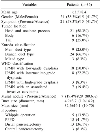
Table 1. Clinicopathologic features of 36 patients who under- went resection of the IPMN
Variables Patients (n=36) Mean age
Gender (Male/Female) Symptom (Presence/Absence) Tumor location
Head and uncinate process Body
Tail
Kuroda classification Main duct type Branch duct type Mixed type WHO classification
IPMN with low-grade dysplasia IPMN with intermediate-grade dysplasia
IPMN with high-grade dysplasia IPMN with an associated invasive carcinoma
Mural nodule (Presence/Absence) Duct size (diameter, mm) Mass size (mm)
Procedure
Whipple operation PPPD
Distal pancreatectomy Central pancreatectomy
63.5±8.4 21 (58.3%)/15 (41.7%) 21 (58.3%)/15 (41.7%)
21 (58.3%) 6 (16.7%) 9 (25.0%) 9 (25.0%) 24 (66.7%) 3 (8.3%) 18 (50.0%) 8 (22.2%) 3 (8.3%) 7 (19.4%) 7 (19.4%)/29 (80.6%)
4.9±3.7 (1.0-14.2) 32.5±16.1 (10-70)
5 (13.9%) 15 (41.7%) 13 (36.1%) 3 (8.3%) for the presence of symptoms, tumor location, tumor size,
maximum diameter of the main pancreatic duct (MPD), and presence of a mural nodule.
The diameter of main duct dilatation and the size of measurable mass lesions were used as independent con- tinuous variables. According to the World Health Organi- zation classification, the 36 resected IPMNs were patho- logically described as non-invasive IPMN (IPMN with low-grade dysplasia, with intermediate-grade dysplasia, with high-grade dysplasia) and invasive IPMN (IPMN with an associated invasive carcinoma).
Statistical analysis of the data was performed using SPSS version 18.0. The difference in clinicopathological factors between non-invasive IPMNs and invasive IPMNs were analyzed by Student's t-test, chi-square test, or Fisher’s exact test. A multivariate analysis was performed to determine the predictors of invasive IPMN using binary logistic regression. Receiver operating characteristic (ROC) curve analysis was used to analyze the sensitivity and specificity of possible cut-off values for the diameter of the main pancreatic duct and for mass size. ROC curves for the diameter of the main pancreatic duct were analyzed for 13 patients excluding pure branch-duct IPMNs. A similar ROC analysis was done for the size of the mass lesion among 26 patients excluding pure main duct IPMNs.
RESULTS
Clinicopathologic characteristics of 36 patients with IPMNs
The mean age of the 36 patients with IPMN was 63.5±8.4 (range: 42-77) years. There were 21 males (58.3%) and 15 females (41.7%). Twenty one patients (58.3%) were symptomatic at presentation. A majority of patients (81%) presented with abdominal discomfort and pain. The mean size of the mass lesions was 32.5 mm
ciated invasive carcinoma) (Table 1).
Eight patients (22%) with IPMNs were associated with extrapancreatic neoplasms. Benign neoplasms were pres- ent in 5 cases (adrenal adenoma, 2; uterine myoma, 2;
ampullary adenoma, 1); malignancies were present in 5 cases (gastric cancer [n=2], colon cancer [n=1], thyroid cancer [n=1], and cervical cancer [n=1]). Pancreatoduo- denectomy was the most common operation and was per- formed in 20 cases (55.6%), 15 of which were pylo- rus-preserving operations. Distal pancreatectomy was per- formed in 13 cases (36.1%). Three patients (8.3%) under- went central pancreatectomy (Table 1).
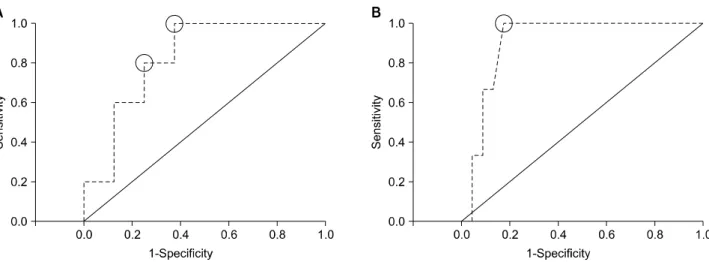
Fig. 1. Receiver operating characteristic curve analysis for cut-off values of the diameter of the main pancreatic duct and mass size. Using 7 mm as a cut-off level for the main duct diameter yielded a sensitivity of 100% and a specificity of 63%; 8 mm yielded a sensitivity of 80% and a specificity of 75% (A). Using 35 mm as a cut-off level for the size of the mass yielded a sensitivity of 100% and a specificity of 82.6% (B).
Receiver operating characteristic curve analysis for cut-off values for the diameter of the main pancreatic duct and mass size ROC curve analysis for cut-off values of the diameter of the main pancreatic duct and mass size was performed using data from the different patients group. ROC curve analysis for duct dilatation was done in 13 patients with duct dilatation excluding pure branch duct type IPMN.
Using 7 mm as a cut-off level for the main duct diameter yielded a sensitivity of 100% and a specificity of 63%, whereas 8 mm yielded a sensitivity of 80% and a specifi city of 75% (Fig. 1A).
For the size of the mass lesion, ROC curve analysis was done in 26 patients with any measurable lesion dilata- tion excluding pure main duct type IPMN. Using 35 mm as a cut-off level for the size of the mass yielded a sensi- tivity of 100% and a specificity of 82.6% (Fig. 1B).
Univariate analysis between non-invasive IPMN and invasive IPMN
IPMN lesions were categorized as non-invasive IPMN and invasive IPMN by the WHO classification. There were 29 non-invasive IPMNs (80.6%) and 7 invasive IPMNs (19.4%). The mean age of the patients with non-invasive IPMN was 64.4±8.8 years, whereas the mean age of patients with invasive IPMN was 59.9±5.7 years.
There were no significant differences between the 2 groups with regard to age, sex and symptoms. The mean
size of the mass lesions of invasive IPMNs was larger than that of non-invasive IPMNs but the difference fell just short of significance (30.3±15.3 mm vs 47.0±15.0 mm; p=0.051). The mean diameter of the main pancreatic duct in invasive IPMN patients was significantly larger than that in non-invasive IPMN patients (4.1±3.2 mm vs 8.2±3 mm; p=0.007). A mural nodule was seen in pre- operative imaging in 7 patients, Three (3 of 29 [10.3%]) patients had non-invasive IPMN and 4 (4 of 7 [57.1%]) invasive IPMN. The relationship between the presence of a mural nodule and invasive IPMN was significant (p=0.016) (Table 2).
ROC curve analysis yielded optimal cut-off values for the diameter of the main pancreatic duct and mass size.
Since an 8 mm diameter of the main pancreatic duct yielded high sensitivity (80%) and specificity (75%), we utilized 8 mm as the cutoff to divide patients into two groups. Nine patients had a main pancreatic duct diameter of 8 mm or greater including 4 patients out of 29 non-in- vasive IPMN patients (13.8%) and 5 patients out of 7 in- vasive IPMN patients (71.4%). A main pancreatic duct di- ameter of 8mm or greater was significantly more frequent in invasive IPMN (p=0.006). Similarly, since the 35 mm of mass size yielded high sensitivity (100%) and specific- ity (82.6%), we utilized 35 mm as a criterion to divide the patients into two groups. Nine patients had a mass size of 35mm or greater including 5 patients among the 26 non-invasive IPMN patients (19.2%) and 4 patients
Table 2. Clinicopathologic comparison between non-invasive IPMN and invasive IPMN
Non-invasive (n=29)
Invasive (n=7)
Univariate analysis
p-value Mean age
Gender Male Female Symptom Presence Absence Tumor location Head and uncinate process
Body Tail Mural nodule Presence Absence
Duct size (diameter, mm)
<8 ≥8
Mass size (mm) <35
≥35 Unmeasured
64.4±8.8 16 13 15 14 14 6 9 3 26 4.1±3.2
25 4 30.3±15.3
21 5 3
59.9±5.7 4 2 6 1 7 0 0 4 3 8.2±3.9
2 5 47.0±15.0
0 4 3
0.221
0.674
0.200
0.036
0.016 0.007
0.006 0.051 0.002
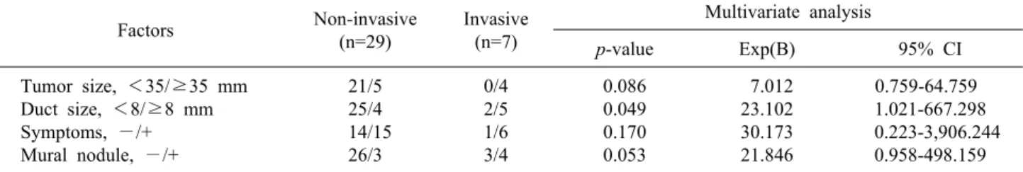
Table 3. Multivariate analysis results for predictive factors of invasive IPMN
Factors Non-invasive
(n=29)
Invasive (n=7)
Multivariate analysis
p-value Exp(B) 95% CI
Tumor size, <35/≥35 mm Duct size, <8/≥8 mm Symptoms, −/+
Mural nodule, −/+
21/5 25/4 14/15
26/3
0/4 2/5 1/6 3/4
0.086 0.049 0.170 0.053
7.012 23.102 30.173 21.846
0.759-64.759 1.021-667.298 0.223-3,906.244
0.958-498.159 of tumor size (<35 versus ≥35 mm), main duct dilata-
tion (<8 versus ≥8 mm), symptoms (absent versus pres- ent), and mural nodules (absent versus present) were put into the binary logistic regression model. It showed that
Recent advances in diagnostic imaging have resulted in an increased frequency of diagnosis for cystic mucin-pro- ducing pancreatic neoplasms. According to previous re- ports, IPMN represent about 1% of the pancreas exocrine tumors and about 12% of the pancreas cystic tumors.4 Two-thirds of IPMN patients are men. The peak age is the sixth decade. Despite the more frequent reporting of IPMN, the natural history of this disease is not well understood. How to manage patients with IMPNs, espe- cially when it comes to timing of the surgical inter- vention, remains controversial.
Sonh et al.5 reported a lag-time of approximately 5 years from the time of development of an IPMN adenoma to the progression to IPMN with an associated invasive carcinoma. The incidence of invasive IPMN has been re- ported to be 0-35%.6
If an invasive component is present in IPMNs, the prognosis becomes unfavorable even after curative resection. The overall 5-year survival for patients with an invasive IPMN has been reported to range from 26 to 60%, compared with 90-95% in patients with a non-in- vasive component.7 Therefore, it is important to accurately differentiate malignant from benign IPMNs and select the appropriate treatment strategy.
The preoperative diagnosis and classification of IPMN is based upon imaging. In spite of advances in diagnostic imaging, it remains difficult to predict malignancy cor- rectly4,8-11 and the accuracy is reported to be about
58-82%. The incidence of malignancy without malignant predicting factors in preoperative imaging was reported to be 10%.12 Thus, it is important to evaluate various clinical factors suggesting malignancy. Because prognosis of in- vasive IPMN is very poor, examination of predictive fac- tors for invasive carcinoma is most important.
According to previous reports, tumor size, presence of a mural nodule, related symptoms, and dilatation of the main pancreatic duct have been reported to be important in predicting malignancy13-19; a mural nodule (≥6.3 mm) in the main pancreatic duct and a solid mass in pancreatic parenchyma were reported to be associated with invasive disease.11,20-22
In the present study, we tried to determine the sensi- tivity and specificity for invasive IMPNs of the diameter of the main pancreatic duct and the size of mass lesions using ROC curve analysis because these factors are con- tinuous variables and have been reported as significant factors in previous reports. A cutoff of 8 mm for the main duct diameter and of 35 mm for the mass lesion size yielded both high sensitivity and specificity. We use these cutoff values as criteria to divide patients into groups for univariate and multivariate analysis.
Univariate analysis was performed to determine pre- dictors of invasive IPMN. It showed that tumor location (p=0.036), mural nodule (p=0.016), and main duct dilata- tion (≥8 mm) (p=.006), and the size of mass lesion (≥35 mm) were statistically significant.
However, multivariate analysis showed that main ductal dilatation (≥8 mm) was the only independent predictive factor for invasive IPMN (p=0.049); tumor size and mural nodule fell just short of statistical significance. Failure of these two important factors, tumor size and mural nodule, to reach statistical significance may be related to the small number of cases in this study. Further studies enlarging the number of cases are required to determine the true sig- nificance in future studies.
In conclusion, IPMN is a premalignant lesion. It is im- portant to distinguish invasive carcinoma from non-in- vasive IPMN. Our study showed that main duct dilatation
≥8 mm can be a useful indicator for predicting invasive IPMN.
REFERENCES
1. Kloppel G, Solcia E, Longnecker DS, et al. World Health Organization International Histological Classification of Tum- ours: Histological Typing of Tumors of the Exocrine Pancreas.
2nd ed. Berlin; Springer-Verlag, 1996.
2. Cho KR, Vogelstein B. Genetic alterations in the adenoma--carci- noma sequence. Cancer 1992;70(6 Suppl):1727-1731.
3. Wilentz RE, Hruban RH. Pathology of cancer of the pancreas.
Surg Oncol Clin N Am 1998;7:43-65.
4. Taouli B, Vilgrain V, O'Toole D, Vullierme MP, Terris B, Menu Y. Intraductal papillary mucinous tumors of the pancreas: fea- tures with multimodality imaging. J Comput Assist Tomogr 2002;26:223-231.
5. Sohn TA, Yeo CJ, Cameron JL, et al. Intraductal papillary muci- nous neoplasms of the pancreas: an updated experience. Ann Surg 2004;239:788-797.
6. Klimstra DS. Cystic, mucin-producing neoplasms of the pancreas:
the distinguishing features of mucinous cystic neoplasms and in- traductal papillary mucinous neoplasms. Semin Diagn Pathol 2005;22:318-329.
7. Bosman FT, Carneiro F, Hruban RH, et al. WHO Classification of Tumours of the Digestive System: Tumours of the Pancreas.
4th ed. Berlin; Springer, 2010.
8. Baba T, Yamaguchi T, Ishihara T, et al. Distinguishing benign from malignant intraductal papillary mucinous tumors of the pan- creas by imaging techniques. Pancreas 2004;29:212-217.
9. Chiu SS, Lim JH, Lee WJ, et al. Intraductal papillary mucinous tumour of the pancreas: differentiation of malignancy and benig- nancy by CT. Clin Radiol 2006;61:776-783.
10. Sahani DV, Kadavigere R, Blake M, et al. Intraductal papillary mucinous neoplasm of pancreas: multi-detector row CT with 2D curved reformations--correlation with MRCP. Radiology 2006;
238:560-569.
11. Ogawa H, Itoh S, Ikeda M, Suzuki K, Naganawa S. Intraductal papillary mucinous neoplasm of the pancreas: assessment of the likelihood of invasiveness with multisection CT. Radiology 2008;248:876-886.
12. Jang JY, Kim SW, Ahn YJ, et al. Multicenter analysis of clin- icopathologic features of intraductal papillary mucinous tumor of the pancreas: is it possible to predict the malignancy before sur- gery? Ann Surg Oncol 2005;12:124-132.
13. Tanaka M, Chari S, Adsay V, et al; International Association of Pancreatology. International consensus guidelines for manage- ment of intraductal papillary mucinous neoplasms and mucinous cystic neoplasms of the pancreas. Pancreatology 2006;6:17-32.
14. Pelaez-Luna M, Chari ST, Smyrk TC, et al. Do consensus in- dications for resection in branch duct intraductal papillary muci- nous neoplasm predict malignancy? A study of 147 patients. Am J Gastroenterol 2007;102:1759-1764.
15. Kobayashi G, Fujita N, Noda Y, et al. Mode of progression of intraductal papillary-mucinous tumor of the pancreas: analysis of patients with follow-up by EUS. J Gastroenterol 2005;40:744- 751.
16. Yamaguchi T, Baba T, Ishihara T, et al. Long-term follow-up of intraductal papillary mucinous neoplasm of the pancreas with ultrasonography. Clin Gastroenterol Hepatol 2005;3:1136-1143.
17. Schmidt CM, White PB, Waters JA, et al. Intraductal papillary mucinous neoplasms: predictors of malignant and invasive pathology. Ann Surg 2007;246:644-651.
18. Lee CJ, Scheiman J, Anderson MA, et al. Risk of malignancy in resected cystic tumors of the pancreas < or =3 cm in size:
is it safe to observe asymptomatic patients? A multi-institutional