Descriptive Epidemiology of Patients Undergoing Total Hip Arthroplasty in Korea with Focus on Incidence of
Femoroacetabular Impingement: Single Center Study
We analyzed the causes leading to total hip arthroplasty (THA), aimed to clarify the incidence of femoroacetabular impingement (FAI) among the causes, and compared the incidence in Korea with those in other countries. From January 2000 to December 2014, 1,206 hips of 818 patients who underwent primary THA at our institute were reviewed retrospectively in terms of radiographs and electronic charts. The radiographs and radiographic parameters were reviewed and measured by 2 of the authors, who are orthopedic surgeons. Patients were categorized in terms of the causes leading to THA as primary osteoarthritis (OA), rheumatoid arthritis (RA), posttraumatic arthritis, post infectious arthritis, avascular necrosis (AVN) of the femoral head, fracture of the femoral head or neck, ankylosing spondylitis (AS), developmental dysplasia of the hip (DDH), Legg- Calvé-Perthes disease (LCPD), FAI, and others. There were 32 patients (3.91%) in the primary OA group, 41 (5.01%) in the RA group, 84 (10.27%) in the posttraumatic arthritis group, 39 (4.77%) in the post infectious arthritis group, 365 (44.62%) in the AVN group, 39 (4.77%) in the fracture group, 21 (2.57%) in the AS group, 52 (6.36%) in the DDH group, 71 (8.68%) in the LCPD group, 52 (6.36%) in the FAI group, and 22 (2.69%) in the
‘other’ group. The causes leading to THA in Korea differ from those in Western countries.
FAI could be causes of severe secondary OA that requires THA in Korea, therefore symptomatic FAI should not be neglected.
Keywords: Hip; Osteoarthritis; Total Hip Arthroplasty; Femoroacetabular Impingement;
Incidence Woo-Yong Lee, Deuk-Soo Hwang,
and Chang-Kyun Noh
Department of Orthopaedic Surgery, Chungnam National University School of Medicine, Daejeon, Korea
Received: 10 October 2016 Accepted: 18 December 2016 Address for Correspondence:
Deuk-Soo Hwang, MD
Department of Orthopaedic Surgery, Chungnam National University School of Medicine, 282 Munhwa-ro, Jung-gu, Daejeon 35015, Republic of Korea
E-mail: [email protected]
Funding: This research was supported by the Chungnam National University Hospital (CNUH) research fund (No. 2016- CF-008).
https://doi.org/10.3346/jkms.2017.32.4.581 • J Korean Med Sci 2017; 32: 581-586
INTRODUCTION
Total hip arthroplasty (THA) is an effective intervention for pa- tients with severe hip disease and has been shown to lead to marked improvement in health outcomes (1). Because of the significant benefits realized with THA, utilization rates for this procedure have been increasing in most Western and Asian countries, including Korea (2,3). Thus, many Western epidemi- ological studies have examined the incidence of THA and re- ported osteoarthritis (OA) as the most common underlying con- dition for THA in Western countries (2-5). However, Western epidemiological data cannot be applied to Korea, and most of these studies only determined the prevalence and trends of THA or were limited to THA for patients with OA. The pathogenesis of coxarthrosis leading to THA is complex, with many risk fac- tors (6,7). Many conditions that can lead to THA include prima- ry OA, secondary OA resulting from femoroacetabular impinge- ment (FAI), dysplasia and peripheral joint involvement of sero- negative spondylitis, such as ankylosing spondylitis (AS) and diffuse idiopathic skeletal hyperostosis, avascular necrosis (AVN),
and fractures. Although the ratios of primary OA have previous- ly been considered high, morphological deformities around the hip may cause OA over several decades (8,9). Recently, several studies have reported that FAI results in early OA and hip OA pathology related to FAI (10-12). To our knowledge, no previous study has evaluated the epidemiology of causes leading to THA in Koreans, particularly with a focus on the incidence of FAI.
The purposes of the present study were 1) to analyze the causes leading to THA, 2) to clarify the incidence of FAI among these causes, and 3) to compare the incidence in Korea with those in other countries.
MATERIALS AND METHODS Data selection
From January 2000 to December 2014, 1,206 hips of 818 patients who underwent primary THA at our institute were reviewed retrospectively in terms of radiographs and electronic charts.
Clinical records, surgical records, and preoperative simple ra- diographs were evaluated to determine the causes leading to
THA. The radiographs and radiographic parameters were re- viewed and measured by 2 of the authors, who are orthopedic surgeons. The radiographs were also reviewed again, 4 weeks later, by the same observers. Exclusion criteria were as follows:
1) absent or insufficient radiographs or electronic charts to de- termine the cause, 2) revision of THA, and 3) conversion from hemiarthroplasty to THA.
Patients were categorized according to the causes leading to THA: primary OA, rheumatoid arthritis (RA), posttraumatic ar- thritis, post infectious arthritis, AVN of the femoral head, frac- ture of the femoral head or neck, AS, developmental dysplasia of the hip (DDH), Legg-Calvé-Perthes disease (LCPD), FAI, and other. The FAI group was subcategorized into cam, pincer, and mixed types.
Primary OA cases were defined as those with no systemic dis- ease, no history of hip disease, and no remarkable deformity of the proximal femur. Atlas photographs illustrating the individu- al radiographic features of OA were used to standardize the read- ings (13,14). The following radiographic parameters were also applied to determine the primary OA cases: center-edge angle
> 20°, sharp angle < 45°, and acetabular roof obliquity < 15° (15).
FAI was defined by the obvious presence of a bony prominence in the anterolateral head-neck junction (cam type) and/or an acetabular abnormality, such as retroversion, coxa profunda, or acetabular protrusion (pincer type) (15). The alpha angle was considered to have been increased if > 50°, and anterior femur offset was considered to have been decreased if < 8 mm; these were classified as cam type (16). Acetabular retroversion is a form of hip dysplasia in which the alignment of the acetabulum does not face the normal anterolateral direction but is inclined more posterolaterally (17). In simple radiography, crossover of the anterior wall of acetabulum over the posterior wall (cross- over sign, figure-8 sign) was considered to be acetabular retro- version, the adjoining of the acetabular fossa and ilioischial line to be coxa profunda, and the femur head passing the ilioischial
to be protrusion acetabuli (18). These were classified as the pin- cer type. Cases having both types were classified as the mixed type. ‘Others’ were defined as cases that could not be catego- rized or cases with very small numbers, such as synovial chon- dromatosis and neurofibromatosis.
Statistical analysis
Two blinded reviewers reviewed the radiographs independent- ly; there was no communication between the reviewers. Radio- graphs were presented to the reviewers 2 times in random or- der at intervals of 3 weeks. Intraobserver and interobserver reli- abilities of the 5 measured radiological parameters (center-edge angle, sharp angle, acetabular roof obliquity, alpha angle, ante- rior femur offset) were assessed using intraclass correlation co- efficients. And intraobserver and interobserver reliabilities of the causes leading to primary THA were assessed using Kappa coefficient. The SPSS software (ver. 19.0; SPSS Korea, Seoul, Ko- rea) was used for statistical analysis.
Ethics statement
This retrospective study was approved by the Institutional Re- view Board (IRB) of Chungnam National University Hospital (IRB No. CNUH 2015-07-011). Informed consent was waived by the IRB.
RESULTS
In total, 1,206 hips in 818 patients underwent THA at out insti- tute between January 2000 and December 2014. There were 477 male and 341 female patients. The numbers of cases undergo- ing THA has increased consistently since 2000 (Fig. 1). The chan-
Fig. 1. The graph shows that the procedural number of total hip arthroplasties from 2000 to 2014 increased consistently.
2000 2001 2002 2003 2004 2005 2006 2007 2008 2009 2010 2011 2012 2013 2014 140
120 100 80 60 40 20 0
Number of primary total hip arthroplasty
0%
10%
20%
30%
40%
50%
60%
70%
80%
90%
100%
2000 - 2002 2003 - 2005 2006 - 2008 2009 - 2011 2012 - 2014 Avascular necrosis of femoral head Postraumatic arthritis
Legg-Calvé-Perthes disease Developmental dysplasia of the hip Femoroacetabular impingement Rheumatoid arthritis
Fracture of femoral head or neck Postinfectious arthritis
2000-2002 2003-2005 2006-2008 2009-2011 2012-2014 100
90 80 70 60 50 40 30 20 10 0 (%)
0%
10%
20%
30%
40%
50%
60%
70%
80%
90%
100%
2000 - 2002 2003 - 2005 2006 - 2008 2009 - 2011 2012 - 2014 Avascular necrosis of femoral head Postraumatic arthritis
Legg-Calvé-Perthes disease Developmental dysplasia of the hip Femoroacetabular impingement Rheumatoid arthritis
Fracture of femoral head or neck Postinfectious arthritis 0%
10%
20%
30%
40%
50%
60%
70%
80%
90%
100%
2000 - 2002 2003 - 2005 2006 - 2008 2009 - 2011 2012 - 2014 Avascular necrosis of femoral head Postraumatic arthritis
Legg-Calvé-Perthes disease Developmental dysplasia of the hip Femoroacetabular impingement Rheumatoid arthritis
Fracture of femoral head or neck Postinfectious arthritis AVN of femoral head
LCPD FAI
Fracture of femoral head or neck
Postraumatic arthritis DDH
RA
Postinfectious arthritis
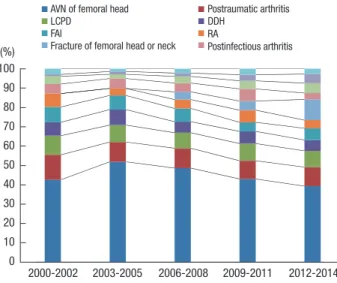
Fig. 2. The graph shows the changes of causes leading to THA.
THA = total hip arthroplasty, AVN = avascular necrosis, LCPD = Legg-Calvé-Perthes disease, DDH = developmental dysplasia of the hip, FAI = femoroacetabular impinge- ment, RA = rheumatoid arthritis.
ges of causes leading to THA showed that AVN has been decre- ased since the interval between 2003 and 2005 and fracture has been increased (Fig. 2).
There were 32 patients (3.91%; 13 males, 19 females) in the primary OA group (Fig. 3), 41 (5.01%; 12 males, 29 females) in the RA group, 84 (10.27%; 43 males, 41 females) in the posttrau- matic arthritis group, 39 (4.77%; 17 males, 22 females) in the post infectious arthritis group, 365 (44.62%; 270 males, 95 fe- males) in the AVN group, 39 (4.77%; 17 males, 22 females) in the fracture group, 21 (2.57%; 16 males, 5 females) in the AS
group, 52 (6.36%; 16 males, 36 females) in the DDH group, 71 (8.68%; 52 males, 19 females) in the LCPD group, 52 (6.36%; 13 males, 39 females) in the FAI group, and 22 (2.69%; 8 males, 14 females) in the ‘others’ group. In the FAI group, there were 36 patients (69.23%; 9 males, 27 females) of the cam type (Fig. 4), 15 (28.85%; 3 males, 12 females) of the pincer type (Fig. 5), and 1 (1.92%; 1 male) of the mixed type (Table 1).
Intraobserver and interobserver correlations for causes and the combination of all measurements were found to be repro- ducible and reliable among observers (Tables 2 and 3).
A B
Fig. 3. Anteroposterior radiographs illustrate the case of a 62-year-old female patient who had primary OA of both hips. (A) The radiograph was taken before THA. Lateral cen- ter-edge angle, sharp angle, and acetabular roof obliquity of right hip was 37.2°, 35.8°, and 8.6°, respectively. Left hip was 43.5°, 35.2°, and 7.0°, respectively. (B) The radio- graph was taken after THA to treat primary OA.
THA = total hip arthroplasty, OA = osteoarthritis.
R
ap
R
ap
Fig. 4. Anteroposterior radiographs illustrate the case of an 85-year-old female patient who had secondary OA caused by Cam type FAI of left hip. (A) Preoperative radiograph shows typical pistol grip deformity on left hip. (B) The radiograph was taken after THA.
THA = total hip arthroplasty, FAI = femoroacetabular impingement, OA = osteoarthritis.
A B
RT RT
Fig. 5. Anteroposterior radiographs illustrate the case of a 76-year-old female patient who had secondary OA caused by Pincer type FAI of left hip. (A) Preoperative radiograph shows typical acetabular protrusion on right hip. (B) The radiograph was taken after THA.
THA = total hip arthroplasty, FAI = femoroacetabular impingement, OA = osteoarthritis.
A B
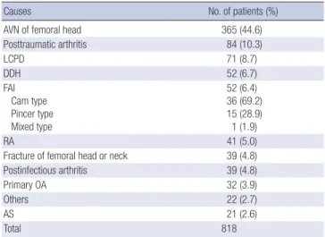
Table 1. Causes leading to primary THA
Causes No. of patients (%)
AVN of femoral head 365 (44.6)
Posttraumatic arthritis 84 (10.3)
LCPD 71 (8.7)
DDH 52 (6.7)
FAI Cam type Pincer type Mixed type
52 (6.4) 36 (69.2) 15 (28.9) 1 (1.9)
RA 41 (5.0)
Fracture of femoral head or neck 39 (4.8)
Postinfectious arthritis 39 (4.8)
Primary OA 32 (3.9)
Others 22 (2.7)
AS 21 (2.6)
Total 818
THA = total hip arthroplasty, AVN = avascular necrosis, LCPD = Legg-Calvé-Perthes disease, DDH = developmental dysplasia of the hip, FAI = femoroacetabular impinge- ment, RA = rheumatoid arthritis, OA = osteoarthritis, AS = ankylosing spondylitis.
Table 2. Intraclass intraobserver and intraobserver reliabilities of radiographic para- meters
Measuring parameters Intraobserver repeatability Interobserver reproducibility
Center-edge angle 0.91 0.89
Sharp angle 0.90 0.85
Acetabular roof obliquity 0.86 0.84
Alpha angle 0.92 0.90
Anterior femur offset 0.87 0.86
Table 3. Intraobserver and intraobserver reliabilities of the causes leading to primary THA
Causes Intraobserver
repeatability Interobserver reproducibility
AVN of femoral head 0.92 0.89
Posttraumatic arthritis 0.92 0.88
LCPD 0.86 0.84
DDH 0.83 0.82
FAI Cam type Pincer type Mixed type
0.87 0.83 0.89 0.82
0.83 0.82 0.84 0.81
RA 0.89 0.88
Fracture of femoral head or neck 0.99 0.98
Postinfectious arthritis 0.94 0.91
Primary OA 0.81 0.81
Others 0.83 0.82
AS 0.86 0.84
THA = total hip arthroplasty, AVN = avascular necrosis, LCPD = Legg-Calvé-Perthes disease, DDH = developmental dysplasia of the hip, FAI = femoroacetabular impinge- ment, RA = rheumatoid arthritis, OA = osteoarthritis, AS = ankylosing spondylitis.
DISCUSSION
Primary THA has increased globally, including Korea. Yoon et al. (3) reported that both the number and the rate of primary THAs increased substantially between 2007 and 2011 in Korea:
7,229 hips in 2007, 7,829 hips in 2008, 8,124 hips in 2009, 8,652 hips in 2010 and 8,926 in 2011 underwent primary THAs. Our study showed similar results between 2000 and 2014 in our in- stitution. The most common cause will be OA because OA is a
leading cause of disability (2-5). The 2 main categories of hip OA are primary and secondary. Although the precise propor- tion of each category remains controversial, primary hip OA is commonly believed to account for the majority of all hip OA cases in Western populations (19). Western epidemiological studies showed that more than 75% of primary THAs have been performed for OA (20). In the United States, hip OA affects ap- proximately 5% of those over the age of 60 years, and approxi- mately 360,000 hip replacement surgeries are performed annu- ally (21). However, Korean epidemiological studies have report- ed that primary OA accounted for only 18% of THA cases in Ko- rea, and the most common indication for primary THA in Ko- rea was AVN (5), similar to our findings. This result suggests that severe hip OA is much rarer in Korea than in Western countries.
This discrepancy between the Western and Korean incidence of hip OA and primary THA may be attributed partially to life- style factors, such as frequent kneeling or squatting, which are common in daily living in Asia and may protect against hip OA (5).
Many studies have reported that FAI could be a cause of car- tilage or acetabular labrum lesions, ultimately resulting in de- generative arthritis (9,11). It has also become clear that so called primary OA of the hip is often FAI-positive. Harris (8) reported that most cases of primary OA of the hip had mild dysplasia and/
or a pistol-grip deformity, with true primary OA being extreme- ly rare. Tanzer and Noiseux (27) reported that the etiology of OA in 125 of 200 consecutive patients undergoing THA was idio- pathic arthritis, but all 125 patients also had a pistol-grip defor- mity. Takeyama et al. (15) reported that 6 (0.6%) and 693 (73.3%) of 946 hips that underwent primary surgery for OA or other dis- eases of the hip were associated with FAI and DDH in Japan, respectively, and only 11 (1.2%) hips were classified as primary OA of unknown etiology. The exact reason as to why the inci- dence of FAI is low in Japan remains unknown, but it was sug- gested that the normal Japanese acetabulum may be more dys- plastic than that of Caucasians, and the antero-posterior size of the proximal femur was smaller in Japanese subjects, decreas- ing impingement between the acetabulum and femur (15). In this study, 52 (6.36%) of 818 patients who underwent primary THA were associated with FAI and 52 (6.36%) of the patients were associated with DDH in Korea. Only 32 (3.91%) patients were associated with primary OA. The rate of FAI in Korea was higher than that in Japan. These results may be due to lifestyle.
It has been suggested, for example, that frequent squatting or a crossed-legged position, particularly on the floor, common to daily life in Korea, may increase impingement.
This study has some limitations. First, there was patient bias due to geographical or institutional predominance, potentially influencing the epidemiological results. Second, this was a sin- gle center study. Thus, the sample size was small for a cohort study. However, to our knowledge, no reported study has evalu-
ated the epidemiology of the causes leading to THA in Koreans, particularly, with a focus on the incidence of FAI.
In conclusion, the causes leading to THA in Korea differ from those in Western countries. In particular, the incidence of pri- mary OA as a factor leading to THA in Korea is much lower than that in Western countries, whereas the incidence of AVN in Ko- rea is much higher than that in Western countries. Also, FAI could be cause of severe secondary OA that requires THA in Korea, therefore symptomatic FAI should not be neglected.
DISCLOSURE
The authors have no potential conflicts of interest to disclose.
AUTHOR CONTRIBUTION
Conceptualization: Lee WY, Hwang DS. Data curation: Lee WY, Noh CK. Investigation: Lee WY, Hwang DS, Noh CK. Writing - original draft: Lee WY. Writing - review & editing: Lee WY, Hwang DS.
ORCID
Woo-Yong Lee http://orcid.org/0000-0001-8706-6026 Deuk-Soo Hwang http://orcid.org/0000-0003-1009-3784 Chang-Kyun Noh http://orcid.org/0000-0002-6327-467X
REFERENCES
1. Chang RW, Pellisier JM, Hazen GB. A cost-effectiveness analysis of total hip arthroplasty for osteoarthritis of the hip. JAMA 1996; 275: 858-65.
2. Cram P, Lu X, Callaghan JJ, Vaughan-Sarrazin MS, Cai X, Li Y. Long-term trends in hip arthroplasty use and volume. J Arthroplasty 2012; 27: 278- 285.e2.
3. Yoon PW, Lee YK, Ahn J, Jang EJ, Kim Y, Kwak HS, Yoon KS, Kim HJ, Yoo JJ. Epidemiology of hip replacements in Korea from 2007 to 2011. J Kore- an Med Sci 2014; 29: 852-8.
4. Singh JA. Epidemiology of knee and hip arthroplasty: a systematic review.
Open Orthop J 2011; 5: 80-5.
5. Kim HA, Koh SH, Lee B, Kim IJ, Seo YI, Song YW, Hunter DJ, Zhang Y. Low rate of total hip replacement as reflected by a low prevalence of hip os- teoarthritis in South Korea. Osteoarthritis Cartilage 2008; 16: 1572-5.
6. Bowler DJ, Flandry F. Prevalence of femoroacetabular impingement in younger patients undergoing total hip arthroplasty. J Surg Orthop Adv 2012; 21: 122-5.
7. Lehmann TG, Engesaeter IØ, Laborie LB, Lie SA, Rosendahl K, Engesaeter LB. Total hip arthroplasty in young adults, with focus on Perthes’ disease and slipped capital femoral epiphysis: follow-up of 540 subjects reported to the Norwegian Arthroplasty Register during 1987–2007. Acta Orthop 2012; 83: 159-64.
8. Harris WH. Etiology of osteoarthritis of the hip. Clin Orthop Relat Res 1986:
20-33.
9. Tönnis D, Heinecke A. Acetabular and femoral anteversion: relationship
with osteoarthritis of the hip. J Bone Joint Surg Am 1999; 81: 1747-70.
10. Crawford JR, Villar RN. Current concepts in the management of femoro- acetabular impingement. J Bone Joint Surg Br 2005; 87: 1459-62.
11. Ganz R, Parvizi J, Beck M, Leunig M, Nötzli H, Siebenrock KA. Femoroac- etabular impingement: a cause for osteoarthritis of the hip. Clin Orthop Relat Res 2003: 112-20.
12. Ito K, Minka MA 2nd, Leunig M, Werlen S, Ganz R. Femoroacetabular im- pingement and the cam-effect. A MRI-based quantitative anatomical study of the femoral head-neck offset. J Bone Joint Surg Br 2001; 83: 171-6.
13. Lane NE, Nevitt MC, Genant HK, Hochberg MC. Reliability of new indi- ces of radiographic osteoarthritis of the hand and hip and lumbar disc degeneration. J Rheumatol 1993; 20: 1911-8.
14. Altman RD, Gold GE. Atlas of individual radiographic features in osteoar- thritis, revised. Osteoarthritis Cartilage 2007; 15 Suppl A: A1-56.
15. Takeyama A, Naito M, Shiramizu K, Kiyama T. Prevalence of femoroace- tabular impingement in Asian patients with osteoarthritis of the hip. Int Orthop 2009; 33: 1229-32.
16. Nötzli HP, Wyss TF, Stoecklin CH, Schmid MR, Treiber K, Hodler J. The contour of the femoral head-neck junction as a predictor for the risk of anterior impingement. J Bone Joint Surg Br 2002; 84: 556-60.
17. Reynolds D, Lucas J, Klaue K. Retroversion of the acetabulum. A cause of hip pain. J Bone Joint Surg Br 1999; 81: 281-8.
18. Cooperman D. What is the evidence to support acetabular dysplasia as a cause of osteoarthritis? J Pediatr Orthop 2013; 33 Suppl 1: S2-7.
19. Hartofilakidis G, Karachalios T. Idiopathic osteoarthritis of the hip: inci-
dence, classification, and natural history of 272 cases. Orthopedics 2003;
26: 161-6.
20. Zhan C, Kaczmarek R, Loyo-Berrios N, Sangl J, Bright RA. Incidence and short-term outcomes of primary and revision hip replacement in the Unit- ed States. J Bone Joint Surg Am 2007; 89: 526-33.
21. Felson DT, Zhang Y. An update on the epidemiology of knee and hip os- teoarthritis with a view to prevention. Arthritis Rheum 1998; 41: 1343-55.
22. Jones CA, Voaklander DC, Johnston DW, Suarez-Almazor ME. Health re- lated quality of life outcomes after total hip and knee arthroplasties in a community based population. J Rheumatol 2000; 27: 1745-52.
23. Ethgen O, Bruyère O, Richy F, Dardennes C, Reginster JY. Health-related quality of life in total hip and total knee arthroplasty. A qualitative and sys- tematic review of the literature. J Bone Joint Surg Am 2004; 86-A: 963-74.
24. Cornell CN, Salvati EA, Pellicci PM. Long-term follow-up of total hip re- placement in patients with osteonecrosis. Orthop Clin North Am 1985;
16: 757-69.
25. Xenakis TA, Beris AE, Malizos KK, Koukoubis T, Gelalis J, Soucacos PN.
Total hip arthroplasty for avascular necrosis and degenerative osteoar- thritis of the hip. Clin Orthop Relat Res 1997: 62-8.
26. Kim JO, Park BJ, Cho HM, Kim JH. The results of cementless total hip ar- throplasty for primary osteoarthritis compared with avascular necrosis of the femoral head. J Korean Hip Soc 2011; 23: 192-9.
27. Tanzer M, Noiseux N. Osseous abnormalities and early osteoarthritis. Clin Orthop Relat Res 2004: 170-7.