pISSN 2288-0585⋅eISSN 2288-6850
Bacterial Endocarditis Caused by Abiotrophia defectiva in a Healthy Adult: A Case Report with Literature Review
Hyunggon Je1, Duyeal Song2, Chulhun L. Chang2 Departments of 1Thoracic Surgery and 2Laboratory Medicine, Pusan National University Yangsan Hospital, Yangsan, Korea
Infective endocarditis caused by Abiotrophia defectiva is rarely encountered. A 67-year-old male transferred from a local hospital presented with severe dyspnea and pulmonary edema. Preoperative transthoracic echocardiography revealed severe mitral regurgitation with large vegetation. Blood cultures grew A. defecti- va, a gram positive, nutritionally deficient strepto- coccus variant. Emergent mitral valve replacement through right thoracotomy was performed, and after completing six weeks of antibiotic combination ther- apy (vancomycin, ampicillin, and gentamicin), the pa-
tient recovered fully. Because of the need for prompt surgical treatment and long-term antibiotic therapy and lack of laboratory experience with the organism, physicians and laboratory workers should pay close attention to the possibility of A. defectiva infective en- docarditis when gram positive cocci are detected in blood cultures. (Ann Clin Microbiol 2019;22:23-27) Key Words: Abiotrophia defectiva, Infective endocardi-
tis, Nutritionally variant streptococci
23
Received 12 July, 2018, Revised 7 August, 2018, Accepted 8 August, 2018
Correspondence: Chulhun L. Chang, Department of Laboratory Medicine, Pusan National University Yangsan Hospital, 20 Geumo-ro, Mulgum-eup, Yangsan 50612, Korea. (Tel) 82-55-360-1877, (Fax) 82-55-360-1880, (E-mail) [email protected]
ⓒ The Korean Society of Clinical Microbiology.
This is an Open Access article distributed under the terms of the Creative Commons Attribution Non-Commercial License (http://creativecommons.org/licenses/by-nc/4.0) which permits unrestricted non-commercial use, distribution, and reproduction in any medium, provided the original work is properly cited.
INTRODUCTION
Abiotrophia defectiva is a nutritionally variant streptococcus (NVS) and is found not infrequently in infective endocarditis patients with a negative blood culture, and thus, other methods like polymerase chain reaction are required to detect this organ- ism [1]. A. defectiva was firstly identified by Frenkel and Hirsch [2] in 1961 in a case of sub-acute infectious endocarditis.
Because A. defectiva is primarily isolated from the oral cavity or intestinal and genitourinary tracts, it can harm normal valves in the absence of any underlying cardiac or immunosuppressive illness or previous dental manipulation. However, the bacterium affects diseased valves more frequently, by causing embolic complications and valvular destructions [3,4]. It has been re- ported infective endocarditis attributable to A. defectiva ac- counted for ∼5% of all microbial endocarditis cases [5], but its incidence appears to be decreasing. Furthermore, the bacterium rarely involve intact valves, so physicians and laboratory work- ers may not familiar with this organism. Here we report a case
of infective endocarditis due to A. defectiva in an otherwise healthy adult and provide a review of recent literature.
CASE REPORT
A 67-year old male with a complaint of aggravating dyspnea of three months duration was transferred to Pusan National University Yangsan Hospital under suspicion of infective endocarditis. He had not undergone any recent surgical or dental procedure. Physical examination revealed; body temperature 36.1°C, heart rate 86 beats/min, a hypotensive status (90/60 mmHg), and an oxygen saturation of 98% on an oxygen supply of 3 L/min via a nasal cannula. Cardiac auscultation revealed a regular rate and rhythm with a pansystolic murmur at the apex, and coarse crepitation in both lungs. No Janeway’s lesions, Osler’s nodes or Roth’s spot were observed. Chest X-ray showed diffuse bilateral thoracic haziness with suspicion of pul- monary edema. Transesophageal echocardiography showed se- vere mitral regurgitation with resting pulmonary hypertension
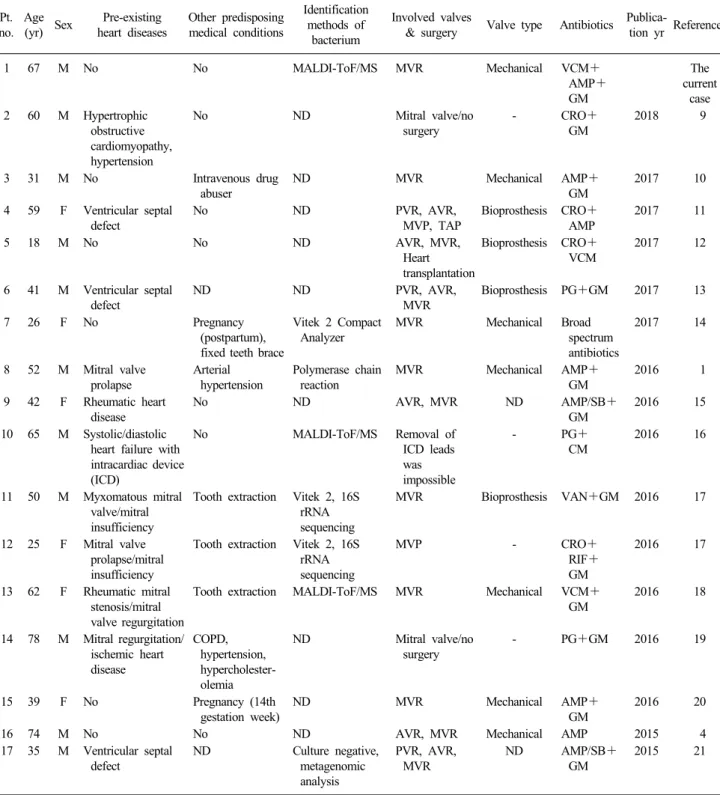
Table 1. Summary of reported cases of Abiotrophia defectiva infective endocarditis
Pt.
no.
Age
(yr) Sex Pre-existing heart diseases
Other predisposing medical conditions
Identification methods of bacterium
Involved valves
& surgery Valve type Antibiotics Publica-
tion yr References
1 67 M No No MALDI-ToF/MS MVR Mechanical VCM+
AMP+
GM
The
current case 2 60 M Hypertrophic
obstructive cardiomyopathy, hypertension
No ND Mitral valve/no
surgery
- CRO+
GM
2018 9
3 31 M No Intravenous drug
abuser
ND MVR Mechanical AMP+
GM
2017 10
4 59 F Ventricular septal defect
No ND PVR, AVR,
MVP, TAP
Bioprosthesis CRO+
AMP
2017 11
5 18 M No No ND AVR, MVR,
Heart transplantation
Bioprosthesis CRO+
VCM
2017 12
6 41 M Ventricular septal defect
ND ND PVR, AVR,
MVR
Bioprosthesis PG+GM 2017 13
7 26 F No Pregnancy
(postpartum), fixed teeth brace
Vitek 2 Compact Analyzer
MVR Mechanical Broad
spectrum antibiotics
2017 14
8 52 M Mitral valve prolapse
Arterial hypertension
Polymerase chain reaction
MVR Mechanical AMP+
GM
2016 1
9 42 F Rheumatic heart disease
No ND AVR, MVR ND AMP/SB+
GM
2016 15
10 65 M Systolic/diastolic heart failure with intracardiac device (ICD)
No MALDI-ToF/MS Removal of
ICD leads was impossible
- PG+
CM
2016 16
11 50 M Myxomatous mitral valve/mitral insufficiency
Tooth extraction Vitek 2, 16S rRNA sequencing
MVR Bioprosthesis VAN+GM 2016 17
12 25 F Mitral valve prolapse/mitral insufficiency
Tooth extraction Vitek 2, 16S rRNA sequencing
MVP - CRO+
RIF+
GM
2016 17
13 62 F Rheumatic mitral stenosis/mitral valve regurgitation
Tooth extraction MALDI-ToF/MS MVR Mechanical VCM+
GM
2016 18
14 78 M Mitral regurgitation/
ischemic heart disease
COPD, hypertension, hypercholester- olemia
ND Mitral valve/no
surgery
- PG+GM 2016 19
15 39 F No Pregnancy (14th
gestation week)
ND MVR Mechanical AMP+
GM
2016 20
16 74 M No No ND AVR, MVR Mechanical AMP 2015 4
17 35 M Ventricular septal defect
ND Culture negative,
metagenomic analysis
PVR, AVR, MVR
ND AMP/SB+
GM
2015 21
Abbreviations: ND, not described; MALDI-ToF/MS, matrix-assisted laser desorption/ionization time-of-flight mass spectrometry; COPD, chronic obstructive pulmonary disease; AVR, aortic valve replacement; MVR, mitral valve replacement; MVP, mitral valve plasty; PVR, pulmonic valve replacement; TAP, tricuspid annuloplasty; AMP, ampicillin; CRO, ceftriaxone; CM, chloramphenicol; GM, gentamicin; PG, penicillin G; RIF, rifampin; SB, sulbactam; VCM, vancomycin.
and vegetation (1.0×2.6 cm sized) on anterior and posterior mi- tral leaflets. Blood testing revealed anemia (Hb 9.6 g/dL; refer- ence range 13.5-17.5 g/dL) and a normal leukocyte count (8,640
cells/mm3; reference range 6,510-13,320 cells/mm3). Serum C-reactive protein (7.75 mg/dL; reference range 0-0.5 mg/dL) and B-type natriuretic peptide (681 pg/mL; reference range
0-100 pg/mL) were elevated. In the absence of any neurological symptom, preoperative brain MRI (magnetic resonance imaging) showed multiple diffuse restriction foci in both cerebral hemi- spheres and left cerebellum, and subarachnoid hemorrhage (SAH) along both parietal and right occipital sulci. Blood cul- tures were requested and ceftriaxone and vancomycin were started empirically. To prevent further embolism by the cardiac vegetation, emergent mitral valve replacement was conducted through right mini-thoracotomy. Intraoperative findings showed massive destruction of anterior and posterior mitral valve leaf- lets with huge vegetation, which extended to posterior medial annulus of the mitral valve and to posterior left atrial endocardium. After massive debridement of all infected tissues, the mitral valve was replaced with a Carpentier-Edwards Perimount Magna mitral valve bioprosthesis (Edwards Lifesciences, Irvine, CA, USA). The preoperative blood culture revealed Gram positive cocci in three sets of culture bottles. The organism was identified as A. defectiva by MALDI-ToF/MS (matrix-assisted laser desorption/ionization time-of-flight mass spectrometry; bioMérieux, Marcy-l'Étoile, France), and E-testing showed susceptibility to penicillin and vancomycin (bioMérieux, Durham, NC, USA). Accordingly, vancomycin, ampicillin and gentamicin antibiotic treatment was continued for 6 weeks. His postoperative recovery course was uncomplicated and resulted in complete disease resolution. At the time of writing the patient had been followed uneventfully for 4 months.
DISCUSSION
A. defectiva endocarditis cases have been continuously re- ported since the bacterium was first identified as a cause of sub-acute infectious endocarditis in 1961 [2]. Roberts et al. [5]
reported A. defectiva, which was originally called Streptococcus mitior or vitamin B6-dependent streptococcus, accounted for 5-6% of all microbial endocarditis cases during the periods 1944 to 1955 and 1970 to 1978. Subsequently the incidence of A. de- fectiva associated infective endocarditis seemed to decrease. For example, Brouqui and Raoult [6] reported in 2001 that 4.3% of cases of streptococcal endocarditis, that is, not all cases of mi- crobial endocarditis, were caused by Abiotrophia spp., and Raoult et al. [7] reported in 2005 that only 2 of 348 micro- biologically confirmed endocarditis were caused by A. defectiva.
Recently, Doig et al. [8] reported Abiotrophia spp. was the eti- ology in 4 of 112 (3.6%) cases of infective endocarditis.
Since 2015, 17 cases (including our case) of A. defectiva en-
docarditis have been reported in the English literature (Table 1) [1,4,9-21], and these cases show a male predominance (11:6) and a mean age of 48.5±18.1 years. Approximately 2/3 had a pre-existing heart disease and of 15 with other medical con- ditions, eight had a history of some specific event like tooth ex- traction or pregnancy. Thus, only one case, two including the current case, did not have a pre-existing heart problem or medi- cal condition. Among the 17 cases, the mitral valve was most frequently involved. Fortunately, all cases were successfully treated with appropriate antibiotics and/or surgery, although car- diac transplantation was needed in one case [12].
The microbiological aspects of this organism are of concern.
A. defectiva requires specific growth factors, including vitamin B6, and is rarely isolated from clinical specimens, as is demon- strated by the literature [5-8]. Accordingly, because it is only rarely detected laboratory workers are likely to be unfamiliar with the microorganism. However, modern automated blood cul- ture and MALDI-ToF/MS made it easier to cultivate and identi- fy this bacterium. Actually, our literature review showed three of six cases, in which identification methods were specified, were identified by MALDI-ToF/MS (Table 1). Antimicrobial susceptibility data for A. defectiva is also limited. Generally speaking, A. defectiva associated infective endocarditis is less susceptible to penicillin, synergistically responds to beta-lactams or vancomycin with aminoglycosides and requires long-term combination therapy (4 to 6 weeks) [22]. As noted in Table 1, a combination of beta-lactams or vancomycin and aminoglyco- sides were administered to 12 of 15 cases, in which treatment regimens were specified.
In conclusion, A. defectiva infective endocarditis is rarely encountered. Because of the needs for urgent surgery and long-term antibiotic therapy and likely lack of laboratory experi- ence of the organism, physicians and laboratory workers should pay close attention to possible cases with a Gram positive cocci blood culture result.
ACKNOWLEDGMENTS
This work was supported by the annual clinical research grant from Pusan National University Yangsan Hospital.
REFERENCES
1. Giannakopoulos K, Zompolou C, Behnes M, Elmas E, Borggrefe M, Akin I. Infective endocarditis-a word of caution on non-typical bacteria. Eur Rev Med Pharmacol Sci 2016;20:4782-5.
2. Frenkel A and Hirsch W. Spontaneous development of L forms of streptococci requiring secretions of other bacteria or sulphydryl compounds for normal growth. Nature 1961;191:728-30.
3. Lainscak M, Lejko-Zupanc T, Strumbelj I, Gasparac I, Mueller-Premru M, Pirs M. Infective endocarditis due to Abiotrophia defectiva: a report of two cases. J Heart Valve Dis 2005;14:33-6.
4. Carleo MA, Del Giudice A, Viglietti R, Rosario P, Esposito V.
Aortic valve endocarditis caused by Abiotrophia defectiva: case report and literature overview. In Vivo 2015;29:515-8.
5. Roberts RB, Krieger AG, Schiller NL, Gross KC. Viridans streptococcal endocarditis: the role of various species, including pyridoxal-dependent streptococci. Rev Infect Dis 1979;1:955-66.
6. Brouqui P and Raoult D. Endocarditis due to rare and fastidious bacteria. Clin Microbiol Rev 2001;14:177-207.
7. Raoult D, Casalta JP, Richet H, Khan M, Bernit E, Rovery C, et al. Contribution of systematic serological testing in diagnosis of infective endocarditis. J Clin Microbiol 2005;43:5238-42.
8. Doig F, Loewenthal M, Lai K, Mejia R, Iyengar A. Infective endocarditis: a Hunter New England perspective. Intern Med J 2018;48:1109-16.
9. Chowdhury S and German ML. Rare but not infrequent: infective endocarditis caused by Abiotrophia defectiva. Case Rep Infect Dis 2018;2018:5186520.
10. Rudrappa M and Kokatnur L. Infective endocarditis due to Abiotrophia defectiva and its feared complications in an immunocompetent person: rare, but real. J Glob Infect Dis 2017;9:79-81.
11. Planinc M, Kutlesa M, Barsic B, Rudez I. Quadruple-valve infective endocarditis caused by Abiotrophia defectiva. Interact Cardiovasc Thorac Surg 2017;25:998-9.
12. Escarcega E, Trovato C, Idahosa O, Gillard J, Stankewicz H.
Abiotrophia defectiva endocarditis: an easy miss. Clin Pract Cases Emerg Med 2017;1:229-31.
13. Bhattacharya P, Mohammed A, Mizrahi E. Aorto-right ventricular fistula: a rare complication of Abiotrophia endocarditis. Oxf Med Case Reports 2017;2017:omx035.
14. Birlutiu V and Birlutiu RM. Endocarditis due to Abiotrophia defectiva, a biofilm-related infection associated with the presence of fixed braces: a case report. Medicine (Baltimore) 2017;96:
e8756.
15. Bozkurt I, Coksevim M, Cerik IB, Gulel O, Tanyel E, Leblebicioglu H. Infective endocarditis with atypical clinical feature and relapse by Abiotrophia defectiva. J Saudi Heart Assoc 2017;29:136-8.
16. van Roeden S, Hartog H, Bongers V, Thijsen S, Sankatsing S.
(18)F-FDG-PET scanning confirmed infected intracardiac device- leads with Abiotrophia defectiva. Case Rep Cardiol 2016;2016:
6283581.
17. Rhodes HM, Hirigoyen D, Shabnam L, Williams DN, Hansen GT.
Infective endocarditis due to Abiotrophia defectiva and Granulicatella spp. complicated by infectious intracranial cerebral aneurysms: a report of three cases and review of the literature. J Med Microbiol 2016;65:493-9.
18. Park S, Ann HW, Ahn JY, Ku NS, Han SH, Hong GR, et al. A case of infective endocarditis caused by Abiotrophia defectiva in Korea. Infect Chemother 2016;48:229-33.
19. Mouyis K, Metaxa S, Missouris C. Abiotrophia defectiva endocarditis complicated by ventricular tachycardia. J Heart Valve Dis 2016;25:114-5.
20. Botta L, Merati R, Vignati G, Orcese CA, De Chiara B, Cannata A, et al. Mitral valve endocarditis due to Abiotrophia defectiva in a 14th week pregnant woman. Interact Cardiovasc Thorac Surg 2016;22:112-4.
21. Fukui Y, Aoki K, Okuma S, Sato T, Ishii Y, Tateda K.
Metagenomic analysis for detecting pathogens in culture-negative infective endocarditis. J Infect Chemother 2015;21:882-4.
22. Sinner SW and Tunkel AR. Viridans Streptococci, Nutritionally Variant Streptococci, Groups C and G Streptococci, and Other Related Organisms. In: Bennet JE, Dolin R, Blaser MJ, eds.
Mandell, Douglas, and Bennett's Principles and Practice of Infectious Diseases. 8th ed. Philadelphia, PA: Elsevier Saunders, 2015:2349-61.
=국문초록=
건강 성인에서의
Abiotrophia defectiva
에 의한 심내막염과 문헌고찰양산부산대학교병원 1흉부외과, 2진단검사의학과
제형곤1, 송두열2, 장철훈2
Abiotrophia defectiva 심내막염은 매우 드물다. 심한 호흡곤란과 폐부종을 호소하는 67세 남자가 지역 병원에서 전원되어 왔다. 술 전에 시행한 경흉벽 심에코검사에서 심한 승모판 역류와 큰 증식증을 보였다. 혈액배양에서 그람양성 영양요구 성 사슬알균인 A. defectiva가 자랐다. 환자는 응급으로 승모판 치환술을 받고 6주간의 복합 항균제(vancomycin, ampicillin, and gentamicin) 치료로 완전히 회복되었다. 이와 같은 환자는 응급 수술과 장기간의 항균제 치료가 필요하지만 임상 의 사와 검사실 근무자가 이 세균에 대한 경험이 적으므로, 혈액배양에서 그람양성 세균이 나오면 이 세균일 가능성을 주의 깊게 검토해야 한다. [Ann Clin Microbiol 2019;22:23-27]
교신저자 : 장철훈, 50612, 경남 양산시 물금읍 금오로 20 양산부산대학교병원 진단검사의학과 Tel: 055-360-1877, Fax: 055-360-1880 E-mail: [email protected]