Mechanical Antiallodynic Effect of Intrathecal Nefopam in a Rat Neuropathic Pain Model
Nefopam has a pharmacologic profile distinct from that of opioids or other anti-
inflammatory drugs. Several recent studies demonstrate that nefopam has a mechanism of action similar to those of anti-depressants and anticonvulsants for treating neuropathic pain. The present study investigates the mechanical antiallodynic effect of nefopam using immunohistochemical study and western blot analysis in a rat neuropathic pain model.
Twenty-eight male Sprague-Dawley rats were subjected to left fifth lumbar (L5) spinal nerve ligation and intrathecal catheter implantation, procedures which were not performed on the 7 male Sprague-Dawley rats in the sham surgery group (group S).
Nefopam, either 10 or 100 μg/kg (group N10 or N100, respectively), and normal saline (group C) were intrathecally administered into the catheter every day for 14 days. The mechanical allodynic threshold of intrathecal nefopam was measured using a dynamic plantar aesthesiometer. Immunohistochemistry targeting cluster of differentiation molecule 11b (CD11b) and glial fibrillary acidic protein (GFAP) was performed on the harvested spinal cord at the level of L5. Extracellular signal-regulated kinase 1/2 (ERK 1/2) and cyclic adenosine monophosphate response element binding protein (CREB) were measured using western blot analysis. The N10 and N100 groups showed improved mechanical allodynic threshold, reduced CD11b and GFAP expression, and attenuated ERK 1/2 and CREB in the affected L5 spinal cord. In conclusion, intrathecal nefopam reduced mechanical allodynia in a rat neuropathic pain model. Its mechanical antiallodynic effect is associated with inhibition of glial activation and suppression of the transcription factors’
mitogen-activated protein kinases in the spinal cord.
Keywords: Mechanical Allodynia; Nefopam; Neuropathic Pain; Spinal Nerve Ligation Kyung-Hoon Kim,1 Gyeong-Jo Byeon,1
Hee-Young Kim,1 Seung-Hoon Baek,1 Sang-Wook Shin,1 and Sung-Tae Koo2
1Department of Anesthesia and Pain Medicine, Pusan National University School of Medicine;
Research Institute for Convergence of biomedical science and technology Pusan National University Yangsan Hospital, Yangsan; 2Division of Meridian and Structural Medicine, Pusan National University School of Korean Medicine, Yangsan, Korea Received: 20 October 2014
Accepted: 29 April 2015 Address for Correspondence:
Gyeong-Jo Byeon, MD
Department of Anesthesia and Pain Medicine, Pusan National University Yangsan Hospital, Geumo-ro 20, Yangsan 626-770, Korea
Tel: +82.55-360-2129, Fax: +82.55-360-2149 E-mail: byeongj@pusan.ac.kr
Funding: This study was supported by Research institute for Convergence of biomedical science and technology (30-2014- 002) Pusan National University Yangsan Hospital.
http://dx.doi.org/10.3346/jkms.2015.30.8.1189 • J Korean Med Sci 2015; 30: 1189-1196
INTRODUCTION
Neuropathic pain is defined as pain resulting from damage or dysfunction of peripheral nerves and as a result of injury or dis- ease of the somatosensory system. Many drugs such as opioids, anticonvulsants, and antidepressants offer relatively effective relief from neuropathic pain (1-5).
Nefopam has been used to control postoperative pain since 1976, and several animal and human clinical studies demon- strate its analgesic activity. It has also been demonstrated to in- duce a rapid and strong depression of the nociceptive flexion reflex in humans (6). In a study conducted using a rat model, Nefopam showed a pre-emptive analgesic effect on chronic constriction injury (CCI) of the sciatic nerve, which involves the activation of N-methyl-D-aspartate (NMDA) receptors (7). Re- cently, nefopam is suspected to induce analgesia using a mech- anism similar to those of triple-receptor (serotonin, norepine- phrine, and dopamine) reuptake inhibitors (8). Thus, it may prove beneficial in treating neuropathic pain, in addition to its
effect on nociceptive pain. However, few studies investigate the efficacy of nefopam in alleviating neuropathic pain.
Unilateral ligation of the fifth and sixth lumbar spinal nerves in rats produces signs that are representative of neuropathic pain, including mechanical allodynia, hyperalgesia, and spon- taneous pain (9). The procedure causes a significant increase in transcription factors’ mitogen-activated protein (MAP) kinase such as extracellular signal-regulated kinase 1/2 (ERK 1/2), and cyclic AMP response element binding (CREB) and activation of spinal glial cells, which are molecular indicators of allodynia (10-12).
Therefore, the present study evaluates the efficacy of intra- thecal nefopam in attenuating mechanical allodynia and char- acterizes its mechanism of action in a spinal nerve ligation (SNL) rat model. We conducted immunohistochemistry targeting clus- ter of differentiation molecule 11b (CD11b) and glial fibrillary acidic protein (GFAP) which are molecular indicators of micro- glial and astrocytic activation and assessed by western blot anal- ysis using ERK 1/2 and CREB in a SNL rat model.
MATERIALS AND METHODS Animals
Twenty-eight male Sprague-Dawley rats, initially weighing 150- 250 g, were used. Two rats per cage were housed under stan- dard laboratory conditions under controlled room temperature (23 ± 2°C), humidity (55% ± 5%), and a 12-h light/dark cycle.
Rats had free access to food and water. Body weight was record- ed on each experimental day. Rats were divided randomly into 4 experimental groups: sham surgery group (S group, n = 7), in- trathecal normal saline group (C group, n = 7), intrathecal ne- fopam (10 μg/kg) group (N10 group, n = 7), and intrathecal ne- fopam (100 μg/kg) group (N100 group, n = 7).
Fifth lumbar (L5) spinal nerve ligation and intrathecal catheter implantation
All surgical procedures were performed under inhalational an- esthesia with isoflurane in 100% oxygen, induced at 2 vol% and maintained at 1.5 vol%. Rats were anesthetized and placed on a surgical apparatus in a prone position. A dorsal midline incision was performed, the left paraspinal muscles were separated from the spinous processes at L4-S1, and the left L5 transverse pro- cess was removed. The left L5 spinal nerves were identified and tightly ligated with a 6-0 silk suture, except in the S group rats, which did not have. Then, the incision was closed. After a 7-day postoperative period, rats in the C, N10, and N100 groups were implanted under anesthesia with a sterilized 32-gauge polyeth- ylene intrathecal catheter (CR3212 Cth RSR 32G, RecathCo, LLC, Allison Park, PA, USA) connected to an 8.5 cm Tygon external tubing (Saint-Gobain Performance Plastics, Akron, OH, USA).
The catheter was inserted at the cisterna magna, pushed cau- dally to the spinal cord lumbar enlargement, and finally exter- nalized through the skin. Proper location of the catheter was confirmed by inducing a temporary motor block of both hind- limbs after injection of 2% lidocaine 5 µL, followed by saline.
Only rats that showed no evidence of neurologic deficit or pa- ralysis after surgery and lidocaine injection were studied. All rats of 4 experimental groups were showed no evidence of neu- rologic deficit or paralysis.
Drug administration
Nefopam and normal saline were both injected intrathecally every day for 14 days after intrathecal catheter implantation. In group N10 or N100, nefopam was dissolved in normal saline to a concentration of either 10 or 100 μg/kg, and 5 µL was injected, followed by a 10 µL normal saline injection to flush the catheter.
In group C, 5 µL of normal saline injected and followed 10 µL normal saline injection to flush the catheter.
Behavioral assessments
All rats were allowed about 10 min of adaptation time in the trans-
parent acrylic box under the wire mesh bench. Withdrawal thre- shold to hind-paw pressure was measured using a dynamic plan- tar aesthesiometer (Ugo Basile, Comerio, Italy) and expressed in grams. A metal filament (0.5-mm diameter) was pushed against the hind paw with increasing force, from 0 to 50 g over a 10 s period. When the rat withdrew its hind paw, the mechanical stimulus stopped automatically, and the force was recorded to the nearest 0.1 g. A maximal cut-off value of 50 g was set to pre- vent tissue injury. Withdrawal responses were averaged from four non-consecutive trials at 10-s intervals, and each paw with- drawal threshold was averaged from four measurements. Each animal was trained for this test for 3 days prior to spinal nerve ligation. On day 1 after intrathecal catheter implantation, the time course of withdrawal threshold after first intrathecal injec- tion of nefopam was recorded at 30-min intervals until reach- ing 180 min. Before intrathecal injection of nefopam, the change in withdrawal threshold was recorded before SNL (Initial), on day 3 after SNL (Post-SNL), on day 1 after surgical intrathecal catheter implantation (Post-ICI), and on days 7 and 14 after in- trathecal injection of nefopam every day (Day-7 and Day-14) (Fig. 1).
Immunohistochemistry
After behavioral assessments were finished, all rats of the four experimental groups were sacrificed for immunohistochemistry.
In the present study, monoclonal antibodies targeting CD11b (ab52478, Abcam plc., Cambridge, MA, USA) and GFAP (ab7260, Abcam plc., Cambridge, MA, USA) were used as primary anti- bodies. These were used at a dilution ratio of 1:2,000 and 1:5,000, respectively. Biotinylated anti-mouse and anti-rabbit immuno- globulin were used as secondary antibodies.
The rats were deeply anesthetized with sodium pentobarbi- tal (50 mg/kg, intraperitoneal) and intracardially administered 20 mL of potassium-free phosphate-buffered saline (K+-free PBS; pH 7.4), followed by 50 mL of 4% paraformaldehyde solu- tion. The L5 spinal cord region was harvested, sectioned, fixed for 3 hr in paraformaldehyde, and cryo-protected overnight in 25% sucrose dissolved in 0.01 M phosphate-buffered saline (pH 7.4). The tissues were fast-frozen in cryo-embedding compound on a mixture of ethanol and dry ice and stored at -80°C.
The preserved tissues were cut transversely at 10-µm thick on a freezing microtome, thaw-mounted onto silane-coated glass slides, and air-dried overnight at room temperature. Slides were incubated overnight at 4°C in PBS-diluted primary anti- bodies against CD11b and GFAP. Then, the slides were incubat- ed in PBS-diluted secondary antibodies (biotinylated anti-mouse IgG, goat anti-rabbit IgG, 1:100) for 2 hr at room temperature and washed with PBS. The slides were exposed to avidin-biotin peroxidase complex for 1 hr at room temperature, washed with PBS, and stained with 0.05% 3, 3-diaminobenzidine tetrahydro- chloride (Sigma Chemical Co., St. Louis, OK, USA). The tissue
sections were posted on gelatin coated slides, and dried for 2 hr at room temperature. The slides were then washed with distilled water for 10 min, dehydrated with alcohol, and rinsed with xylene.
Glial responses were assessed in three randomly chosen L5 spinal cord segments. The specimens were examined with an optical microscope, and the appearance and fluorescence in- tensities of glial cells in the spinal dorsal horn (lamina I-III) were assessed. The same spinal area was also photographed with a digital camera attached to the microscope set at 40 × magnifi- cation. The images were calibrated using Adobe Photoshop 5.0 (Adobe Systems Incorporated, USA), and the relative mean op- tical density was measured in the spinal cord dorsal horn. The data were expressed as the relative mean optical density (MOD) and calculated by the following method (13):
Relative mean optical density ratio = corrected MOD of left gray horn/corrected MOD of right gray horn (corrected MOD
= MOD of area of interest-MOD of background).
Western blot analysis
All rats of the four experimental groups were sacrificed for west- ern blot analysis. The ipsilateral L5 spinal cord segment was re- moved and immediately frozen on dry ice. Spinal cord tissue was homogenized in buffer (250 mM sucrose, 20 mMTris-HCL [pH 7.4], 1.5 mM Na-EDTA, 1.5 mM Na-EGTA, 1 mM MgCl2, 1 mM DTT [Sigma-Aldrich, St. Louis, OK, USA]), 20 mM KCl, and centrifuged. The L5 spinal cord segment proteins were applied to a 10% SDS-polyacrylamide gel. The phospho-extracellular
signal-regulated kinase 1/2 (ERK1/2, CEL-4376S, Cell signaling Technology, Inc., Danvers, MA, USA), total-ERK1/2 (CEL-9102S), phospho-cyclic adenosine monophosphate response element binding protein (CREB, CEL-9198S), and total-CREB (CEL-9197S) were used as primary antibodies. The membrane was blocked with 2% BSA in Tris-buffered saline containing 0.1% Tween (TB- ST) at room temperature for 1 hr and then incubated overnight at 4°C with antibody against β-actin and cytochrome c. After washing with TBST, peroxidase affinipure goat anti-rabbit IgG (JA-111-035-003, Jackson Immuno Research Laboratories Inc., West Grove, PA, USA) was used as a secondary antibody (1:2,000 dilution in 2% BSA in TBST, 1 hr 30 min incubation), and the antigen-antibody complexes were visualized using an enhanced chemiluminescence detection reagent. Bands were scanned using a densitometer (GS-700; Bio-Rad Laboratories), and quan- tified with Multi-Analyst 1.0.2 software (Bio-Rad Laboratories, Hercules, CA, USA). The data were expressed as immunoblot reactive band intensities (%). Immunoblot reactive band inten- sities were measured and compared to the densities of the cor- responding loading control (β-actin).
Statistical analysis
The mean ± standard error was calculated and the data analyz- ed using the SPSS Statistics 16.0 for Windows (SPSS Inc, Chica- go, IL, USA). Comparisons of behavioral assessments between the groups at identical time points were performed using one- way analysis of variance (ANOVA), followed by Tukey test for Fig. 1. Behavioral assessments. (A) Schematic representation of behavioral assessments after intrathecal injection of nefopam. (B) Time course change of hind limb mechanical sensitivity to a dynamic plantar aesthesiometer after intrathecal injection of nefopam. (C) The change in withdrawal threshold following intrathecal injection of nefopam (10 or 100 μg/kg) every day or normal saline in rats receiving spinal nerve ligation (SNL). *P < 0.05 compared to control by one-way analysis of variance (ANOVA); †P < 0.05 com- pared to baseline by repeated measure ANOVA. S: sham surgery group, C: normal saline group, N10: 10 μg/kg nefopam group, N100: 100 μg/kg nefopam group, Initial: before SNL, Post-SNL: 3rd day after SNL, Post-ICI: 1st day after surgical intrathecal catheter insertion, Day-7: 7th day after intrathecal nefopam injection, Day-14: 14th day after intra- thecal nefopam injection.
Behavioral assessments
Spinal nerve ligation Intrathecal catheter implantation
Initial Post-SNL
Day: 0 1 2 3 4 5 6 7 8 9 10 11 12 13 14 15 16 17 18 19 20 21 22 Drug administration
Post-ICI Day-7 Day-14
Withdrawal threshold (g)
Time (min)
Initial 0 30 60 90 120 150 180 40
30
20
10
0
S C N10 N100
*
*
*
* *
* *
*
Withdrawal threshold (g)
Time (min)
Initial Post-SNL Post-ICI Day-7 Day-14 40
30
20
10
0
S C N10 N100
† †
A
B C
multiple post hoc analysis. Intragroup comparisons of post-treat- ment and baseline values were made using repeated measure ANOVA, followed by Tukey test for multiple post hoc analysis.
Comparisons of immunohistochemical study and western blot analysis between the groups were performed using Kruskal-Wal- lis test. Values of P < 0.05 were considered significant. Specific calculated significance levels are indicated in the figures.
Ethics statement
The experimental protocol was approved by the institutional animal care and use committee of Pusan National University (Approval number: PNU-2012-0046).
RESULTS
All the rats maintained good health and continued to gain weight through the experimental period. No motor dysfunction was
observed in rats that were injected nefopam and normal saline.
No significant side effects, such as ascites, weight loss, or alope- cia were observed during the study.
Nefopam reduced mechanical antiallodynic threshold in the SNL rat model
The withdrawal threshold decreased significantly relative to baseline withdrawal on day 3 after SNL, confirming the devel- opment of mechanical allodynia. On day 1 after intrathecal cath- eter implantation, intrathecal injection of nefopam (10-100 µg/
kg) produced antiallodynic effects in a dose dependent manner (P < 0.05), and the peak effect was observed at 30 min after in- jection. Following intrathecal catheter implantation, the with- drawal thresholds in the N10 and N100 groups were higher than in group C at 7 and 14 days after intrathecal injection of nefo- pam, and a significant mechanical antiallodynic effect occurred (P < 0.05) (Fig. 1).
Fig. 2. Photographs (40 × magnification) illustrating the effect of nefopam on spinal immunoreactivity to cluster of differentiation molecule 11b (CD11b) after left L5 spinal nerve ligation. The immunohistochemical intensity of CD11b in the L5 segment decreased in a dose dependent manner following nefopam administration. (A) sham surgery group, (B) normal saline group, (C) 10 μg/kg of nefopam group, (D) 100 μg/kg of nefopam group.
N10 N100
S A C
C
B
D
Nefopam suppressed microglial and astrocytic activation in the SNL rat model
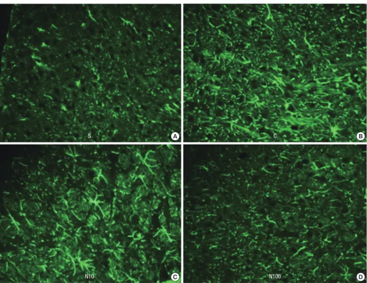
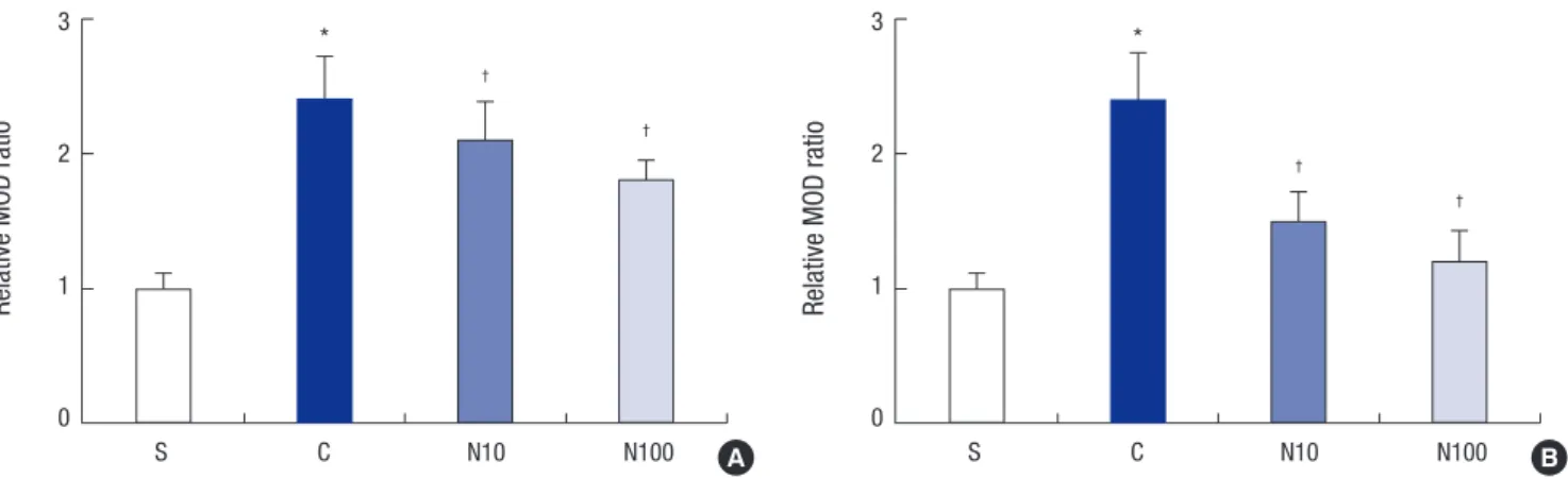
Fourteen days after intrathecal injection of nefopam, the CD11b and GFAP immunofluorescence intensities decreased on the ipsilateral L5 spinal cord region in groups N10 and N100 (Fig. 2 and 3). No group showed significant change in the contralateral spinal cord region (Figs not shown). The relative MOD ratio of CD11b and GFAP in the ipsilateral dorsal horn showed mark- edly intense CD11b and GFAP immunoreactivity, indicating that microglia and astrocytes had been activated. Intrathecal nefopam dose-dependently attenuated the microglial and as- trocytic activation in the left L5 dorsal horn, indicated by the decreased relative MOD ratio of CD11b and GFAP (Fig. 4).
Nefopam suppressed the transcription factors’ MAP kinases, such as ERK 1/2, and CREB, induced by spinal nerve ligation
Fourteen days after surgery, the ipsilateral L5 spinal cord seg-
ment was removed, and the amount of ERK 1/2 and CREB were quantified by western blot analysis. Analysis revealed a signifi- cantly increased ERK 1/2 and CREB on the ipsilateral side of the L5 SNL when compared to the S group. However, the amount of ERK 1/2 and CREB induced to the L5 SNL were attenuated by administration of intrathecal nefopam (Fig. 5).
DISCUSSION
The purpose of the present study was to evaluate the antiallo- dynic effect of intrathecal nefopam in a rat neuropathic pain model. In this study, intrathecal nefopam in a SNL rat model blocked mechanical allodynia, suppressed microglial and as- trocytic activation, and attenuated the transcription factors’ MAP kinases.
Nefopam has a unique profile distinct from opioids or other anti-inflammatory drugs. Several animal studies demonstrate the analgesic activity of nefopam. Daily administration of intra-
Fig. 3. Photographs (40 × magnification) illustrating the effect of nefopam on spinal immunoreactivity to glial fibrillary acidic protein (GFAP) after left L5 spinal nerve ligation.
The immunohistochemical intensity of GFAP in the L5 segment decreased in a dose dependent manner following nefopam. (A) sham surgery group, (B) normal saline group, (C) 10 μg/kg nefopam group, (D) 100 μg/kg nefopam group.
S C
N10 N100
A
C
B
D
Fig. 4. Changes in the relative mean optical density (MOD) ratio for (A) CD11b and (B) GFAP after left L5 spinal nerve ligation. The relative MOD ratio increased in the spinal cord side ipsilateral to the spinal nerve ligation in rats. The relative MOD ratio decreased in a dose-dependent manner in rats administered intrathecal nefopam (N10 and N100) compared to the ratio in rats administered intrathecal saline (group C). *P < 0.05 compared to the S group; †P < 0.05 compared to the C group. S: sham surgery group, C: nor- mal saline group, N10: 10 μg/kg nefopam group, N100: 100 μg/kg nefopam group.
Relative MOD ratio
S C N10 N100
3
2
1
0
*
†
†
Relative MOD ratio
S C N10 N100
3
2
1
0
*
†
†
A B
Fig. 5. The expression of extracellular signal-regulated kinase 1/2 (ERK 1/2) and cyclic adenosine monophosphate response element-binding (CREB) measured by immunoblot- ting. (A) Representative immunoblot images for ERK 1/2 and CREB protein in spinal cord from the S, C, N10, and N100 groups. (B, C) Densitometric quantifications of ERK 1/2 and CREB band intensities. *P < 0.05 compared to the S group; †P < 0.05 compared to the C group. S: sham surgery group, C: normal saline group, N10: 10 μg/kg nefopam group, N100: 100 μg/kg nefopam group.
Immunoblot relative band intensities (%)
S C N10 N100
250 200 150 100 50 0
*
†
†
Immunoblot relative band intensities (%)
S C N10 N100
250 200 150 100 50 0
* †
†
p-CREB
CREB
p-ERK1/2
ERK 1/2
β-actin
S C N10 N100 A
B
C
peritoneal nefopam attenuated pain behavior in a dose-depen- dent manner in a chronic constriction injury (CCI) model of neuropathic pain in rats (14). It also induced a rapid and strong depression of the nociceptive reflex in humans (6). Reuptake inhibition of monoamines, such as serotonin, norepinephrine and dopamine, which mediate descending inhibitory pain mod- ulation, has been suggested as a mechanism for nefopam-in- duced antinociception (15, 16). In addition, nefopam acts as a voltage-gated sodium channel blocker, which may mediate its antinociceptive effects partially or completely (17). Most nefo- pam studies were performed to gauge its antinociceptive effect;
its efficacy in treating neuropathic pain and its underlying phar- macological actions remain unclear. Furthermore, few studies
examine the molecular biological change occurring in the chron- ic neuropathic pain pathway.
In the present study, nefopam at 10 or 100 μg/kg and normal saline were injected intrathecally every day for 14 days after sur- gical implantation of an intrathecal catheter in rats. A significant, dose-dependent mechanical antiallodynic effect was caused by intrathecal nefopam and observed on days 7 and 14. Nefopam is administered by the intramuscular or intravenous route in clinical settings at present; the pharmacologic effects of intra- thecally administered nefopam need to be evaluated due to the important role of spinal cord in the pain pathway. After periph- eral nerve injury, glial activation and cell signaling pathways are induced within the spinal cord. This region also serves as a site
of action for monoamines that mediate descending inhibitory pain modulation (18, 19). There have been few studies on the effect of nefopam at this level. Some studies suggest that anti- nociceptive effects of nefopam have both spinal and supraspi- nal targets (20, 21).
In another study, intrathecal nefopam produced an antinoci- ceptive effect in formalin-induced pain behavior during both phases of the formalin test while interacting with intrathecal morphine synergistically in phase 1 and additively in phase 2 (22). The current study was demonstrated that intrathecal nefo- pam alone also has an antiallodynic effect, and its mechanism was proven through immunohistochemical study and western blot analysis.
Peripheral nerve injury induces profound changes in microg- lia and astrocytes. After peripheral nerve injury, the central ter- minals of primary sensory neurons may release neurotransmit- ters, such as glutamate and substance P, that may activate spi- nal microglia and astrocytes, and result in the production and release of pronociceptive mediators, such as interleukin-1β, tu- mor necrosis factor-α, prostaglandin E2, and adenosine triphos- phate (23). Thereafter, microglia and astrocytes form a positive feedback loop and release pronociceptive mediators continu- ously (24). These mediators may facilitate pain processing by enhancing either presynaptic release of neurotransmitters or postsynaptic excitability (23). In the process, glial activation main tains a chronic pathological state experienced as persistent pain. Microglia respond early in the process, while astrocytes may continue to respond chronically beyond 7 days (25, 26).
Blocking microglial and astrocytic activity and inhibiting prolif- eration are both effective in preventing or delaying neuropathic pain (27). In the present study, intrathecal nefopam suppressed the microglial and astrocytic activation induced by L5 spinal- nerve ligation, indicated by the decreased in CD11b and GFAP, respectively.
There are a few studies on the cell-signaling pathway (CNP) of neuropathic pain in rats. The activation of ERK pathways con- tributes to neuropathic pain in CCI rats, and the function of pho- spho-ERK may be accomplished in part through CREB depen- dent gene expression (11). Increased CREB phosphorylation, induced by SNL, may be one of the key molecular events lead- ing to synaptic alterations and persistent pain in the SNL model of neuropathic pain (12). In the CNP of injured rats, activated kinase, such as phospho-ERK 1/2, is upregulated, which does not occur in injured rats that fail to develop a CNP. Activated CREB is also upregulated following spinal cord injury (28). In the present study, the amounts of ERK 1/2 and CREB protein induced by L5 SNL were attenuated by treatment with intrathe- cal nefopam.
In conclusion, intrathecal nefopam reduced mechanical al- lodynia in a rat neuropathic pain model. Its mechanical antial- lodynic effect may be associated with the inhibition of microg-
lial and astrocytic activation and suppression of the transcrip- tion factors’ MAP kinases in the spinal cord. Therefore, intrathe- cal administration of nefopam may be a promising therapeutic intervention to treat peripheral nerve injury-induced neuropa- thy. Nevertheless, further studies are required to determine the efficacy of intrathecal nefopam for treatment of other forms of neuropathic pain, such as thermal allodynia and hyperalgesia, as well as its safety in a clinical setting.
DISCLOSURE
All authors declare no conflict of interest.
AUTHOR CONTRIBUTION
Conception and design of the manuscript: Kim KH, Byeon GJ, Baek SH, Shin SW. Acquisition of data: Kim HY. Interpretation of data: Byeon GJ. Writing of the manuscript: Kim KH, Byeon GJ. Drafting the article: Kim HY, Byeon GJ, Koo ST. Final approv- al of the version for publishing: Shin SW. Revision of the article for important intellectual content: Koo ST.
ORCID
Gyeong-Jo Byeon http://orcid.org/0000-0001-5333-3894 Sung-Tae Koo http://orcid.org/0000-0001-7773-3905 REFERENCES
1. Lee HJ, Shin SW, Jang HJ. The combined antiallodynic effect of gabapen- tin and milnacipran in a rat neuropathic pain model. Korean J Pain 2007; 20: 8-14.
2. Sindrup SH, Jensen TS. Efficacy of pharmacological treatments of neu- ropathic pain: an update and effect related to mechanism of drug ac- tion. Pain 1999; 83: 389-400.
3. Hunter JC, Gogas KR, Hedley LR, Jacobson LO, Kassotakis L, Thomp- son J, Fontana DJ. The effect of novel anti-epileptic drugs in rat experi- mental models of acute and chronic pain. Eur J Pharmacol 1997; 324:
153-60.
4. Idänpään-Heikkilä JJ, Guilbaud G. Pharmacological studies on a rat model of trigeminal neuropathic pain: baclofen, but not carbamaze- pine, morphine or tricyclic antidepressants, attenuates the allodynia-like behaviour. Pain 1999; 79: 281-90.
5. Iyengar S, Webster AA, Hemrick-Luecke SK, Xu JY, Simmons RM. Effi- cacy of duloxetine, a potent and balanced serotonin-norepinephrine reuptake inhibitor in persistent pain models in rats. J Pharmacol Exp Ther 2004; 311: 576-84.
6. Guirimand F, Dupont X, Bouhassira D, Brasseur L, Chauvin M. Nefo- pam strongly depresses the nociceptive flexion (R(III)) reflex in humans.
Pain 1999; 80: 399-404.
7. Biella GE, Groppetti A, Novelli A, Fernández-Sánchez MT, Manfredi B, Sotgiu ML. Neuronal sensitization and its behavioral correlates in a rat
model of neuropathy are prevented by a cyclic analog of orphenadrine. J Neurotrauma 2003; 20: 593-601.
8. Gregori-Puigjané E, Setola V, Hert J, Crews BA, Irwin JJ, Lounkine E, Mar- nett L, Roth BL, Shoichet BK. Identifying mechanism-of-action targets for drugs and probes. Proc Natl Acad Sci U S A 2012; 109: 11178-83.
9. Kim SH, Chung JM. An experimental model for peripheral neuropathy produced by segmental spinal nerve ligation in the rat. Pain 1992; 50:
355-63.
10. Zhuang ZY, Gerner P, Woolf CJ, Ji RR. ERK is sequentially activated in neurons, microglia, and astrocytes by spinal nerve ligation and contrib- utes to mechanical allodynia in this neuropathic pain model. Pain 2005;
114: 149-59.
11. Song XS, Cao JL, Xu YB, He JH, Zhang LC, Zeng YM. Activation of ERK/
CREB pathway in spinal cord contributes to chronic constrictive injury- induced neuropathic pain in rats. Acta Pharmacol Sin 2005; 26: 789-98.
12. Ma W, Quirion R. Increased phosphorylation of cyclic AMP response ele- ment-binding protein (CREB) in the superficial dorsal horn neurons fol- lowing partial sciatic nerve ligation. Pain 2001; 93: 295-301.
13. Kang G, Choi KY, Lee MS, Ahn YJ, Kang SS, Cheong IY, Chun W, Kim SS. Minocycline attenuates the development of allodynia: an immuno- histochemical study on CD11b, GFAP and c-Fos in the spinal dorsal horn in SD rat. Korean J Pathol 2004; 38: 311-8.
14. Saghaei E, Moini Zanjani T, Sabetkasaei M, Naseri K. Enhancement of antinociception by co-administrations of nefopam, morphine, and nime- sulide in a rat model of neuropathic pain. Korean J Pain 2012; 25: 7-15.
15. Fuller RW, Snoddy HD. Evaluation of nefopam as a monoamine uptake inhibitor in vivo in mice. Neuropharmacology 1993; 32: 995-9.
16. Rosland JH, Hole K. The effect of nefopam and its enantiomers on the uptake of 5-hydroxytryptamine, noradrenaline and dopamine in crude rat brain synaptosomal preparations. J Pharm Pharmacol 1990; 42: 437-8.
17. Verleye M, André N, Heulard I, Gillardin JM. Nefopam blocks voltage- sensitive sodium channels and modulates glutamatergic transmission in
rodents. Brain Res 2004; 1013: 249-55.
18. Ren K, Dubner R. Descending modulation in persistent pain: an update.
Pain 2002; 100: 1-6.
19. Millan MJ. Descending control of pain. Prog Neurobiol 2002; 66: 355-474.
20. Fasmer OB, Berge OG, Jørgensen HA, Hole K. Antinociceptive effects of (+/-)-, (+)- and (-)-nefopam in mice. J Pharm Pharmacol 1987; 39: 508- 11.
21. Piercey MF, Schroeder LA. Spinal and supraspinal sites for morphine and nefopam analgesia in the mouse. Eur J Pharmacol 1981; 74: 135-40.
22. Cho SY, Park AR, Yoon MH, Lee HG, Kim WM, Choi JI. Antinociceptive effect of intrathecal nefopam and interaction with morphine in forma- lin-induced pain of rats. Korean J Pain 2013; 26: 14-20.
23. Marchand F, Perretti M, McMahon SB. Role of the immune system in chronic pain. Nat Rev Neurosci 2005; 6: 521-32.
24. Mollace V, Colasanti M, Muscoli C, Lauro GM, Iannone M, Rotiroti D, Nistico G. The effect of nitric oxide on cytokine-induced release of PGE2 by human cultured astroglial cells. Br J Pharmacol 1998; 124: 742-6.
25. Kreutzberg GW. Microglia: a sensor for pathological events in the CNS.
Trends Neurosci 1996; 19: 312-8.
26. Raghavendra V, Tanga F, Rutkowski MD, DeLeo JA. Anti-hyperalgesic and morphine-sparing actions of propentofylline following peripheral nerve injury in rats: mechanistic implications of spinal glia and proin- flammatory cytokines. Pain 2003; 104: 655-64.
27. Ledeboer A, Sloane EM, Milligan ED, Frank MG, Mahony JH, Maier SF, Watkins LR. Minocycline attenuates mechanical allodynia and proin- flammatory cytokine expression in rat models of pain facilitation. Pain 2005; 115: 71-83.
28. Crown ED, Ye Z, Johnson KM, Xu GY, McAdoo DJ, Hulsebosch CE. In- creases in the activated forms of ERK 1/2, p38 MAPK, and CREB are cor- related with the expression of at-level mechanical allodynia following spinal cord injury. Exp Neurol 2006; 199: 397-407.