INTRODUCTION
Choledochal cysts, although rare in adulthood, are now cor- rectly diagnosed more frequently due to recent improvement in biliary imaging techniques (1). There have been numer- ous descriptions and classifications based on the location and anatomical structure of choledochal cyst. Among these, the most practical classification is proposed by Alonso-Lej et al.
(2), which is modified by Todani et al. (3) in 1977. The major point of this classification is that the gallbladder is usually not distended and arises from a small cystic duct, usually not at the upper end of the cyst (4). Initial reports suggested that the following treatments provided satisfactory results: T-tube choledochocystomy with internal or external drainage, cys- tojejunostomy, modified cystojejunostomy with T-tube exter- nal drainage, cyst excision and hepaticoduodenostomy, cys- toduodenostomy, jejunal segment interposition with cysto- duodenostomy, and jejunal segment interposition with hep- aticojejunostomy. However, it is now recognized that these procedures are associated with an unacceptably high rate of late complications. Late complications of internal cyst drainage
may result in a high rate of cholangiocarcinoma arising from the cyst (1), as well as the development of cholangitis and biliary stone disease (1, 5-8). Surgical excision of cyst and hepaticojejunostomy are now considered as the treatment of choice for choledochal cysts (1, 5-8). In this paper, we describe our experience with 72 patients with choledochal cysts.
MATERIALS AND METHODS
Seventy-two patients (59 females and 13 males) with ages ranging from 15 to 63 (mean age=31) yr were treated for choledochal cyst by the Department of Surgery, the second Affiliated Hospital of Harbin Medical University, between January 1, 1985 and December 31, 2002. The medical records of all 72 patients were reviewed. Choledochal cysts were clas- sified according to Alonso-Lej et al. (2) and Todani’s modified classification system (2, 3). Follow-ups were done on all of the patients until December 31, 2002. We analyzed the clin- ical outcome in patients who underwent the following treat- ments: cyst excision with hepatojejunostomy (Fig. 1), cyst
Long-Xian Zheng, Hong-Bo Jia, De-Quan Wu, Hong Shang*, Xiang-Yu Zhong, Qiu-Shi Wang, Wen-Xue Zhou, Zhen-Hua Sun
Department of Surgery, the Second Affiliated Hospital of Harbin Medical University; Department of Clinical Laboratory*, the Second Affiliated Hospital of Harbin Medical University, Harbin, China
Address for correspondence Long-Xian Zheng, M.D., Ph.D.
Department of Surgery, the Second Affiliated Hospital of Harbin Medical University, 157 Baojian Road, Harbin, HeiLongJiang PR China 150086 Tel : +86.451-8660-5117, Fax : +86.451-8666-6546 E-mail : [email protected]
842
Experience of Congenital Choledochal Cyst in Adults: Treatment, Surgical Procedures and Clinical Outcome in the Second Affiliated Hospital of Harbin Medical University
This study was undertaken to analyze and evaluate the diagnosis and principal treatment methods for congenital choledochal cyst, focusing on various surgical procedures and clinical outcome. A comprehensive, retrospective study was con- ducted on 72 adult patients who presented with choledochal cyst from 1985 to 2002. Surgical procedures were cyst excision with hepaticojejunostomy in 25 cases for type I or type IV-B, extrahepatic cyst excision with hepaticojejunostomy in 8 cases for type IV-A, extrahepatic cyst excision with modified hepaticojejunostomy in 2 cases for type IV-B, non-cyst excision with or without hepaticojejunostomy in 27 cases for types I, II, IV-A, IV-B. The early postoperative morbidity and mortality rate were 16.1% (9/62) and 6.5% (4/62) respectively, and the complication rate related to surgical procedure was 30.6% (19/62). The incidence of cholangiocarci- noma with non-cyst excision or non-operated congenital choledochal cyst was 10.8% (4/37). One patient died of primary hepatocellular carcinoma after cyst exci- sion with hepatojejunostomy. In conclusion, our results showed that complete exci- sion of choledochal cyst for types I, II, and IV-B and complete excision of extra- hepatic choledochal cyst from the hepatic hilum in type IV-A with hepaticojejunos- tomy or modified hepaticojejunostomy are the treatment of choice for choledochal cyst in adult patients.
Key Words : Choledochal Cyst; Surgical Procedures, Operative; Biliary Tract; Anastomosis Roux-en-Y; Adult;
Disease Management
Received : 17 April 2004 Accepted : 5 July 2004
excision with modified hepatojejunostomy (Fig. 2), and non- cyst excision and without hepatojejunostomy.
RESULTS
A total of 72 adult patients were identified with chole- dochal cysts. There were 49 (68.1%) Type I or Type IV-B, one (1.3%) Type II, 21 (29.3%) Type IV-A, and one (1.3%) Type V choledochal cysts. Among the patients, the most common symptom was abdominal pain, which occurred in 64 of the 72 patients (88.9%). Jaundice was present in 40 patients (55.6%), and abdominal mass was present in 21 patients (29.2%). Only eight patients (11.1%) presented with the classic triad of abdominal pain, jaundice, and abdom- inal mass. The biochemical measurement showed anomalies in AST, which appeared in 22 of the 72 patients (30.6%), ALT in 48.6%, -GT in 31.9%, and total bilirubin in 34.7%
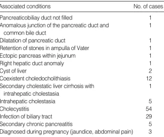
of all the patients. One or more of the following test proce- dures established the diagnosis of choledochal cyst: ultra- sonography (US) (n=49), magnetic resonance imaging (MRI) (n=4), computed tomography (CT) (n=13) and endoscopic retrograde cholangiopancreatography (ERCP) (n=14). The choledochal cysts were associated with other diseases includ- ing the pancreaticobiliary duct not filled in one case, anoma- lous junction of the pancreatic duct and common bile duct in one case, dilatation of pancreatic duct in one case, reten- tion of stones in ampulla of Vater in one case, ectopic pancreas within jejunum in one case, right hepatic duct anomaly in one case, cyst of liver in two cases, coexistent choledocholithi- asis in 12 cases, secondary cholestatic liver cirrhosis, which occurred in one of the 5 cases of intrahepatic cholestasia, chole- cystitis in 54 cases, infection of biliary tract in 29 cases and secondary chronic pancreatitis in 5 cases. Women with chole-
dochal cyst were diagnosed during pregnancy (jaundice, abdo- minal pain) in 3 cases (Table 1).
The definite surgical procedures were cyst excision with hepaticojejunostomy in 25 cases for type I or type IV-B and complete excision of extrahepatic choledochal cyst from the hepatic hilum with hepaticojejunostomy in 8 cases for type IV-A, extrahepatic cyst excision with modified hepaticoje- junostomy in 2 cases for type IV-A, choledochocystomy with internal or external drainage in 3 cases for type I or type IV- B and 1 case in type IV-A, cyst excision and the biliary tract reconstruction by jejunal segment interposition with hep- aticojejunostomy (Fig. 3) in 3 cases for type I or type IV-B and extrahepatic cyst excision and the biliary tract reconstruc- tion by jejunal segment interposition with hepaticojejuno- stomy in 2 cases for type IV-A, the biliary tract reconstruc-
Associated conditions No. of cases
Pancreaticobiliay duct not filled 1
Anomalous junction of the pancreatic duct and 1 common bile duct
Dilatation of pancreatic duct 1
Retention of stones in ampulla of Vater 1
Ectopic pancreas within jejunum 1
Right hepatic duct anomaly 1
Cyst of liver 2
Coexistent choledocholithiasis 12
Secondary cholestatic liver cirrhosis with 1 intrahepatic cholestasia
Intrahepatic cholestasia 5
Cholecystitis 54
Infection of biliary tract 29
Secondary chronic pancreatitis 5
Diagnosed during pregnancy (jaundice, abdominal pain) 3 Table 1.The associated conditions in 72 patients with chole- dochal cyst
Fig. 1.Cyst excision with hepatojejunos- tomy.
Fig. 2.Cyst excision with modified hepa- tojejunostomy.
Fig. 3.Cyst excision, jejunal segment in- terposition with hepatojejunostomy.
Tied
in 2 cases for type I or type IV-B and 1 case in type IV-A, cyst excision and hepaticoduodenostomy in 2 cases for type I or type IV-B and 1 case for type II, cystojejunostomy in 4 cases for type I or type IV-B and 5 cases in type IV-A, modi-
cases were performed non-cyst excision with or without hep- aticojejunostomy in type I, II, IV-A, IV-B. Because of chole- dochal cyst adhered with surrounding tissue extensively, we can not dissect choledochal cyst from the round ligament in 27 cases. Ten out of the 72 patients did not receive operation.
The early postoperative morbidity and mortality rates were 16.1% (9/62) and 6.5% (4/62) respectively. The complica- tion rate related to the surgical procedure was 30.6% (19/62).
The incidence of carcinoma was 6.9% (5/72), while the inci- dence of cholangiocarcinoma with non-cyst excision or non- operated congenital choledochal cyst was 10.8% (4/37). Early postoperative complications included wound infection, sep- tic shock after cystojejunostomy in one case, acute septic peri- tonitis after cyst excision with hepatojejunostomy in one case, abdominal pain, pancreatic ductal dilatation with retrograde cholangitis after cyst excision with hepatojejunostomy in one case, which was resolved after a few days with conservative antibiotics treatment.
Two cases required reoperative revision in the early post- operative period due to anastomotic leakage after cyst exci- sion with Roux-en-Y hepaticojejunostomy. Unfortunately, one patient died of brain infection within one week and the other suffered from anastomotic leakage and intra-abdomi- nal hematoma formation after cystoduodenostomy and even- tually died of multiorgan failure within one week. During the immediate postoperative period, one patient with fistu- la formation after cystoduodenostomy died of hepatic failure within 72 hr. In two cases, a 44 yr old male patient and a 37
Type Surgical procedures No. of
cases I or IV-B Cyst excision with hepaticojejunostomy 25
Cyst excision, and jejunal segment interposition 3 with hepaticojejunostomy
Jejunal segment interposition with 2 cystoduodenostomy
Cystoduodenostomy 2 Cyst excision and hepaticoduodenostomy 2 Cystojejunostomy 4 Choledochocystomy with internal or external drainage 3 IV-A Extrahepatic cyst excision with hepaticojejunostomy 8 Extrahepatic cyst excision with modified 2
hepaticojejunostomy
Choledochocystostomy with internal or external 1 drainage
Extrahepatic cyst excision, and jejunal segment 2 interposition with hepaticojejunostomy
Cystoduodenostomy 1
Cysto-jejunostomy 5
Modified cystojejunostomy with T-tube external 1 drainage
II Cyst excision and hepaticoduodenostomy 1 Table 2.The surgical procedures according to the choledochal cyst type in 62 out of 72 patients
Type Sex Initial operation Complication Final operation Prognosis
(Survival time) Patients
No. Age (yr)
1 56 I or IV-B F Cyst excision, hepatojejunostomy Acute septic peritonitis Non-operation Survived (12 yr) 2 36 I or IV-B F Cyst excision, hepatojejunostomy Leakage of anastomosis Non-operation Survived (5 yr) 3 18 IV-A F Cyst excision, hepatojejunostomy Primary hepatocellular carcinoma Exploratory operation Died (10 yr)
4 37 I or IV-B F Cyst excision, hepatojejunostomy Cholangiocarcinoma Died (6 month)
5 44 I or IV-B M Cyst excision, hepatojejunostomy Cholangiocarcinoma Died (6 month)
6 34 I or IV-B F Cyst excision, hepatojejunostomy Brain infection Exploratory operation Died (1 week) 7 32 I or IV-B F Cyst excision, hepatojejunostomy Retrograde cholangitis Non-operation Survived (5 yr) 8 26 I or IV-B F Cyst excision, hepatojejunostomy Retrograde cholangitis Exploratory biopsy Died (10 yr)
Cholangioadenocarcinoma
9 29 IV-A F Choledochocystomy with Retention of stones Extrahepatic cyst excision, Survived (2 yr) external drainage in ampulla of Vater hepatojejunostomy
10 20 I or IV-B F Cyst excision, jejunal segment Anastomosis stricture Modified hepatojejunostomy Survived (14 yr) interposition with hepatojejunostomy
11 20 I or IV-B F Cystoduodenostomy Intra-abdominal hematoma Died (1 week)
multiorgan failure
12 33 IV-A F Cystoduodenostomy Fistula Died (3 hr)
13 17 IV-A F Cystoduodenostomy Retrograde cholangitis Cyst excision, hepatojejunostomy Survived (14 yr) 14 25 I or IV-B F Cystoduodenostomy Retrograde cholangitis Cyst excision, hepatojejunostomy Survived (9 yr) 15 32 I or IV-B F Cystojejunostomy Cyst sclerosing cholangitis Cyst excision, hepatojejunostomy Survived (9 yr)
16 27 IV-A F Cystojejunostomy Septic shock Died (25 days)
17 28 IV-A F Cystojejunostomy Cholangiocarcinoma Cyst excision, hepatojejunostomy Died (12 yr) Table 3.The treatment and post-operative complications in 72 patients with choledochal cyst
yr old female, both with type I or type IV-B, were admitted to the hospital with (postoperative) diagnosis of well-differ- entiated cholangiocarcinoma. Within sixth month following cyst excision with hepaticojejunostomy, both patients died.
For one pregnant 27 yr old (type IV-A), a Roux-en-Y cysto- jejunostomy with T-tube external drainage was performed.
However, primigravida presented at 20 weeks’ gestation, and the patient died of septic shock and multiorgan failure on postoperative day 25.
The long-term complications: Of the 6 patients who were re-hospitalized, two patients developed retrograde cholangitis after cystoduodenostomy with internal drainage. These pati- ents were treated cyst excision with hepatojejunostomy. Sec- ond patient suffered from recurrent retrograde cholangitis and subsequently developed secondary sclerosing cholangitis 10 yr after cystojejunostomy; this was resolved by cyst exci- sion with hepatojejunostomy. One patient presented with pro- gressive obstructive jaundice, bile duct stones, and strictures after T-tube choledochocystostomy with external drainage but was cured by cyst excision and hepatojejunostomy. One case presented with anastomotic stricture after jejunal seg- ment interposition with cystoduodenostomy and successfully treated by cyst excision and modified hepatojejunostomy.
Another patient presented with postoperative abdominal pain as well as retrograde cholangitis 5 yr after cystojejunostomy, which was treated with cyst excision with hepatojejunosto- my. One 25 yr old female patient with type IV originally received cyst excision with hepaticojejunostomy, but devel- oped primary hepatocellular carcinoma and died eight years after the original procedure. A 26 yr old multiparous woman presented at 28 weeks’ gestation with choledochal cyst with a type I or type IV-B. This patient received Roux-en-Y cys- tojejunostomy after a vertex delivery by induced labor at 28 weeks’ gestation. She developed intermittent retrograde cho- langitis within 10 yr, and ultimately died of well-differenti- ated congenital cholangioadenocarcinoma within 1 month after re-operation with exploratory biopsy at the age of 36 yr.
One patient received cystojejunostomy for type IV-A with development of cholangiocarcinoma in 12 yr after operation.
She received cyst excision with Rox-en-Y hepaticojejuno- stomy, but died after one year at age 60 yr. Finally, one patient with type IV-A died of carbon monoxide toxicosis within six years following cystojejunostomy. Out of 72 patients, only 10 cases did not receive operation. Over the course of the study, two patients with type I or type IV-B died of natural causes (Table 3).
DISCUSSION
Long after the first pathological description of a choledochal cyst by Vatero in 1723, and the first clinical report by Dou- glas in 1852, the aetiology of this anomaly remains contro- versial (9). It is unclear whether the choledochal cyst is con-
genital or acquired. Two main theories emerged. In 1936, Yotsuyanagi (9) suggested that choledochal cysts arise from inequality in the vacuolization of the biliary tract in early embryonic life. The common channel theory proposed by Babbitt (9) in 1969 is presently the most widely accepted idea. This theory is based on an anomaly of the pancreatico- biliary junction and the formation of an abnormally long common channel (greater than 15 mm) outside the control of the sphincters of Boyden. Thus configuration permits pan- creatic enzymes to reflux into the common bile duct. The pan- creatic enzymes then lead to constant inflammation, epithelial denudation, thinning of the bile duct wall and distal obstruc- tion, and eventually leading to cyst formation. Although the incidence of choledochal cysts was reported to be low (from 1 in 13,000 to 1 in 2 million patients) (1, 9), they are now diagnosed more frequently with the use of improved diag- nostic techniques such as US, CT, and direct cholangiogra- phy (PTC and ERCP).
It is necessary to classify the type of cyst and to recognize the presence of an anomalous pancreaticobiliary duct junction, visualization of both the biliary tree and pancreatic duct. For this purpose, direct cholangiography, especially ERCP, is ben- eficial. However, intraoperative cholangiography is not sen- sitive enough to detect the presence of an anomalous pancre- aticobiliary duct junction. Radiographic visualization of both the biliary tree and pancreatic duct prior to surgery is helpful for the surgical manipulation and complete excision of the cyst (1, 10, 11). In our review, a common channel could not be identified. Out of 14 cases, ERCP was only shown the pan- creaticobiliary duct not filled, an anomalous junction of the pancreatic duct and common bile duct, dilatation of pancre- atic duct and retention of stones in the ampulla of Vater (Table 1). Therefore, no conclusion can be justified concerning the presence or absence of a common channel in our experience.
Other pathologic features of choledochal cysts include acute and chronic mucosal inflammation, mucosal dysplasia, and a relative absence of smooth or elastic fibers. A true mucosal lining may be hard to find. However, it is where present cuboi- dal or columnar and frequently ulcerated. The cyst wall varies from 1 to 10 mm in thickness. Mucus-producing glands are rarely seen. There is usually a large amount of fibrosis, some of which may be involved in luminal stenoses. The bile is often very thick and often less pigmented than normal. Bil- iary calculi are uncommon. Pathologic complications include biliary obstruction, cholangitis, hepatic abscess, rupture, or development of cancer. Cholelithiasis is unusual with chole- dochal cysts (4). However, in our study we observed very dif- ferent preoperative pathologic complications, as listed in Table 1. In our experience, the combination of thorough history takings along with understanding of mechanism of manifes- tation and pathology of disease, it is possible to correctly diag- nose and treat congenital choladochal cyst in adults.
Early reports suggested that internal or external drainage of a choledochal cyst by choledochocystojejunostomy or T-
clear that complications such as suppurative cholangitis, lithi- asis, pancreatitis, secondary biliary cirrhosis, portal hyperten- sion, and intrahepatic abscess may manifest in up to 40 per- cent of cases (9). That compares favorably with our results.
In our review, two cases were resolved by cyst excision with hepatojejunostomy after T-tube choledochocystomy. One case appeared with progressive obstructive jaundice, bile duct stones, and strictures after T-tube choledochocystomy with external drainage, and then re-treated by cyst excision with hepatojejunostomy. The other case presented with anastomo- sis stricture after jejunal segment interposition with cysto- duodenostomy; this patient was successfully treated by cyst excision and modified hepatojejunostomy.
The increased risk of bile duct carcinoma in choledochal cysts is well characterized (9). The reported incidence of bil- iary tract carcinoma in choledochal cysts varies from 2.5% to 17.5%, which is significantly higher than that found in the general population, which ranges from 0.01% to 0.05% (9).
The incidence of cancer in those who have undergone enteric drainage without cyst excision is much higher than that of carcinoma of the bile ducts in the general population. Five patients in our study ultimately developed malignant tumors.
One 44 yr old male and one 37 yr old female with Type I or Type IV-B carried postoperative diagnosis of a well-differen- tiated cholangiocarcinoma. These patients were admitted to the hospital but died within sixth months after cyst excision with hepaticojejunostomy. In these two cases, the problem might have been treated or avoided entirely through early diagnosis and early cyst excision (9). The age-related incidence of cyst-associated cancer has been shown to increase from 0.7% in the first decade of life to 14.3% after age 20. This means the favorable outcome in congenital choledochal cyst patients may be due to earlier detection and treatment (9), which may be the most important issue in the successful treat- ment of congenital choladochal cyst.
Some authors have reported that cyst excision does not com- pletely eliminate the risk of intrahepatic cholangiocarcinoma after the excision of a type I cyst (9, 12-18). In our experi- ence, one 25 yr old female patient with type IV-A received cyst excision with hepaticojejunostomy but developed pri- mary hepatocellular carcinoma within 8 yr of postoperation and died. However, there is insufficient evidence in this one case to suggest whether congenital choledochal cyst was a direct causative factor. One gestation women with type-A after cystojejunostomy showed repetitive intermittent retro- grade cholangitis over a 10 yr period, then developed well- differentiated cholangioadenocarcinoma and died at the age 36 yr. One patient with type IV-A received cystojejunosto- my developed cholangiocarcinoma in within 12 yr postoper- ation then received extrahepatic cyst excision from the hepatic hilum with Roux-en-Y hepaticojejunostomy. He died after a year at age 60 yr. Our results indicated that cysts excision
Kasai et al. (9) were the first to report the increased inci- dence of carcinoma arising in choledochal cysts and to advo- cate primary cyst excision. Moreover, the advances in diag- nostic and therapeutic procedures and increased operative experience have lowered the mortality rate to 0-7% (9). Roux- en-Y choledochojejunostomy has replaced choledochoduo- denostomy as the preferred operative procedure due to the high morbidity rate from cholangitis and the frequent need for re-operation (9). In our review, we also noted the same complications; the patients received cyst excision with hep- atojejunostomy.
Excision of type I, II and IV choledochal cysts is now a widely accepted procedure because of the lowered incidence of postoperative complications. In contrast to cyst enteros- tomy, cyst excision with hepaticojejunostomy has demon- strated satisfactory results (Table 3). In type I cysts, total cyst excision showed ideal long-term follow-up results except for one patient who developed hepatolithiasis secondary to steno- sis of the hepaticojejunostomy. All other patients had an une- ventful postoperative course. Although the occurrence of intra- hepatic cholangiocarcinoma after the excision of a type I cyst has been reported (12, 13), cyst excision remains the primary treatment of choice for type I cyst. Type III cyst requires ade- quate drinage and generally can be managed by endoscopic sphincterotomy. If this procedure cannot be undertaken, oper- ative sphincteroplasty with transduodenal cyst excision may be attempted (1). Sphincteroplasty done either endoscopically or surgically are a satisfactory method according to Venu et al.
(14).
Treatment for type IV cyst is still controversial. Either exci- sion of the extrahepatic cyst alone (1) or total cyst excision including hepatectomy (15) has been recommended. We have performed excision of the cyst with hepatojejunostomy in 23 cases for type I or IV-B and excision of the extrahepat- ic cyst with hepatojejunostomy in 8 cases for type IV-A, extra- hepatic cyst excision and modified hepatojejunostomy in 2 cases in type IV-A (Table 2, 3). Although we did not perform hepatectomy in type IV-A cases for complete excision of intra- hepatic cyst, the intrahepatic cyst did not developed to cholan- giocarcinoma in our review, only two cases due to leakage of anastomosis after cyst excision with Roux-en-Y hepaticoje- junostomy were performed reoperation in the early postop- erative days. And among one case, the patient died from brain infection within one week. Other three cases died of non-cyst excision with hepatojejunostomy or non-cyst excision with modified hepatojejunostomy.
In our experience, the early postoperative morbidity and mortality rate were 16.1% (9/62), and 6.5% (4/62). The inci- dence of carcinoma was 6.9% (5/72), but the incidence of biliary tract carcinoma with non-cyst excision or non-oper- ated congenital choladochal cyst was 10.8% (4/37). It has carried considerable number of complications as well as inci-
dence of biliary tract carcinoma. However, in our experience, we noted that the incidence of complications and incidence of biliary tract carcinoma appeared higher in non-cyst exci- sion with or non-hepatojejunostomy than in cyst excision with hepatojejunostomy or cyst excision with modified hepa- tojejunostomy.
Complete excision of the extrahepatic bile duct from the hepatic hilum to the pancreaticobiliary duct junction is the treatment choice for type IV cysts and I (16-19). However, it should be mentioned that since complete excision seems to be difficult in some cases, pancreaticoduodenectomy or hepatic resection should also be considered. In such cases, the distal choledochus is resected just above the pancreaticobil- iary duct junction with the aid of preoperative ERCP and intraoperative US to avoid injury to the pancreatic duct. In the hepatic hilum, the hepatic duct must be resected at the hilum, and hepaticojejunostomy with a wide opening by plas- tic of both the hepatic ducts is necessary.
In conclusion, the surgical strategy should be selected based on the type of cyst. We recommend that 1) complete exci- sion of choledochal cyst for types I, II, IV-B and 2) complete excision of extrahepatic choledochal cysts from the hepatic hilum in type IV-A with hepaticojejunostomy or modified hepaticojejunostomy.
REFERENCES
1. Scudamore CH, Hemming AW, Teare JP, Fache JS, Erb SR, Watkin- son AF. Surgical management of choledochal cysts. Am J Surg 1994;
167: 497-500.
2. Alonso-Lej F, Rever WB Jr, Pessagno DJ. Congenital choledochal cyst, with a report of 2, and an analysis of 94 cases. Int Abstr Surg 1959; 108: 1-30.
3. Todani T, Watanabe Y, Narusue M, Tabuchi K, Okajima K. Congeni- tal bile duct cysts: Classification, operative procedures, and review of thirty-seven cases including cancer arising from choledochal cyst.
Am J Surg 1977; 134: 263-9.
4. Meyers WC, Jones RS. Textbook of liver and biliary surgery. J.B.
Lippincott company 1990; 312-8.
5. Liu CL, Fan ST, Lo CM, Lam CM, Poon RT, Wong J. Choledochal
cysts in adults. Arch Surg 2002; 137: 465-8.
6. Chijiiwa K. Hazard and outcome of retreated choledochal cyst patients.
Int Surg 1993; 78: 204-7.
7. Chijiiwa K, Koga A. Surgical management and long-term follow-up of patients with choledochal cysts. Am J Surg 1993; 165: 238-42.
8. Chen HM, Jan YY, Chen MF, Wang CS, Jeng LB, Hwang TL, Chen SC, Chao TC. Surgical treatment of choledochal cyst in adults: results and long-term follow-up. Hepatogastroenterology 1996; 43: 1492-9.
9. Benhidjeb T, Munster B, Ridwelski K, Rudolph B, Mau H, Lippert H. Cystic dilatation of the common bile duct: surgical treatment and long-term results. Br J Surg 1994; 81: 433-6.
10. Sugiyama M, Atomi Y, Kuroda A. Pancreatic disorders associated with anomalous pancreaticobiliary junction. Surgery 1999; 126: 492-7.
11. Sugiyama M, Atomi Y. Anomalous pancreaticobiliary junction with- out congenital choledochal cyst. Br J Surg 1998; 85: 911-6.
12. Kinoshita H, Nagata E, Hirohashi K, Sasaki K, Kobayashi Y. Car- cinoma of the gallbladder with an anomalous connection between the choledochus and the pancreatic duct. Report of 10 cases and review of the literature in Japan. Cancer 1984; 54: 762-9.
13. Todani T, Watanabe Y, Toki A, Urushihara N, Sato Y. Reoperation for congenital choledochal cyst. Ann Surg 1988; 207: 142-7.
14. Venu RP, Geenen JE, Hogan WJ, Dodds WJ, Wilson SW, Stewart ET, Soergel KH. Role of endoscopic retrograde cholangiopancre- atography in the diagnosis and treatment of choledochocele. Gas- troenterology 1984; 87: 1144-9.
15. Todani T, Narusue M, Watanabe Y, Tabuchi K, Okajima K. Man- agement of congenital choledochal cyst with intrahepatic involve- ment. Ann Surg 1978; 187: 272-80.
16. Hara H, Morita S, Ishibashi T, Sako S, Otani M, Tanigawa N. Sur- gical treatment for congenital biliary dilatation, with or without intra- hepatic bile duct dilatation. Hepatogastroenterology 2001; 48: 638- 41.
17. Gigot JF, Metairie S, Etienne J, Horsmans Y, van Beers BE, Sem- poux C, Deprez P, Materne R, Geubel A, Glineur D, Gianello P. The surgical management of congenital liver cysts. Surg Endosc 2001;
15: 357-63.
18. Kouraklis G, Misiakos E, Glinavou A, Karatzas G, Gogas J, Skalkeas G. Cystic dilatations of the common bile duct in adults. HPB Surg 1996; 10: 91-4 .
19. Bose SM, Lobo DN, Singh G, Wig JD. Bile duct cysts: presentation in adults. Aust N Z J Surg 1993; 63: 853-7.