저작자표시-비영리-변경금지 2.0 대한민국 이용자는 아래의 조건을 따르는 경우에 한하여 자유롭게 l 이 저작물을 복제, 배포, 전송, 전시, 공연 및 방송할 수 있습니다. 다음과 같은 조건을 따라야 합니다: l 귀하는, 이 저작물의 재이용이나 배포의 경우, 이 저작물에 적용된 이용허락조건 을 명확하게 나타내어야 합니다. l 저작권자로부터 별도의 허가를 받으면 이러한 조건들은 적용되지 않습니다. 저작권법에 따른 이용자의 권리는 위의 내용에 의하여 영향을 받지 않습니다. 이것은 이용허락규약(Legal Code)을 이해하기 쉽게 요약한 것입니다. Disclaimer 저작자표시. 귀하는 원저작자를 표시하여야 합니다. 비영리. 귀하는 이 저작물을 영리 목적으로 이용할 수 없습니다. 변경금지. 귀하는 이 저작물을 개작, 변형 또는 가공할 수 없습니다.
Synergistic effects of simvastatin and
bone marrow-derived mesenchymal
stem cells on hepatic fibrosis
Sung Hoon Kim
Department of Medicine
The Graduate School, Yonsei University
[UCI]I804:11046-000000516225
[UCI]I804:11046-000000516225
[UCI]I804:11046-000000516225
[UCI]I804:11046-000000516225
Synergistic effects of simvastatin and
bone marrow-derived mesenchymal
stem cells on hepatic fibrosis
Sung Hoon Kim
Department of Medicine
Synergistic effects of simvastatin and
bone marrow-derived mesenchymal
stem cells on hepatic fibrosis
Directed by Professor Kyung Sik Kim
The Doctoral Dissertation
submitted to the Department of Medicine
the Graduate School of Yonsei University
in partial fulfillment of the requirements for the degree
of Doctor of Philosophy
Sung Hoon Kim
This certifies that the Doctoral
Dissertation of Sung Hoon Kim is
approved.
---
Thesis Supervisor : Kyung Sik Kim
---
Thesis Committee Member#1 : Hyun Ok Kim
---
Thesis Committee Member#2 : Soon Koo Baik
---
Thesis Committee Member#3: Ki Taek Nam
---
Thesis Committee Member#4: Ja Kyung Kim
The Graduate School
Yonsei University
ACKNOWLEDGEMENTS
First of all, this thesis would not have been possible without
the guide of my mentor, Prof. Kyung Sik Kim.
He gave me surgical skills and knowledge in field of HBP
disease. He has always been a great teacher and a role model
as a doctor, surgeon, and human being.
I would like to express my sincere gratitude to my thesis
committee members: Prof. Hyun Ok Kim, Prof. Soon Koo
Baik, Prof Ki Taek Nam, and Prof. Ja Kyung Kim, for the
academic supports and thoughtful comments. Especially, I
really thank Prof. Soon Koo Baik that he gave me a chance to
collaborate with him and Prof. Yoon Ok Jang.
I am deeply grateful to Prof. Yoon Ok Jang for helping me to
proceed with my thesis work. If I had not her help, I could
not finish my thesis work. She is always kind and helpful for
me.
I also would like to give gratitude to my lovely wife Mi Lye
Na for her enormous support and great patience at all times.
She spent most of her times on taking care of our lovely
daughter and son, Ji Won Kim and Jeong Yoon Kim. I also
want to express my appreciation to my daughter and son
because I could not spend enough time with them.
<TABLE OF CONTENTS>
ABSTRACT ··· 1
I. INTRODUCTION ··· 3
1. Background ··· 3
2. Hypothesis ··· 5
3. Purpose ··· 5
II. MATERIALS AND METHODS ··· 6
1. Experimental animals ··· 6
2. Collection and isolation of human BM-derived MSCs ··· 6
3. BM-MSC immunophenotyping and differentiation assays ··· 7
4. Administration of simvastatin and MSCs in rat model of
TAA-induced cirrhosis ··· 9
5. Histomorphological and immunohistochemical analysis ··· 11
6. Measurement of hepatic hydroxyproline content ··· 13
7. Biochemical parameter analysis ··· 13
8. Quantitative real-time polymerase chain reaction analysis ··· 14
9. Western blot analysis ··· 16
10. Cell culture ··· 17
11. Statistical analysis ··· 18
III. RESULTS ··· 18
1. Validation of BM-MSC immunophenotypes and differentiation
potentials ··· 18
2. Treatment with simvastatin and MSCs reverses histological
changes and biochemical parameters of hepatic fiborsis ··· 18
3. Simvastatin in combination with MSCs suppresses hepatic
fibrosisby inhibiting TGF-
signaling ··· 23
4. Simvastatin and BM-MSCs-medicated regulation of TGF-
/Smad
signaling in HSCs ··· 27
5. Each or combination effect of simvastatin and BM-MSCs of
expression of TGF-β1 and collagen-1 in HSCs··· 29
IV. DISCUSSION ··· 31
V. CONCLUSION ··· 35
REFERENCES ··· 36
LIST OF FIGURES
Figure 1. Immunophenotyping and differentiation potentials of
BM-MSCs. ··· 8
Figure 2. Schematic representation of regimens for different
studied groups. ··· 10
Figure 3. Treatment with simvastatin and BM-MSCs reverses
histological changes associated with hepatic fibrosis. ··· 20
Figure 4. Simvastatin and BM-MSCs ameliorate biochemical
indicators of liver injury. ··· 22
Figure 5. Simvastatin and BM-MSCs suppress hepatic fibrosis.
··· 24
Figure 6. Simvastatin and BM-MSCs ameliorate hepatic fibrosis
by inhibiting TGF-
signaling. ··· 26
Figure 7. Effects of simvastatin and BM-MSCs on TGF-
/
Smad signaling in HSCs. ··· 28
Figure 8. Effects of simvastatin or MB-MSCs on TGF-
/collagen-1 in HSCs ··· 30
Figure 9. Schematic diagram of simvastatin and BM-MSCs’
LIST OF TABLES
Table 1. Laennec scoring system for staging fibrosis in liver
specimens. ··· 11
Table 2. Primer sequences for quantitative PCR. ··· 15
1
ABSTRACT
Synergistic effects of simvastatin and bone marrow-derived
mesenchymal stem cells on hepatic fibrosis
Sung Hoon Kim
Department of Medicine
The Graduate School, Yonsei University
(Directed by Professor Kyung Sik Kim)
The beneficial effects of simvastatin on fibrosis in various organs have been reported. In addition, bone marrow (BM)-derived mesenchymal stem cells (MSCs) have been suggested as an effective therapy for hepatic fibrosis and cirrhosis. Recent evidence suggests that pharmacological treatment devoted to regulating stem cell function is a potential new therapeutic strategy that is drawing nearer to clinical practice. The aim of this study was to determine whether the combination treatment of simvastatin plus MSCs (Sim-MSCs) could have a synergistic effect on hepatic fibrosis in a thioacetamide (TAA)-induced cirrhotic rat model and hepatic stellate cells (HSCs). Cirrhotic livers from rats treated with Sim-MSCs exhibited histological improvement compared to those treated with simvastatin alone. Sim-MSCs combination treatment decreased hepatic collagen distribution, lowered the hydroxyproline content, and rescued liver function impairment in rats with TAA-induced
2
cirrhosis. These protective effects were more potent with Sim-MSCs than with simvastatin alone. The upregulation of collagen-1, α-smooth muscle actin (α-SMA), transforming growth factor (TGF)-1, and phospho-Smad3 in cirrhotic livers was prevented by the administration of Sim-MSCs. Intriguingly, Sim-MSCs inhibited both TGF-/Smad3 signaling and α-SMA in HSCs. The Sim-MSCs combination treatment exeed strong protective effects against hepatic fibrosis by suppressing TGF-/Smad signaling. Simvastatin could act synergistically with MSCs as an efficient therapeutic approach for intractable cirrhosis.
---
Key words: Simvastatin; Mesenchymal stem cells; Hepatic fibrosis;
Liver regeneration
3
Synergistic effects of simvastatin and bone marrow-derived
mesenchymal stem cells on hepatic fibrosis
Sung Hoon Kim
Department of Medicine
The Graduate School, Yonsei University
(Directed by Professor Kyung Sik Kim)
I. INTRODUCTION 1. Background
Cirrhosis is the late stage of progressive hepatic fibrosis, which is characterized by distortion of the hepatic construction and the composition of regenerative nodules, angiogenesis, and shunts.1-6 Since hepatic fibrosis is a common development in a variety of chronic liver diseases, its therapy is of great significance. Liver transplantation has been the only treatment for patients with advanced liver diseases. However, liver transplantation has critical limitations that have not yet been overcome.
Transforming growth factor (TGF)-β1 is a key mediator of fibrogenesis, and the TGF-β1 signaling pathway contributes to liver fibrosis progression.7 More importantly, TGF-β1 mediates its biological functions via the canonical Smad pathway by activating the transmembrane receptors that stimulate the cytoplasmic Smad proteins, which in turn activate collagen transcription.8
4
Therefore, the TGF-β1 activated Smad3 signaling pathway is critical for the formation of hepatic fibrosis, and TGF-β signaling pathways are potential therapeutic targets for liver fibrosis.
Statins, 3-hydroxy-3-methylglutaryl coenzyme A (HMG-CoA) reductase inhibitors, have garnered attention for their pleiotropic effects. It has been reported that statins have beneficial effects independent of their ability to reduce cholesterol, including enhancement of endothelial dysfunction, increased nitric oxide bioavailability, immunomodulatory properties, antioxidant effects, and anti-inflammatory activity.9 Furthermore, statins attenuate TGF-β1 signaling by inhibiting the Rho/ROCK pathway, which results in reduced expression of growth factors such as CTGF, reduced collagen transcription, and less extensive collagen contraction.10 Therefore, they can be potent therapeutic agents for fibrotic disease. However, a previous study reported an insufficient anti-fibrotic effect in a patient with chronic liver disease.11 Hence, the clinical suitability of statins is still being critically evaluated.
Stem cell transplantation has been proposed as an alternative therapy for liver disease. Mesenchymal stem cells (MSCs) have many practical advantages in regenerative medicine, including their low immunogenicity, multipotent differentiation capacity, and minimal ethical problems.12-14 In addition, we have previously demonstrated that bone marrow (BM)-derived MSC therapy improves hepatic fibrosis in vitro, in vivo, and in clinical studies.15-17 However, major limitations to the efficacy of cell therapy are the low survival rates and
5
short duration of survival of the transplanted cells. A previous study showed that MSC numbers gradually reduced and disappeared at 2 weeks after injection in fibrotic rat livers.16 Hence, the functional improvement of stem cell therapy may require an important strategic advancement in regenerative medicine. We recently used MSCs in combination with decorin-expressing adenovirus to treat hepatic fibrosis and demonstrated that this combination therapy significantly prevented hepatic fibrosis to a greater extent than either therapy alone while also augmenting MSC viability and tissue repair.18 Furthermore, recent reports have shown that statins could modulate the biological characteristics and functions of various stem cells and thus could be an effective method to facilitate stem cell therapy.19
2. Hypothesis
We hypothesized that a combination treatment consisting of MSCs with simvastatin (Sim-MSCs) could be used as a synergistic therapy and that this system would exhibit improved efficacy and safety compared with simvastatin therapy alone.
3. Purpose
We explored the synergistic effect of a combination treatment consisting of Sim-MSCs on hepatic fibrosis in a rat model of thioacetamide (TAA)-induced cirrhosis and hepatic stellate cells (HSCs). Moreover, we investigated the
6
underlying mechanisms for this process.
II. MATERIALS AND METHODS
1. Experimental animals
Male Sprague-Dawley (SD) rats (7 weeks old) were purchased from Orient Bio Inc. (Seongnam, Korea) and maintained at room temperature (RT) (25°C) with a 12/12-h light/dark cycle. Rats were provided free access to food and water. Hepatic fibrosis was induced in SD rats by intraperitoneal injection of TAA (Sigma-Aldrich, St. Louis, MO, USA; 300 mg/kg body weight) twice a week for 12 weeks. All animal experimental protocols and procedures were approved by the Institutional Animal Care and Use Committee of Yonsei University Wonju College of Medicine.
2. Collection and isolation of human BM-derived MSCs
Human MSCs were obtained from healthy persons who voluntarily donated their BM stem cells. Approximately 20 mL of BM was aspirated from the posterior iliac crests of humans under local anesthesia. BM mononuclear cells were isolated through density-gradient centrifugation (Histopaque-1077, Sigma-Aldrich) as previously described.18 All protocols and procedures involving human subjects were approved by the Institutional Review Board of Yonsei University Wonju Severance Hospital (CR109021) and were conducted
7
according to the principles of the Declaration of Helsinki. All participants provided written informed consent before study participation.
3. BM-MSC immunophenotyping and differentiation assays
The immunophenotypes of the MSCs (identified with the cell surface markers cluster of differentiation (CD)14, CD34, CD45, CD73, and CD105) were analyzed on the day of injection, and their differentiation potentials were also identified (osteogenic and adipogenic; Fig. 1).
8
Fig. 1. Immunophenotyping and differentiation potentials of BM-MSCs. (A) Immunophenotype analysis of MSCs. The expression levels of various cell surface antigens (CD14, CD34, CD45, CD73, and CD105) were evaluated by flow cytometry. (B) Alkaline phosphatase staining. MSCs subjected to osteogenic differentiation in osteogenic medium exhibited positive staining for endogenous alkaline phosphatase activity. In contrast, MSCs incubated in control medium did not exhibit any staining (magnification: × 40). (C) Lipid droplet staining. MSCs subjected to adipogenic differentiation in adipogenic medium exhibited positive staining for lipid droplets. In contrast, MSCs incubated in control medium did not exhibit any staining (× 200). Abbreviations: BM, bone marrow; MSCs, mesenchymal stem cells; Diff., differentiation; FITC, fluorescein isothiocyanate; PE, phycoerythrin.
9
conjugated to fluorescein isothiocyanate (FITC) or phycoerythrin (PE): CD14-FITC, CD34-FITC, CD45-FITC, CD73-PE, and CD105-PE (BD Biosciences, San Jose, CA, USA) as previously described18,20,21.
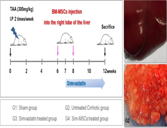
4. Administration of simvastatin and MSCs in rat model of TAA-induced cirrhosis
Animals were randomly allocated to four groups (each group, n = 10) as follows: group I (G1, sham group); group II (G2, untreated cirrhotic group), which received the TAA injections; group III (G3, simvastatin-treated group), which received both the TAA injections and the simvastatin treatment; and group IV (G4, simvastatin plus MSCs-treated group), which received both the TAA injections and the Sim-MSCs treatment.
G3 and G4 were given simvastatin (Sigma-Aldrich) at 10 mg·kg-1·day-1 via drinking water for 5 weeks. Daily water consumption was monitored to adjust the dose of simvastatin delivered daily. Rats were anesthetized by intramuscular injection of a mixture of Zoletil (Virbac Laboratories, Carros, France) and Rompun (Bayer Korea, Seoul, Korea). Using aseptic techniques, a 1-cm incision was made caudal to the costal arch on the right flank to expose the right lobe of the liver. With a syringe, 1 × 10⁶ MSCs were injected directly into the right lobe of the liver at 6 and 8 weeks during the 12-week course of TAA administration (Fig. 2).
10
Fig. 2. Schematic representation of regimens for different studied groups. Experimental procedure. Hepatic fibrosis was induced in Sprague-Dawley rats by intraperitoneal injection of TAA (300 mg/kg) twice a week for 12 weeks. Simvastatin was administered for 7 weeks. MSCs were injected directly into the right liver lobe at weeks 6 and 8 of the 12-week course of TAA administration. Abbreviations: MSCs, mesenchymal stem cells; TAA, thioacetamide.
11
After 12 weeks, blood samples were taken, and the rats were sacrificed. Liver tissue specimens were collected, fixed, immediately frozen, and stored at -80°C for analysis.
5. Histomorphological and immunohistochemical analysis
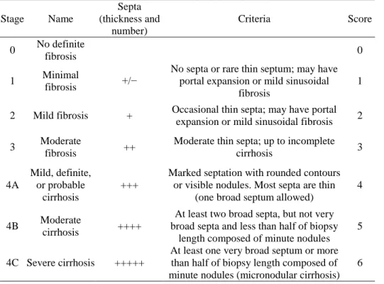
Thick sections (5 µm) of paraffin-embedded liver tissue were prepared and stained with hematoxylin and eosin (H&E), Picrosirius red, and Masson’s trichrome (MTC). The extent of fibrosis was evaluated by using the Laennec fibrosis scoring system (Table 1).
Table 1. Laennec scoring system for staging fibrosis in liver specimens
Stage Name Septa (thickness and number) Criteria Score 0 No definite fibrosis 0 1 Minimal fibrosis +/−
No septa or rare thin septum; may have portal expansion or mild sinusoidal
fibrosis
1
2 Mild fibrosis + Occasional thin septa; may have portal expansion or mild sinusoidal fibrosis 2 3 Moderate
fibrosis ++
Moderate thin septa; up to incomplete
cirrhosis 3 4A Mild, definite, or probable cirrhosis +++
Marked septation with rounded contours or visible nodules. Most septa are thin
(one broad septum allowed)
4
4B Moderate
cirrhosis ++++
At least two broad septa, but not very broad septa and less than half of biopsy
length composed of minute nodules 5
4C Severe cirrhosis +++++
At least one very broad septum or more than half of biopsy length composed of minute nodules (micronodular cirrhosis)
12
In this system, the thickness of the predominant type of septa in each specimen is chosen, and the smallest nodule is selected for scoring. The extent of fibrosis was scored by a liver pathologist blind to the data. The Laennec fibrosis scoring system was used because it incorporates three subclasses of cirrhosis, thereby enabling a more detailed estimation of the effects of the intervention on fibrosis.22,23 To further assess the effects of each treatment on hepatic fibrosis, the fibrotic area in each liver specimen was quantified as a percentage of the total MTC-stained area. The fibrotic area was evaluated in digital photomicrographs by using a computerized image analysis system (Analysis 3.0, Olympus, Tokyo, Japan). To quantify the fibrotic area, fields of vision were selected randomly at a magnification of × 100.
Picrosirius red staining was performed to quantify the total amount of collagen. Thick sections of paraffin-embedded liver tissue were deparaffinized, rehydrated with distilled water, and stained with a Picrosirius red staining kit (Polysciences, Warrington, PA, USA) according to the manufacturer’s instructions. In addition, the amount of collagen (the main component of fibrous tissue) was estimated by determining the percentage of the total area that was stained red. Collagen staining was showed on an Olympus BX51 microscope and quantified using image analysis software (IMT i-solution, Vancouver, BC, Canada). Image artifacts and structural collagen in the large portal tracts and blood vessel walls were omitted from the total collagen area.24
13
acetone, and nonspecific binding sites were blocked by incubation in 10% fetal bovine serum (FBS) for 2 h at RT. Tissue sections were then incubated with monoclonal antibodies against α-smooth muscle actin (α-SMA; Abcam, Cambridge, MA, USA) for 1 h at RT. The slides were washed with PBS and incubated with Alexa Fluor 488 (Thermo Fisher Scientific Life Sciences)-conjugated secondary antibodies for 1 h at RT in the dark. After washing, the slides were mounted with Vectashield mounting medium containing 4′,6-diamino-2-phenylindole (DAPI; Vector Laboratories, Burlingame, CA, USA) for counterstaining of nuclei. Fluorescence images were showed under a laser scanning confocal microscope (TCS SPE, Leica Microsystems GmbH, Wetzlar, Germany).
6. Measurement of hepatic hydroxyproline content
Liver tissue was hydrolyzed with 6 N HCl for 16 h at 120°C. The hydrolysates were then cooled, neutralized with 6 N NaOH, and centrifuged at 13,000 × g for 10 min. Hepatic hydroxyproline content was spectrophotometrically measured using prepared Ehrlich’s solution (dimethylaminobenzaldehyde with perchloric acid and isopropanol) as previously described.18
7. Biochemical parameter analysis
Measurements of alanine transaminase (ALT), aspartate transaminase (AST), albumin, and total bilirubin levels were carried out using commercially
14
available kits (Asan Pharmaceutical, Seoul, Korea) according to the manufacturer’s instructions.
8. Quantitative real-time polymerase chain reaction (PCR) analysis
Total RNA was extracted from liver tissue using TRIzol reagent (Thermo Fisher Scientific Life Sciences) according to the manufacturer’s protocol. RNA purity and concentration were determined using a spectrophotometer (Ultrospec 2100 pro UV/Visible, GE Healthcare Life Sciences, Piscataway, NJ, USA). cDNA was synthesized from total RNA (1 µg) using the GeneAmp RNA PCR Kit (Applied Biosystems, Foster City, CA, USA) with oligo-dT (Applied Biosystems). Transcript levels were measured by real-time PCR using sequence-specific primers for TGF-1, α-SMA, and type 1 collagen (Table 2).
15
Table 2. Primer sequences for quantitative real-time PCR
Gene Forward/reverse Primer sequence
GAPDH Forward 5′-AGACAGCCGCATCTTCTTGT-3′ Reverse 5′-TGATGGCAACAATGTCCACT-3′ TGF-1 Forward 5′-GGACTCTCCACCTGCAAGAC-3′ Reverse 5′-GACTGGCGAGCCTTAGTTTG-3′ Collagen-1 Forward 5′-CATGTTCAGCTTTGTGGACCT-3′ Reverse 5′-GCAGCTGACTTCAGGGATGT-3′ α-SMA Forward 5′-CGAAGCGCAGAGCAAGAGA-3′ Reverse 5′-CATGTCGTCCCAGTTGGTGAT-3′
GAPDH, glyceraldehyde-3-phosphate dehydrogenase; TGF-β1, transforming growth factor-beta 1; collagen-1, type 1 collagen; α-SMA, α-smooth muscle actin
16
Amplification reactions contained SYBR Green PCR Master Mix (Applied Biosystems) and were performed in an ABI PRISM 7900HT Sequence Detection System (Applied Biosystems) according to the manufacturer’s instructions. Data were analyzed using SDS 2.2.2 software (Applied Biosystems). The cycle threshold (Ct) values of the target genes were normalized to those of the endogenous control gene (GAPDH). Relative changes were calculated using the equation 2– ΔΔCt.
9. Western blot analysis
To extract the total protein from liver tissue, samples were homogenized in T-PER protein extraction reagent (Thermo Fisher Scientific Life Sciences) using a TissueLyser II apparatus (Qiagen GmbH, Haan, Germany). The resultant lysates were spun by centrifugation at 13,000 rpm for 15 min at 4°C, and the protein concentrations of the supernatants were determined using a protein assay kit (Bio-Rad Laboratories Inc., Hercules, CA, USA). A total of 30 µg of each liver protein extract was electrophoresed on a 10% sodium dodecyl sulfate-polyacrylamide (SDS-PAGE) gel and then transferred to a polyvinylidene difluoride (PVDF) membrane (EMD Millipore, Billerica, MA, USA). The membranes were blocked with 5% skim milk in Tris-buffered saline containing 0.1% Tween 20 for 1 h at RT, and the membranes were then incubated with primary antibodies at 4°C overnight. The following primary antibodies were used: anti-TGF-1 (Abcam), anti-α-SMA (Abcam), anti-Smad3
17
(Cell Signaling, Danvers, MA, USA), and anti-phospho-Smad3 (Cell Signaling). After washing, membranes were incubated with horseradish peroxidase conjugated secondary antibodies against either mouse IgG (Abcam) or rabbit IgG (Cell Signaling) for 1 h at RT. Immunoreactive bands were visualized using an enhanced chemiluminescence detection kit (GE Healthcare Life Sciences). All membranes were also probed with -actin antibodies (Abcam), and the intensity of each band was normalized to that of -actin.
10. Cell culture
Immortalized human HSCs that were established by retroviral expression of human telomerase reverse transcriptase were used for the majority of the in vitro experiments. Characterization by immunofluorescence staining of α-SMA revealed a purity of > 96%. HSCs were plated in a 75-cm2 flask with DMEM containing 10% FBS and 1% penicillin/streptomycin and cultured at 37°C in a 5% CO2 atmosphere.
For an indirect co-culture system of HSCs and BM-MSCs, cells were seeded at a 1:1 ratio in each well of a 12-well plate or Transwell membranes (12 mm diameter, 0.4 µm pore size, Costar, Corning Inc., Corning, NY, USA), respectively. Subconfluent HSC cultures were treated either with or without simvastatin and treated either with or without BM-MSCs for 48 h. For an indirect co-culture system, HSCs were placed in the lower chamber with BM-MSCs placed on the membrane insert.
18
11. Statistical analysis
All values are presented as means ± SEs. Data were analyzed by the Kruskal-Wallis H test and the Mann–Whitney U test and one-way ANOVA followed by Tukey’s test using SPSS software version 23.0 (IBM, Armonk, NY, USA). For all analyses, p values < 0.05 were considered statistically significant.
III. RESULTS
1. Validation of BM-MSC immunophenotypes and differentiation potentials The CD105, CD73, CD45, CD34, and CD14 immunophenotypes of the cells were determined, and adipogenic or osteogenic differentiation was induced on the day of adenovirus infection (Fig. 1). CD105 and CD73 (positive markers of MSCs) were expressed in more than 99% of the cells, whereas CD45, CD34, and CD14 (negative markers of MSCs) were expressed in less than 1% of the cells. Consequently, the MSCs had successfully differentiated into osteocytes and adipocytes (Fig. 1).
2. Treatment with simvastatin and MSCs reverses histological changes and biochemical parameters of hepatic fibrosis
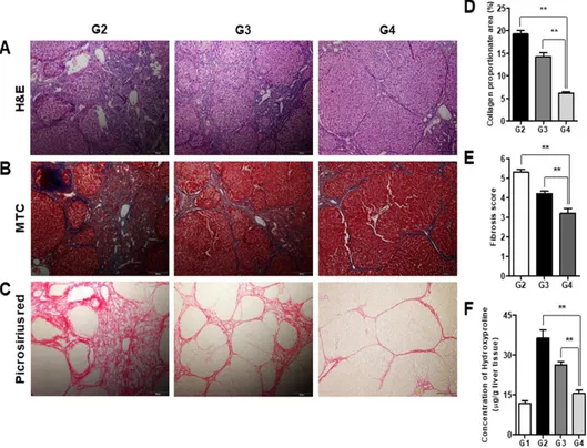
The Laennec fibrosis scores revealed detailed individual changes within the cirrhotic tissue (F4A–F4C, Table 1). Histological analysis was evaluated by H&E and MTC staining (Fig. 3). In the untreated cirrhotic group, the liver
19
sections exhibited strong H&E and MTC staining, revealing definite cirrhosis (stage 4 fibrosis) with regenerating nodules and fibrous septa. Thus, treatment with Sim-MSCs was shown to elicit protective effects against fibrotic changes. These results were further confirmed by Picrosirius red staining to estimate the collagen amount in each sample (Fig. 3). The relative proportions of the Picrosirius red-stained collagen areas were determined using image analysis software. The percentages of the collagen areas were 19.29 ± 0.79, 14.27 ± 0.78, and 6.16 ± 0.29 (p < 0.01) in the untreated cirrhotic group (G2), the simvastatin-treated group (G3), and the Sim-MSCs-treated group (G4), respectively (p < 0.01; Fig. 3D). The Sim-MSCs-treated group had a significantly lower mean score compared with the untreated cirrhotic group (Fig. 3E). The degree of fibrosis was decreased in the Sim-MSCs-treated group compared with the simvastatin-treated group, demonstrating the additional antifibrotic actions of Sim-MSCs (Figs. 3A–3E).
20
Fig. 3. Treatment with simvastatin and BM-MSCs reverses histological changes associated with hepatic fibrosis.
Histological analysis was performed by (A) H&E and (B) MTC staining. The untreated cirrhotic group exhibited clear cirrhosis with broad septae with minute nodules (F4C). The simvastatin-treated group exhibited cirrhosis with thin septation with rounded contours or visible large nodules (F4A), whereas the Sim-MSCs-treated group showed periportal fibrosis (F2) (magnification: × 100). (C) Picrosirius red staining of sections from liver biopsy specimens show the difference in the collagen-stained area (red) between the untreated cirrhotic group and the Sim-MSCs-treated group (magnification: × 100). (D)
21
Quantification of relative Picrosirius red-stained (collagen) areas. Data were obtained using an image analysis program. (E) Histological stage of hepatic fibrosis. (F) Measurement of hepatic hydroxyproline content. A colorimetric assay was used to quantify the hydroxyproline content of each liver sample. Values are expressed as means ± SEs. **, p < 0.01.
G1, sham group; G2, untreated cirrhotic group; G3, simvastatin-treated group; G4, Sim-MSCs-treated group. Abbreviations: BM, bone marrow; MSCs, mesenchymal stem cells; Sim-MSCs, simvastatin and bone marrow-derived mesenchymal stem cells; H&E, hematoxylin & eosin; MTC, Masson’s trichrome; TAA, thioacetamide; G, group.
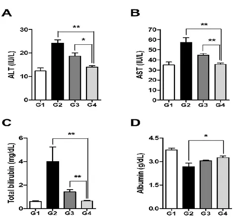
The levels of hydroxyproline in the liver tissue were 11.72 ± 1.16 µg/g, 36.41 ± 3.09 µg/g, 26.25 ± 1.12 µg/g, and 15.46 ± 1.26 µg/g (p < 0.01) in the sham control group (G1), G2, G3, and G4, respectively (Fig. 3F). Compared with the untreated cirrhotic group, the Sim-MSCs-treated group exhibited significantly decreased collagen and lower hepatic hydroxyproline content. The serum levels of AST, ALT, and total bilirubin were significantly increased in the TAA-induced cirrhosis group (G2) compared with G1 (p < 0.01; Figs. 4A–4C). Treatment with Sim-MSCs (G4) markedly reduced the AST, ALT, and bilirubin levels compared with G2 (p < 0.01). Furthermore, the serum level of albumin was increased in G4 compared with G2 (p < 0.05; Fig. 4D).
22
Fig. 4. Simvastatin and BM-MSCs ameliorate biochemical indicators of liver injury.
Levels of (A) ALT, (B) AST, (C) total bilirubin, and (D) albumin after Sim-MSCs administration. G1, sham group; G2, untreated cirrhotic group; G3, simvastatin-treated group; G4, simvastatin plus MSCs-treated group. Values are expressed as means ± SEs. *, p < 0.05, **, p < 0.01. Abbreviations: BM, bone marrow; MSCs, mesenchymal stem cells; ALT, alanine aminotransferase; AST, aspartate aminotransferase.
23
These changes were all greater upon treatment with Sim-MSCs compared with treatment with simvastatin, demonstrating the protective effects of Sim-MSCs against liver injury and hepatic fibrosis.
3. Simvastatin in combination with MSCs suppresses hepatic fibrosis by inhibiting TGF- signaling
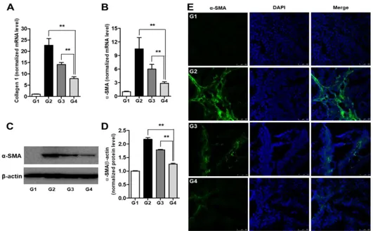
The effects of Sim-MSCs on the expression of α-SMA and collagen-1 in liver tissue were evaluated by quantitative real-time PCR and Western blot analysis. The relative transcript levels of collagen-1 were 1 ± 0.13, 22.63 ± 3.00, 14.17 ± 0.91, and 7.91 ± 0.80 (p < 0.01) in G1, G2, G3, and G4, respectively (Fig. 5A). The relative mRNA levels of α-SMA were 1 ± 0.12, 10.45 ± 2.52, 5.99 ± 1.07, and 2.88 ± 0.31 (p < 0.01), whereas the α-SMA protein levels were 1 ± 0.01, 2.17 ± 0.06, 1.78 ± 0.02, and 1.26 ± 0.03 (p < 0.01) in G1, G2, G3, and G4, respectively (Figs. 5B–5D). The antifibrogenic effects of Sim-MSCs (G4) were consistently higher than those of simvastatin alone (G3). We also showed the expression of α-SMA in the untreated cirrhotic liver group (G2) by immunofluorescence and found that α-SMA was expressed at significantly higher levels compared with G1. However, treatment with Sim-MSCs abolished α-SMA upregulation in livers from rats with TAA-induced cirrhosis (Fig. 5E).
24
Fig. 5. Simvastatin and BM-MSCs suppress hepatic fibrosis.
mRNA expression levels of (A) collagen-1 and (B) α-SMA. Representative Western blot for (C) α-SMA and (D) its densitometric analysis. Values are expressed as means ± SEs. **, p < 0.01. (E) Immunofluorescence staining of α-SMA in liver tissue. The expression of α-SMA in the Sim-MSCs-treated group (G4) was significantly decreased compared with the untreated cirrhotic group (G2) and the simvastatin-treated group (G3). Nuclei were stained with DAPI. Merged immunofluorescence images of α-SMA (green) and DAPI (blue) are shown. Scale bar = 100 µm. Abbreviations: BM, bone marrow; MSCs, mesenchymal stem cells; DAPI, 4’,6-diamino-2-phenylindole; Sim-MSCs, simvastatin and bone marrow-derived mesenchymal stem cells; α-SMA, α-smooth muscle actin.
25
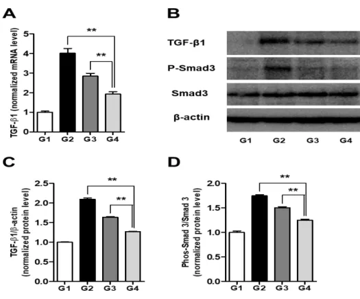
animal model of cirrhosis. We next investigated whether Sim-MSCs-mediated protection against hepatic fibrosis involves TGF- signaling. To this end, the levels of TGF-1 expression in liver tissue from each of the groups were determined by Western blot analysis and quantitative PCR. The relative mRNA levels of TGF-1 were 1 ± 0.06, 4.02 ± 0.24, 2.85 ± 0.14, and 1.93 ± 0.11 (p < 0.01) in G1, G2, G3, and G4, respectively (Fig. 6A). Likewise, the relative protein levels of TGF-1 were 1 ± 0.01, 2.10 ± 0.03, 1.64 ± 0.01, and 1.27 ± 0.01 (p < 0.01) in G1, G2, G3, and G4, respectively (Figs. 6B, 6C). These results show that Sim-MSCs treatment prevents TGF-1 upregulation in the cirrhotic liver, which is important because TGF-1 has both autocrine and paracrine modes of action. Moreover, we revealed that Sim-MSCs treatment significantly reduced the phospho-Smad3, which acts downstream of TGF- receptor activation. The normalized levels of phospho-Smad3 were 1 ± 0.03, 1.75 ± 0.02, 1.50 ± 0.02, and 1.25 ± 0.02 (p < 0.01) in G1, G2, G3, and G4, respectively (Figs. 6B, 6D).
26
Fig. 6. Simvastatin and BM-MSCs ameliorate hepatic fibrosis by inhibiting TGF- signaling.
(A) mRNA expression levels of TGF-1. (B) Representative Western blot and densitometric analysis for (C) TGF-1 and (D) phospho-Smad3. Values are expressed as means ± SEs. **, p < 0.01.
Abbreviation: BM, bone marrow; MSCs, mesenchymal stem cells; mRNA, messenger RNA; TGF- β1, transforming growth factor- β1.
We hypothesize that Sim-MSCs protect against hepatic fibrosis by efficiently blocking TGF--mediated responses. However, it remains to be established
27
whether the antifibrotic effect of Sim-MSCs is due to liberated Sim-MSCs or another factor.
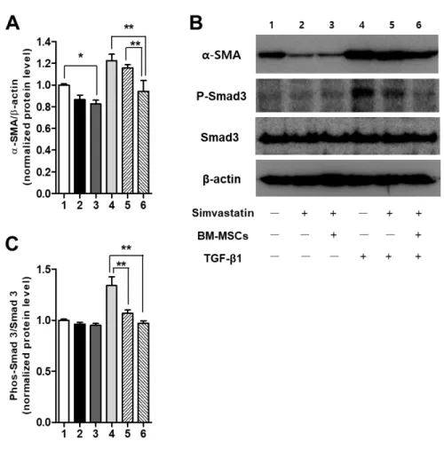
4. Simvastatin and BM-MSCs-mediated regulation of TGF-β1/Smad signaling in HSCs
We next investigated whether Sim-MSCs-mediated protection against hepatic fibrosis involves TGF-/Smad signaling. Sim-MSCs attenuated the protein levels of α‐SMA (Figs. 7A and 7B) and phospho-Smad3 (Figs. 7B and 7C) in HSCs, suggesting that Sim-MSCs ameliorate HSC activation by suppressing TGF-β1/Smad signaling. This result has provided in vitro evidence showing that Sim-MSCs exert synergistic effects on HSCs.
28
Fig. 7. Effects of simvastatin and BM-MSCs on TGF-b/Smad signaling in HSCs.
Western blot analysis of levels of α‐SMA and phospho-Smad3 in immortalized HSCs. (A) Representative Western blot and densitometric analysis for (B) α‐SMA and (C) phospho-Smad3. For the analysis of α-SMA protein levels, HSCs were treated with or without TGF-β1 (1 ng/ml) for 48 h. For the analysis of phospho-Smad3 protein levels, HSCs were treated with or without TGF-β1 (1 ng/ml) for 30 min. The experiment was repeated three times in triplicate with
29
similar results. Values are expressed as means ± SEs. *, p < 0.05, **, p < 0.01. Abbreviations: BM, bone marrow; MSCs, mesenchymal stem cells; Sim-MSCs, simvastatin and bone marrow-derived mesenchymal stem cells; HSCs, hepatic stellate cells; α-SMA, α-smooth muscle actin; TGF- β1, transforming growth factor- β1.
5. Each or combination effect of simvastatin and BM-MSCs of expression of TGF--β1 and collagen-1 in HSCs
We showed that the relative mRNA levels of TGF-β1 (Fig 8A) and collagen-1 (Fig 8B) decreased in the simvastatin treatment group compared with the MSCs treatment group. Moreover, we observed that the relative mRNA levels of TGF-β1 and collagen-1 decreased markedly in the Sim-MSCs-treated group compared with the simvastatin- or MSCs-treated groups. In this study, we suggested that a combination treatment of Sim-MSCs exerts a more potent effect on hepatic fibrosis than treatment with simvastatin and/or MSCs alone.
30
Fig. 8. Simvastatin or BM-MSCs regulation of mRNA of TGF- β1 and collagen-1 in HSCs
(A) mRNA expression level of TGF- β1 (B) collagen-1
Abbreviation: BM, bone marrow; MSCs, mesenchymal stem cells; mRNA, messenger RNA; TGF-1, transforming growth factor-1.
31
IV. DISCUSSION
In this study, we estimated the therapeutic synergistic effects of Sim-MSCs on hepatic fibrosis in a rat model of TAA-induced cirrhosis and HSCs. We also elucidated the fundamental mechanism by which the Sim-MSCs combination treatment ameliorates fibrotic changes. The major findings of our study are as follows: (1) administration of Sim-MSCs into rat livers after TAA-mediated induction of cirrhosis protected against pathogenic fibrosis; (2) Sim-MSCs treatment recovered the impaired liver function through reduces in TGF-β1, α-SMA, and collagen-1 expression; (3) Sim-MSCs exerted strong protective effects against hepatic fibrosis by inhibiting TGF-/Smad signaling; and (4) combined administration with Sim-MSCs had a more potent effect on fibrosis than simvastatin alone.
Previous studies have suggested that statins, such as simvastatin, exert various pleiotropic effects besides their well-known potency to decrease levels of cholesterol, including anticancer and antioxidant effects.25,26
Stem cell-based therapy has been proposed as a promising alternative approach for treating liver disease.27 Several studies have revealed that MSCs exert therapeutic benefits in the context of cirrhosis and hepatic fibrosis. In addition, a recent study has shown that treatment with a combination of MSCs and simvastatin was effective in hepatic fibrosis.28 However, the exact mechanism by which MSCs and simvastatin repair the liver fibrosis has not been investigated. Hence, in this study, we explored the synergistic effect of a
32
combination treatment consisting of Sim-MSCs on hepatic fibrosis. Furthermore, we investigated the underlying mechanisms for this process, evaluating the involvement of the TGF-β/Smad pathway.
In this study, we observed that a combination treatment consisting of Sim-MSCs yielded greater liver function improvement after the development of hepatic fibrosis compared with treatment with simvastatin alone. Through histological H&E, Picrosirius red staining, and MTC, we demonstrated that the administration of Sim-MSCs resulted in dramatic improvements in hepatic fibrosis compared with the untreated cirrhotic group. It was particularly noteworthy that Sim-MSCs exerted significantly more potent preventive effects on hepatic fibrosis compared with simvastatin, implying that the effects of Sim-MSCs are synergistic. In the present study, we used the new Laennec fibrosis scoring system, because it enables finer classification of F4 cirrhosis. This classification approach is desirable because the severity of cirrhosis exhibits clear histological variability. According to the Laennec fibrosis scoring system, histological improvement of hepatic fibrosis was detected most strongly after Sim-MSCs treatment (p < 0.01). These findings were supported by hepatic hydroxyproline content analyses, immunohistochemistry, and Picrosirius red staining.
TGF-1 has been revealed as a key mediator of hepatic fibrosis progression.29 The fibrogenic functions of TGF-1, such as collagen synthesis, are mediated by the canonical Smad pathway via the activation of transmembrane TGF-
33
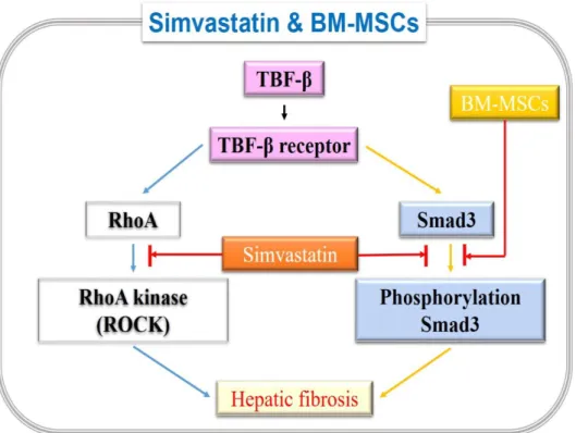
receptors. Moreover, Smad3 may be the primary mediator of TGF-1-associated fibrotic and proinflammatory responses.30 Furthermore, simvastatin inhibits TGF- signaling by inhibiting phospho-Smad3. These findings reveal that the TGF-/Smad signaling pathway could be an attractive therapeutic target for hepatic fibrosis prevention and treatment.31 We showed significant upregulation of TGF-1 and phospho-Smad3 in cirrhotic rat livers, both of which were completely blocked by combination Sim-MSCs treatment. Notably, Sim-MSCs also suppressed both TGF-/Smad3 signaling and α‐SMA in HSCs, demonstrating that Sim-MSCs treatment not only attenuates TGF-1 release but also inhibits Smad signaling cascades. These results clearly illustrate that the liberation of Sim-MSCs contributes to their therapeutic effects against hepatic fibrosis progression. Here, we observed that the administration of Sim-MSCs into the cirrhotic liver ameliorates hepatic fibrosis and that Sim-MSCs act in a synergistic manner. We found the mechanism of Sim-MSCs through the inhibition of Smad signaling cascades (Fig 9).
34
Fig. 9. Schematic diagram of Simvastatin and BM-MSCs synergistic effect on hepatic fibrosis.
Abbreviation: BM, bone marrow; MSCs, mesenchymal stem cells; RhoA, Ras homolog gene family, member A; TGF-b1, transforming growth factor-b1.
However, the clinical application of our approach is somewhat limited because the safety for therapy has not yet been well established in humans. Further studies are necessary to determine whether our approach is clinically viable. Nevertheless, our study clearly demonstrates that combination treatment consisting of Sim-MSCs exerts synergistic effects, a finding that has the potential to affect new therapeutic approaches for treating intractable cirrhosis.
35
Furthermore, the modulation of MSC properties with simvastatin represents a novel field of stem cell-based therapy. A better understanding of the effects of simvastatin on MSCs will likely provide new methods for their clinical use in the treatment of degenerative disorders.
V. CONCLUSION
These findings demonstrated that a combination treatment consisting of Sim-MSCs strongly inhibited the progression of TAA-induced hepatic fibrosis in rats compared with simvastatin alone. Therefore, simvastatin could act synergistically with MSCs as an efficient therapeutic approach for intractable cirrhosis.
36
REFERENCES
1. Friedman SL. Liver fibrosis - from bench to bedside. J Hepatol 2 003;38 Suppl 1:S38-53.
2. Baik SK. Haemodynamic evaluation by Doppler ultrasonography i n patients with portal hypertension: a review. Liver Int 2010;30:14 03-13.
3. Kim G, Lee SS, Baik SK, Cho YZ, Kim MY, Kwon SO, et al. The need for histological subclassification of cirrhosis: a systemati c review and meta-analysis. Liver Int 2016;36:847-55.
4. Kim G, Kim J, Lim YL, Kim MY, Baik SK. Renin-angiotensin s ystem inhibitors and fibrosis in chronic liver disease: a systematic review. Hepatol Int 2016;10:819-28.
5. Jun JH, Choi JH, Bae SH, Oh SH, Kim GJ. Decreased C-reactive protein induces abnormal vascular structure in a rat model of liv er dysfunction induced by bile duct ligation. Clin Mol Hepatol 20 16;22:372-81.
6. Kim G, Kim MY, Baik SK. Transient elastography versus hepatic venous pressure gradient for diagnosing portal hypertension: a syst ematic review and meta-analysis. Clin Mol Hepatol 2017;23:34-41. 7. Gressner AM, Weiskirchen R. Modern pathogenetic concepts of li ver fibrosis suggest stellate cells and TGF-beta as major players a nd therapeutic targets. J Cell Mol Med 2006;10:76-99.
37
8. Friedman SL. Mechanisms of hepatic fibrogenesis. Gastroenterolog y 2008;134:1655-69.
9. Jasinska M, Owczarek J, Orszulak-Michalak D. Statins: a new insi ght into their mechanisms of action and consequent pleiotropic eff ects. Pharmacol Rep 2007;59:483-99.
10. Burke JP, Watson RW, Murphy M, Docherty NG, Coffey JC, O'C onnell PR. Simvastatin impairs smad-3 phosphorylation and modul ates transforming growth factor beta1-mediated activation of intesti nal fibroblasts. Br J Surg 2009;96:541-51.
11. Fernandez G, Spatz ES, Jablecki C, Phillips PS. Statin myopathy: a common dilemma not reflected in clinical trials. Cleve Clin J Med 2011;78:393-403.
12. Suk KT, Yoon JH, Kim MY, Kim CW, Kim JK, Park H, et al. T ransplantation with autologous bone marrow-derived mesenchymal stem cells for alcoholic cirrhosis: Phase 2 trial. Hepatology 2016;6 4:2185-97.
13. Eom YW, Kim G, Baik SK. Mesenchymal stem cell therapy for c irrhosis: Present and future perspectives. World J Gastroenterol 20 15;21:10253-61.
14. Kim G, Eom YW, Baik SK, Shin Y, Lim YL, Kim MY, et al. T herapeutic effects of mesenchymal stem cells for patients with chr onic liver diseases: systematic review and meta-analysis. J Korean
38
Med Sci 2015;30:1405-15.
15. Jang YO, Kim YJ, Baik SK, Kim MY, Eom YW, Cho MY, et al. Histological improvement following administration of autologous bone marrow-derived mesenchymal stem cells for alcoholic cirrhos is: a pilot study. Liver Int 2014;34:33-41.
16. Jang YO, Kim MY, Cho MY, Baik SK, Cho YZ, Kwon SO. Effe ct of bone marrow-derived mesenchymal stem cells on hepatic fib rosis in a thioacetamide-induced cirrhotic rat model. BMC Gastroe nterol 2014;14:198.
17. Jang YO, Jun BG, Baik SK, Kim MY, Kwon SO. Inhibition of h epatic stellate cells by bone marrow-derived mesenchymal stem ce lls in hepatic fibrosis. Clin Mol Hepatol 2015;21:141-9.
18. Jang YO, Cho MY, Yun CO, Baik SK, Park KS, Cha SK, et al. Effect of function-enhanced mesenchymal stem cells infected with decorin-expressing adenovirus on hepatic fibrosis. Stem Cells Tran sl Med 2016;5:1247-56.
19. Xu H, Yang YJ, Yang T, Qian HY. Statins and stem cell modulat ion. Ageing Res Rev 2013;12:1-7.
20. Hu Y, Liao L, Wang Q, Ma L, Ma G, Jiang X, et al. Isolation a nd identification of mesenchymal stem cells from human fetal pan creas. J Lab Clin Med 2003;141:342-9.
39
rtz Z. Proliferation, differentiation, and protein synthesis of human osteoblast-like cells (MG63) cultured on previously used titanium surfaces. Clin Oral Implants Res 1996;7:27-37.
22. Kim MY, Cho MY, Baik SK, Park HJ, Jeon HK, Im CK, et al. Histological subclassification of cirrhosis using the Laennec fibrosi s scoring system correlates with clinical stage and grade of portal hypertension. J Hepatol 2011;55:1004-9.
23. Kim MY, Suk KT, Baik SK, Kim HA, Kim YJ, Cha SH, et al. Hepatic vein arrival time as assessed by contrast-enhanced ultrason ography is useful for the assessment of portal hypertension in co mpensated cirrhosis. Hepatology 2012;56:1053-62.
24. Calvaruso V, Burroughs AK, Standish R, Manousou P, Grillo F, L eandro G, et al. Computer-assisted image analysis of liver collage n: relationship to Ishak scoring and hepatic venous pressure gradie nt. Hepatology 2009;49:1236-44.
25. Blais L, Desgagne A, LeLorier J. 3-Hydroxy-3-methylglutaryl coen zyme A reductase inhibitors and the risk of cancer: a nested case-control study. Arch Intern Med 2000;160:2363-8.
26. Bergman M, Salman H, Djaldetti M, Bessler H. Statins as modula tors of colon cancer cells induced cytokine secretion by human P BMC. Vascul Pharmacol 2011;54:88-92.
40
liver fibrosis. Korean J Intern Med 2015;30:580-9.
28. Motawi TM, Atta HM, Sadik NA, Azzam M. The therapeutic effe cts of bone marrow-derived mesenchymal stem cells and simvastat in in a rat model of liver fibrosis. Cell Biochem Biophys 2014;6 8:111-25.
29. Dooley S, ten Dijke P. TGF-beta in progression of liver disease. Cell Tissue Res 2012;347:245-56.
30. Roberts AB, Russo A, Felici A, Flanders KC. Smad3: a key play er in pathogenetic mechanisms dependent on TGF-beta. Ann N Y Acad Sci 2003;995:1-10.
31. Inagaki Y, Higashiyama R, Higashi K. Novel anti-fibrotic modaliti es for liver fibrosis: molecular targeting and regenerative medicine in fibrosis therapy. J Gastroenterol Hepatol 2012;27 Suppl 2:85-8.
41
ABSTRACT(IN KOREAN)
간섬유화에 대한 simvastatin 및 골수 유래 중간엽 줄기세포의
상승효과
< 지도교수 김경식 >
연세대학교 대학원 의학과
김 성 훈
섬유화에 대한 simvastatin (Sim)의 유익한 효과는 다양한
장기에서
보고
되고
있다.
골수
유래
중간엽
줄기
세포(BM-MSC) 또한 간 섬유화 및 간경변에 대한 효과적인
치료법으로 제안되고 있다. 최근 결과들은 줄기 세포 기능을
조절하는 약리학적 치료가 임상 진료에 적용 가능한 새로운
치료
방법이라
제시하고
있다.
이
연구의
목적은
simavastatin 과 Mesenchymal stem cells (MSCs)의 병합
치료가 thioacetamide (TAA)에 의한 간경변 랫트 모델에서 간
섬유화와 간 성상 세포(hepatic stellate cells, HSCs)에서
시너지
효과를
나타낼
수
있는지를
확인하는
것이다.
Sim-MSCs 로 치료 한 랫트의 간경변은 simvastatin 단독으로
치료한 것과 비교하여 조직학적으로 개선된 것을 보여주었다.
42