• 검색 결과가 없습니다.

Multi-class Classification of Histopathology Images using Fine-Tuning Techniques of Transfer Learning

N/A
N/A
Protected

Academic year: 2021

Share "Multi-class Classification of Histopathology Images using Fine-Tuning Techniques of Transfer Learning"

Copied!
11
0
0

로드 중.... (전체 텍스트 보기)

전체 글

(1)

https://doi.org/10.9717/kmms.2021.24.7.849

Multi-class Classification of Histopathology Images using Fine-Tuning Techniques of Transfer Learning

Kobiljon Ikromjanov

, Subrata Bhattacharjee

††

, Yeong-Byn Hwang

†††

, Hee-Cheol Kim

††††

, Heung-Kook Choi

†††††

ABSTRACT

Prostate cancer (PCa) is a fatal disease that occurs in men. In general, PCa cells are found in the prostate gland. Early diagnosis is the key to prevent the spreading of cancers to other parts of the body.

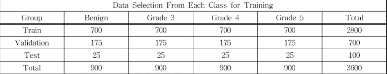
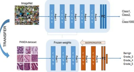
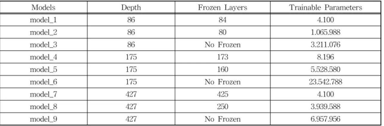
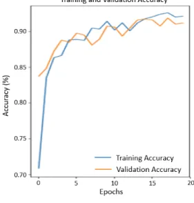
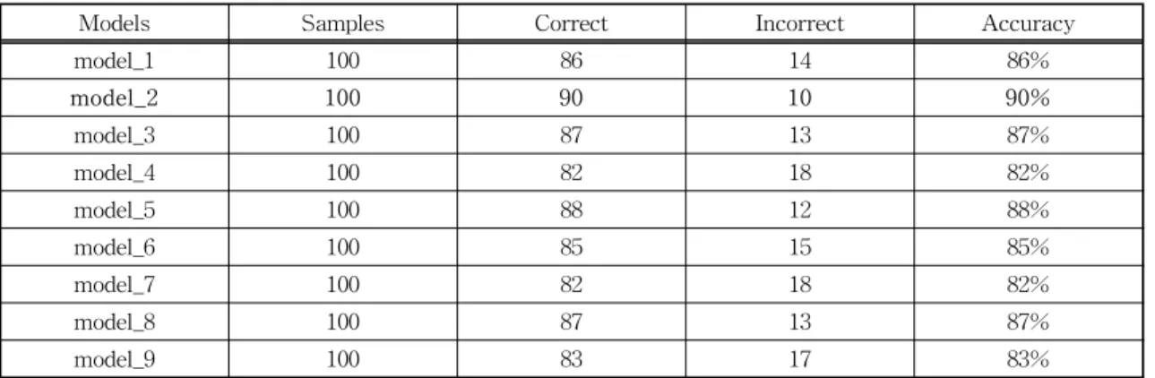
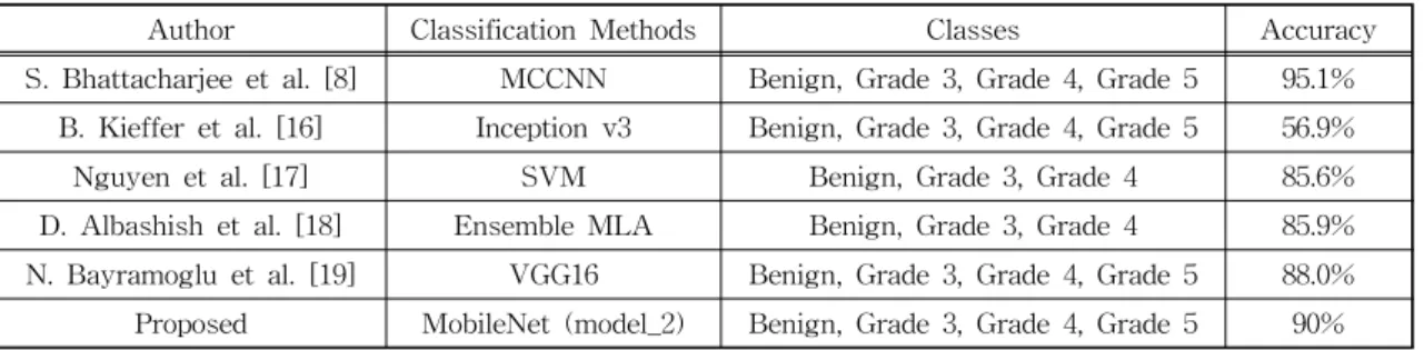
In this case, deep learning-based systems can detect and distinguish histological patterns in microscopy images. The histological grades used for the analysis were benign, grade 3, grade 4, and grade 5. In this study, we attempt to use transfer learning and fine-tuning methods as well as different model architectures to develop and compare the models. We implemented MobileNet, ResNet50, and DenseNet121 models and used three different strategies of freezing layers techniques of fine-tuning, to get various pre-trained weights to improve accuracy. Finally, transfer learning using MobileNet with the half-layer frozen showed the best results among the nine models, and 90% accuracy was obtained on the test data set.

Key words: Transfer Learning, Fine-tuning, Deep Learning, Prostate Cancer

※ Corresponding Author : Heung-Kook Choi, Address:

(50834) 197, Inje-ro, Inje University, Gimhae, Gyeong- nam, Republic of Korea, TEL : E-mail : [email protected]

Receipt date : May 26, 2021, Revision date : Jul. 2, 2021 Approval date : Jul. 12, 2021

††

Dept of Digital Anti-Aging Healthcare, u-AHRC, Inje University (E-mail : [email protected])

††

Dept of Computer Engineering, u-AHRC, Inje Univer- sity (E-mail : [email protected])

†††††

Dept of Computer Engineering, u-AHRC, Inje Uni- versity (E-mail : [email protected])

†††††

Dept of Digital Anti-Aging Healthcare, u-AHRC, Inje University (E-mail : [email protected])

†††††

Dept of Computer Engineering, u-AHRC, Inje Uni- versity (E-mail : [email protected])

※ This work was supported by the National Research Foundation of Korea (NRF) grant funded by the Korea government (MSIT) (No. 2021R1A2C2008576).

1. INTRODUCTION

Cancer begins when cells in the human body start to grow uncontrollably. Cells in almost any part of the body can become cancerous, which can then spread to other areas of the body. PCa is one of the most common cancers after skin cancer in American men. According to the American Cancer Society statistics, approximately 1 in 8 men will be diagnosed with PCa in their lifetime [1].

Histological examination of tissues and the de- tection of cancer by physicians remains the gold standard in cancer diagnosis. The diagnosis of PCa is heavily time-consuming. In addition, it is based

on subjective grading. For example, the study by Ozkan et al. reported that two pathologists dis- agreed about the presence of cancer in 31 of 407 baseline biopsies and that the total concordance of the accessed Gleason score was only 51.7%, de- scribing these challenges in diagnosing the PCa consistently [2]. Therefore, the development of computer-assisted decision support tools is essen- tial for saving time, predicting disease outcomes, and improving precision medicine for pathologists.

There has been considerable interest in the de- velopment of methods based on digital image proc- essing and machine learning. These methods are used to automatically analyze pathological images

***-****-****

(2)

to classify tissues and disease, as well as to im- prove accuracy and diagnostic standard [3-5]. On top of that, recent advances in deep learning re- search have succeeded in increasing the perform- ance of such analytics [6-9]. However, the pro- posed deep learning models often require a sig- nificant amount of annotated data to be properly trained. Because cohort sizes can be small and his- topathological image annotation takes a long time, a concept known as transfer learning, training neural networks with an external dataset, primarily ImageNet [10], and then fine-tuning the model with the dataset in hand, can prove useful. Such a pre-trained model and fine-tuning approach are more effective than training the same neural net- work architecture from scratch in studies involving digital pathological image analysis [11-15]. Trans- fer learning can also be beneficial in adapting areas imaged by different microscopes or staining proce- dures.

B. Kieffer et al. [16] explored the problem of classification within a medical image dataset based on a feature vector extracted from the deepest layer of pre-trained Convolution Neural Network (CNN).

They used feature vectors from several pre-trained structures, including networks with/without trans- fer learning to evaluate the performance of pre- trained deep features versus CNN which have been trained by that specific dataset. Their result shows that pre-trained networks are quite competitive against training from scratch. As well, fine-tuning does not seem to add any tangible improvement for VGG16 to justify additional training while we observed considerable improvement in retrieval and classification accuracy with 56.98% when they fine-tuned the Inception structure.

Nguyen et al. [17] introduced a novel approach to grade prostate malignancy using digitized histo- pathological specimens of the prostate tissue. They have extracted tissue structural features from the gland morphology and co-occurrence texture fea- tures from 82 regions of interest (ROI) with 620

× 550 pixels to classify a tissue pattern into three major categories: benign, grade 3 carcinoma, and grade 4 carcinoma. The authors proposed a hier- archical (binary) classification scheme and ob- tained 85.6% accuracy in classifying an input tissue pattern into one of the three classes.

D. Albashish et al. [18] implemented a new mul- ticlass approach called multi-level (hierarchical) learning architecture (MLA), which addresses the binary classification tasks in the hierarchical strat- egy. It focuses on solving the three-class classi- fication problem in prostate cancer grading, i.e., Grade 3, Grade 4, and Benign. The results also confirmed the high efficiency of the ensemble framework with the MLA scheme in dealing with the multiclass classification problem, which results in 85.9% accuracy.

N. Bayramoglu et al. [19] evaluated perform- ances of convolutional neural network models to classify cell nuclei in hematoxylin and eosin (H&E) stained histopathology images of colorectal adeno- carcinoma. They compared four different CNN ar- chitectures AlexNet, GenderNet, GoogLeNet, and VGG-16 trained on natural images and facial im- ages using transfer learning and fine-tuning and got a maximum of 88.03% accuracy with fine- tuned VGG-16 model.

Subrata Bhattacharjee et al. [8] developed a ma-

chine learning technique to predict the histological

grades in prostate biopsy. To perform a multiclass

classification, an AI-based deep learning algo-

rithm, a multichannel convolutional neural network

(MCCNN) was developed by connecting layers

with artificial neurons inspired by the human brain

system. The histological grades that were used for

the analysis are benign, grade 3, grade 4, and grade

5. An author aimed to classify multiple patterns of

images extracted from the whole slide image (WSI)

of a prostate biopsy based on the Gleason grading

system. The MCCNN model takes three input

channels (Red, Green, and Blue) to extract the com-

putational features from each channel and con-

(3)

(a) (b)

(c) (d)

Fig. 1. Sample images of each class of prostate cancer.

(a) Benign. (b) Grade 3. (c) Grade 4. (d) Grade 5.

Table 1. The arrangement of the dataset for multiclass classification.

Data Selection From Each Class for Training

Group Benign Grade 3 Grade 4 Grade 5 Total

Train 700 700 700 700 2800

Validation 175 175 175 175 700

Test 25 25 25 25 100

Total 900 900 900 900 3600

catenate them for multiclass classification. Stain normalization was carried out for each histological grade to standardize the intensity and contrast lev- el in the image and got an average accuracy of 95.1%.

In this study, MobileNet [20, 21], ResNet50 [22, 23], and DenseNet121 [24, 25] deep convolutional networks were used as a transfer learning frame- work where they were pre-trained on the Image Net dataset. In addition, three different fine-tuning strategies were applied to freeze some of their lay- ers for comparison and increased accuracy. The PANDA dataset, from Kaggle [26] was used for the experiment to classify images into four classes.

In this paper, the classification accuracies of the second model of MobileNet have been visualized using a non-normalized and normalized confusion matrix, giving more accurate results.

2. MATERIALS AND METHODS

2.1 Data Set

In this experiment, we have selected 900 good images for each class, which is a publicly available PANDA dataset on the Kaggle repository. PCa grade assessment (PANDA) [26] started a chal- lenge to develop models for detecting PCa on his- topathology images of prostate tissue samples and estimate the severity of the disease using the most extensive multi-center dataset on Gleason grading.

Our dataset consists of 3600 patched color images of size 256 × 256 pixels that were extracted from WSI. There is a total of four classes to predict, some of the sample images for each class are

shown in Fig. 1. To train the model, 80 percent of data samples were chosen for training (2800 im- ages) and the remaining 20 percent (700 images) for validation. Further, to test the model, a total of 100 unseen data samples were selected (i.e., 25 images per class). Table 1 shows the details of the dataset employed in this work.

2.2 Transfer Learning Methods

Transfer learning is a deep learning technique that stores the knowledge gained while solving one issue and use it to a new but related problem.

Instead of starting the learning process from

scratch, we can start from patterns that have been

learned when solving a different problem. This

way we can build accurate models in a time-saving

way. Many pre-trained models used in transfer

(4)

Fig. 2. Transfer learning and fine-tuning technique.

Table 2. List of the CNN architectures.

Architecture Models Hidden Layers Classification Layer Depth

MobileNet

model_1

84

Global Average Pooling2D

&

Dense

86 model_2

model_3

ResNet50

model_4

173 175

model_5 model_6

DenseNet121

model_7

425 427

model_8 model_9

learning are based on large CNN, which has 2 main parts convolutional base and classifier.

Nine models based on MobileNet, ResNet50, and DenseNet121 were used in this research study.

They are originally trained on the ImageNet data- base which can classify images into thousands of object categories. Due to these advantages, while we were building models for our own needs, we started by removing the original classifier, next we added new classifier layers that fit our purpose, and finally, we had a fine-tuning process on our model according to one of the below mentioned three strategies. Fig. 2 demonstrates one of the transfer learning and fine-tuning techniques that we used

in our experiment. Table 2 demonstrates nine mod- els, feature extraction layers, classification layers, and the depth of each model. The first three models have been applied to 84 MobileNet layers and their pre-trained weights and two additional layers were added to the classification block. While the other three models were pre-trained 173 layers of Res Net50. Furthermore, the remaining models were built-in with 425 layers of DenseNet121. Finally, GlobalAveragePooling2D and Dense layers were used for the multi-class classification.

2.3 Fine Tuning

Fine-tuning is a way of utilizing or applying

(5)

Fig. 3. Three strategies of fine-tuning.

Table 3. List of Frozen layers and trainable parameters.

Models Depth Frozen Layers Trainable Parameters

model_1 86 84 4.100

model_2 86 80 1.065.988

model_3 86 No Frozen 3.211.076

model_4 175 173 8.196

model_5 175 160 5.528.580

model_6 175 No Frozen 23.542.788

model_7 427 425 4.100

model_8 427 250 3.939.588

model_9 427 No Frozen 6.957.956

transfer learning. There are different kinds of fine- tuning techniques, truncating the last layer, using a smaller learning rate, and freezing the weights of the layers are among these methods. In this pa- per, the freezing technique is used to compare each model. Fig. 3 shows three strategies of freezing techniques used with three architectures, while Table 3 demonstrates a list of frozen layers and trainable parameters.

Freezing the convolutional base is the first strategy trained with model_1, model_4 and, mod- el_7. This case illustrates the extreme condition of the train and freezes trade-off. The main idea is

to remove the last fully connected layer, run the pre-trained model as a fixed feature extractor, and then use the resulting features to train a new clas- sifier. We used the pre-trained model as a fixed feature extraction technique (MobileNet, ResNet50, and DenseNet121), which can be useful in case, we have less computational power or a small dataset, and the pre-trained model solves a problem very similar to the one we want to solve.

The second strategy, used for model_2, model_5,

and model_8, is training some layers and keep the

others frozen. Mainly, lower layers mean general

features (problem-independent), while higher lay-

(6)

Fig. 4. Training Accuracy of model_2.

Fig. 5. Training Loss of model_2.

ers indicate specific features (problem-dependent).

Here, we played with that dichotomy by selecting how much we want to modify the weights of the network (a frozen layer does not change during training). Usually, if we have a small dataset and a large number of parameters, we leave more lay- ers frozen to keep away from overfitting. In con- trast, if the dataset is large and the number of pa- rameters is small, we can enhance our model by training more layers to the new task since over- fitting is not an issue. Therefore we have frozen more layers to find the average parameters, as we have a small and different dataset from the pre- trained models’ ImageNet dataset. We froze 80, 160, and 250 layers to get a balance between the number of layers and freeze that gave average parameters on model_2, model_5, and model_8 respectively.

The last strategy, trained with model_3, mod- el_6, and model_9, is to train the entire model. In this case, we implemented the architecture of the pre-trained model and trained it according to our dataset. Our models learned from scratch, so we needed a lot of computational power and more time compared to previous methods.

3. RESULTS AND DISCUSSION

In this section, learning graphs and confusion matrices are used to demonstrate the best result achieved from model_2. For classification, we trained with MobileNet, ResNet50, and DenseNet 121 architectures, which are followed by Global AvaragePooling2D and Dense layers. All layers used ReLU activation functions. When it comes to the output layer, softmax activation was employed.

The optimizer is selected to be Adam [27] with a learning rate of 10

-4

and the loss function is chosen to be categorical cross-entropy. Nine different models were trained with three different archi- tectures, with three models in each: training from scratch (random weight initialization), fine-tuning half layers and parameters, finally fine-tuning all backbone layers on ImageNet pre-trained model

with 20 epochs.

Commonly, above mentioned strategy 1 is used when we have a small dataset and similar to the pre-trained model’s dataset, while strategy 3 is usually applied in case of having a large dataset, but different from the pre-trained model’s dataset.

As we have, a small dataset and different from the

pre-trained model’s ImageNet 1000 class dataset,

strategy 2 is the most suitable for our proposed

model. Fig. 4 and 5 approve that re-trained mod-

(7)

Table 4. Accuracy of test dataset.

Models Samples Correct Incorrect Accuracy

model_1 100 86 14 86%

model_2 100 90 10 90%

model_3 100 87 13 87%

model_4 100 82 18 82%

model_5 100 88 12 88%

model_6 100 85 15 85%

model_7 100 82 18 82%

model_8 100 87 13 87%

model_9 100 83 17 83%

Fig. 6. Non-normalized Confusion Matrix of model_2. Fig. 7. Normalized Confusion Matrix of model_2.

el_2 with only 20 epochs represent a significant in- crease in accuracy and decrease in loss values, showing better performance in early epochs. Com- paring all re-trained models, model_2 used Mobile Net architecture, and pre-trained weights with 80 frozen layers yield the highest accuracy of 93.03%

and 91.32% and the lowest loss of 2.52% and 2.78%

on training and validation sets, respectively. As a result, we prevented overfitting by balancing be- tween the number of layers to train and freeze.

Furthermore, Table 4 demonstrates predictions of 100 unseen test data and their accuracy. On the test set, model_2 predicted 90 images correctly and only making 10 mistakes out of 100 test images, scoring 90% as overall accuracy. Fig. 6 and 7 show

the non-normalized and normalized confusion ma- trices.

In Table 5, we compare the accuracy of different

multi-class classification methods with our pro-

posed model. B. Kieffer et al. [16] got 56.98% accu-

racy on the Inception v3 classification method by

using transfer learning and predicting four differ-

ent classes. Nguyen et al. [17] extracted tissue

structural features from the gland morphology and

co-occurrence texture features and classified three

classes with obtaining 85.6% accuracy. D. Albash-

ish et al. [18] used multi-level learning architecture

(MLA) and focused on solving classification prob-

lems in prostate cancer by categorizing the cases

into three classes. The authors achieved 85.9% ac-

(8)

Table 5. Comparison between the proposed method and other standard methods for the multi-class classification of prostate cancer grading.

Author Classification Methods Classes Accuracy

S. Bhattacharjee et al. [8] MCCNN Benign, Grade 3, Grade 4, Grade 5 95.1%

B. Kieffer et al. [16] Inception v3 Benign, Grade 3, Grade 4, Grade 5 56.9%

Nguyen et al. [17] SVM Benign, Grade 3, Grade 4 85.6%

D. Albashish et al. [18] Ensemble MLA Benign, Grade 3, Grade 4 85.9%

N. Bayramoglu et al. [19] VGG16 Benign, Grade 3, Grade 4, Grade 5 88.0%

Proposed MobileNet (model_2) Benign, Grade 3, Grade 4, Grade 5 90%

curacy that shows the high efficiency of the en- semble framework with MLA. N. Bayramoglu et.

al [19] performed 88.03% accuracy with transfer learning and fine-tuning on VGG16 architecture that changes learning rate according to each epoch.

S. Bhattacharjee et. al [8], on the other hand, devel- oped MCCNN to predict the histological grades in prostate biopsy and achieved an excellent accuracy of 95.1% with analyzing four different classes. In our proposed architecture, we have achieved one of the most satisfying accuracies of 90% by im- plementing MobileNet (model_2) classification me- thod that trains some layers and at the same time, keeps others frozen.

4. CONCLUSION

In this paper, it was found that pre-trained deep learning models on the ImageNet dataset can be fine-tuned to improve the accuracy to classify PCa dataset. Three architectures and their weights were used and applied three different freezing techniques on the layers making them overall nine models. After re-training models with histology image dataset, they are evaluated by predicting 100 unseen test images. As a result, model_2 from MobileNet showed the best accuracy of 90%. It is recommended that this model could be used in re- al-time environments when we do not have enough data.

In conclusion, the proposed fine-tuning techni- ques used in this study is also effective for solving

the over-fitting issue in the model.

REFERENCE

[ 1 ] Key Statistics for Prostate Cancer(2021).

https://www.cancer.org/cancer/prostate-can cer/about/key-statistics.html (accessed April 1, 2021).

[ 2 ] T.A. Ozkan, A.T. Eruyar, O.O. Cebeci, O.

Memik, L. Ozcan, and I. Kuskonmaz, “Inter- observer Variability in Gleason Histological Grading of Prostate Cancer,” Scandinavian Journal of Urology, Vol. 50, No. 6, pp. 420- 424, 2016.

[ 3 ] A. Madabhushi and G. Lee, “Image analysis and machine learning in digital pathology:

Challenges and opportunities,” Medical Image Analysis, Vol. 33, pp. 170-175, 2016.

[ 4 ] A. Madabhushi, “Digital Pathology Image Analysis: Opportunities and Challenges,”

Imaging in Medicine, Vol. 1, No. 1, pp. 7-10, 2009.

[ 5 ] K. Daisuke and I. Shumpei, “Machine Learning Methods for Histopathological Image Anal- ysis,” Computational and Structural Biotech- nology Journal, Vol. 16, pp. 34-42, 2018.

[ 6 ] Z. Hu, J. Tang, Z. Wang, K. Zhang, L. Zhang, and Q. Sun, “Deep Learning for Image-Based Cancer Detection and Diagnosis-A Survey,”

Pattern Recognition, Vol. 83, pp. 134-149, 2018.

[ 7 ] O. Iizuka, F. Kanavati, K. Kato, R. Michael,

(9)

A. Koji, and T. Masayuki, “Deep Learning Models for Histopathological Classification of Gastric and Colonic Epithelial Tumours,”

Scientific Reports, Vol. 10, No. 1504, pp. 1-11, 2020.

[ 8 ] S. Bhattacharjee, D. Prakash, C.-H. Kim, and H.-K. Choi, “Multichannel Convolution Neural Network Classification for the Detection of Histological Pattern in Prostate Biopsy Images,”

Journal of Korea Multimedia Society, Vol. 23, No. 12, pp. 1486-1495, 2020.

[ 9 ] S. Bhattacharjee, R.I. Sumon, K. Ikromjanov, Y.-B. Hwang, H.-C. Kim, and H.-K Choi,

“Unsupervised Classification of Prostate Can- cer Using Deep Learning Based Image Clus- tering Technique,” Proceeding of Internatio- nal Conference on Multimedia Information Technology and Applications, pp. 113-116, 2021.

[10] J. Deng, W. Dong, R. Socher, L.-J. Li, K. Li, and L. Fei-Fei, “Imagenet: A Large-Scale Hierarchical Image Database,” Proceeding of International Conference on Computer Vision and Pattern Recognition, pp. 248-255, 2009.

[11] Shallu and R. Mehra, “Breast Cancer Histology Images Classification: Training from Scratch or Transfer Learning?,” ICT Express, Vol. 4, No. 4, pp. 247-254, 2018.

[12] R. Mormont, P. Geurts, and R. Maree, “Com- parison of Deep Transfer Learning Strategies for Digital Pathology,” Proceedings of Inter- national Conference on Computer Vision and Pattern Recognition Workshops, pp. 2262- 2271, 2018.

[13] B. Kieffer, M. Babaie, S. Kalra, and H.R.

Tizhoosh, “Convolutional Neural Networks for Histopathology Image Classification: Training vs. Using Pre-trained Networks,” Proceeding of International Conference on Image Pro- cessing Theory, Tools and Applications (IPTA), pp. 1-6, 2017.

[14] N. Tajbakhsh, J.Y. Shin, S.R. Gurudu, R.T.

Hurst, C.B. Kendall, M.B. Gotway, et al.,

“Convolutional Neural Networks for Medical Image Analysis: Full Training or Fine Tuning?,”

IEEE Transactions on Medical Imaging, Vol.

35, No. 5, pp. 1299-1312, 2016.

[15] H.-G. Park, S. Bhattacharjee, P. Deekshitha, C.-H. Kim, and H.-K. Choi, “A Study on Deep Learning Binary Classification of Prostate Pathological Images Using Multiple Image Enhancement Techniques,” Journal of Korea Multimedia Society, Vol. 23, No. 4, pp. 539- 548, 2020.

[16] B. Kieffer, M. Babaie, S. Kalra and H.R.

Tizhoosh, “Convolutional Neural Networks for Histopathology Image Classification:

Training vs. Using pre-trained Networks,”

Proceeding of Seventh International Confer- ence on Image Processing Theory, Tools and Application, pp. 1-6, 2017.

[17] K. Nguyen, B. Sabata, and A.K. Jain, “Pros- tate Cancer Grading: Gland Segmentation and Structural Features,” Pattern Recognition Letters, Vol 33, No. 7, pp. 951-961, 2012.

[18] D. Albashish, S. Sahran, A. Abdullah, M.

Alweshah, and A. Adam, “A Hierarchical Classifier for Multiclass Prostate Histopathol- ogy Image Gleason Grading,” Journal of ICT, Vol. 17, No. 2, pp. 323-346, 2018.

[19] N. Bayramoglu and J. Heikkilä, “Transfer Learning for Cell Nuclei Classification in Histopathology Images,” Proceeding of Eur- opean Conference on Computer Vision (ECCV), pp. 532-539, 2016.

[20] H. Pan, Z. Pang, Y. Wang, Y. Wang, and L.

Chen, “A New Image Recognition and Classi- fication Method Combining Transfer Learning Algorithm and MobileNet Model for Welding Defects,” IEEE Access, Vol. 8, pp. 119951- 119960, 2020.

[21] L.G. Falconí, M. Pérez, and W.G. Aguilar,

“Transfer Learning in Breast Mammogram

Abnormalities Classification With Mobilenet

(10)

and Nasnet,” Proceeding of International Con- ference on Systems, Signals and Image Pro- cessing, pp. 109-114, 2019.

[22] K. He, X. Zhang, S. Ren, and J. Sun, “Deep Residual Learning for Image Recognition,"

Proceeding of International Conference on Computer Vision and Pattern Recognition (CVPR), pp. 770-778, 2016.

[23] N.S. Ismail and C. Sovuthy, “Breast Cancer Detection Based on Deep Learning Techni- que,” Proceeding of UNIMAS STEM Inter- national Conference, pp. 89-92, 2019.

[24] G. Huang, Z. Liu, L. Van Der Maaten, and K.

Q. Weinberger, “Densely Connected Convolu- tional Networks,” Proceeding of International Conference on Computer Vision and Pattern

Recognition (CVPR), pp. 2261-2269, 2017.

[25] A. Mahbod, G. Schaefer, C. Wang, R. Ecker, G. Dorffner, and I. Ellinger, “Investigating and Exploiting Image Resolution for Transfer Learning-based Skin Lesion Classification,”

Proceeding of International Conference on Pattern Recognition (ICPR), pp. 4047-4053, 2020.

[26] Prostate Cancer Grade Assessment (PANDA) Challenge(2020). https://www.kaggle.com/c/

prostate-cancer-grade-assessment (accessed March 20, 2021).

[27] D.P. Kingma and J. Ba, “Adam: A Method for

Stochastic Optimization,” Proceeding of In-

ternational Conference for Learning Repre-

sentations, pp. 1-15, 2014.

(11)

Kobiljon Ikromjanov

He is a master student at the Institute of Digital Anti-Aiging Healthcare, Inje University. He has received his Bachelor de- gree in Computer Engineering from South Korea. He has worked in many real-life projects re- lated to artificial intelligence. His research interests in- clude AI, Machine learning, big data analysis, com- puter vision, digital image processing, and Internet of Things.

Subrata Bhattacharjee He received the B.S. degree in information technology (IT) from the University of Derby, UK, in 2016. Currently, he is pursuing an Integrated Masters with a Ph.D. from the Graduate School of Computer Engineering, Inje University, Korea. He is currently a Research Scholar and Teaching Assistant at Medical Image Technology Laboratory (MITL), Inje University, Korea.

His research interests includes multimodal medical imaging, tissue and cell image analysis, digital pathol- ogy, image reconstruction, machine learning and deep learning. He has a number of SCIE paper publications and has participated in International and Domestic conferences. He has served as a reviewer of academic journal IEEE Access and he is also a member of Korea Multimedia Society (KMMS).

Yeong-Byn Hwang

He received his BSc at the de- partment of Computer Engin- eering from Inje University, Korea, in 2021. Currently, he is pursuing his MSc at the depart- ment of Digital Anti-Aging Healthcare, Inje University, Korea.

Also, he is working as a research assistant at Medical Image Technology Laboratory (MITL), Inje University, Korea. He has published 2 papers in domestic and in- ternational and conferences. His interests include digi- tal image processing and analysis, computer vision, AI.

Hee-Cheol Kim

He received his BSc at the de- partment of Mathematics, MSc at the Department of Computer Science in SoGang University Korea, and Ph.D. at Numerical Analysis and Computer Science, Stockholm University in Sweden in 2001. He is a Professor at the Department of Com- puter Engineering and Digital Anti-Aging Healthcare, Inje University, Korea. His research interests include Machine Learning, Text Mining, and Bio Informatics.

Heung-Kook Choi

In 1988 and 1990, he received his Bachelor of Engineering and Master of Engineering, respec- tively, from Linköping University in Sweden. In 1996, he pursued his Ph.D. at the centre for Image Analysis in Computerized Image Analysis from Uppsala University, Sweden. He has been Professor at Inje University from 1997 to 2020.

Currently, he is an Emeritus Professor of Computer

Engineering at Inje University and operates the

Medical Image Technology Laboratory (MITL). He is

the author of eight books, more than 400 articles and

more than 20 inventions. His research interests include

computer graphics, multimedia, image processing and

analysis.

수치

Table 1. The arrangement of the dataset for multiclass classification.
Fig. 2. Transfer learning and fine-tuning technique.
Table 3. List of Frozen layers and trainable parameters.
Fig. 4. Training Accuracy of model_2.
+3

참조

관련 문서

Preliminary Study of IR Sensor Equipped Drone-based Nuclear Power Plant Diagnosis Method using Deep Learning. Ik Jae Jin and

In this study, using concept mapping of a variety of learning techniques in the area of science, especially biology, has a positive effect on learning

The purpose of this study was to examine the effect of shadowing using English movies on listening and speaking skills, learning interest, and attitudes

This study aims to examine the impacts of mentoring functions of corporate field teachers on the transfer of training of learning workers through the

Second, as a result of implementing multiple regression analysis to discover how learning orientation(moderating variable) among small business employees influence

As for learning participation frequency, physical recognition, performance recognition, social recognition, and psychological recognition concerning on

After first field tests, we expect electric passenger drones or eVTOL aircraft (short for electric vertical take-off and landing) to start providing commercial mobility

1 John Owen, Justification by Faith Alone, in The Works of John Owen, ed. John Bolt, trans. Scott Clark, "Do This and Live: Christ's Active Obedience as the