Methionyl-tRNA-synthetase is a Novel Interacting Protein of LRRK2
Hyejung Kim
1, Dong Hwan Ho
1, Ilhong Son
1,2* and Wongi Seol
1*
1
InAm Neuroscience Research Center, Wonkwang University, Sanbonro 321, Gunposi, Gyeonggido 15865, Korea
2
Department of Neurology, Sanbon Medical Center, College of Medicine, Wonkwang University, Sanbonro 321, Gunposi, Gyeonggido 15865, Korea Received October 10, 2017 /Revised November 14, 2017 /Accepted December 15, 2017
Parkinson’s disease (PD) is the most common movement disorder and the second most common neu- rodegenerative disease after Alzheimer’s disease. Approximately 5~10% of PD patients are familial PD cases. Leucine-rich repeat kinase 2 (LRRK2) has been known to be a causal gene of PD when it is mutated. LRRK2 contains the functional kinase and GTPase domains as well as leucine-rich repeat (LRR) and WD40 domains that are known to play critical roles for protein-protein interaction, suggest- ing that LRRK2-interacting proteins are important regulators for PD pathogenesis. In an effort to iden- tify proteins interacting with LRRK2, we carried out co-immunoprecipitation of LRRK2 antibody using extracts of NIH3T3 cells that express LRRK2 at a relatively high level. The mass spectrometry analysis of a precipitated band revealed that the co-precipitated band was methionyl-tRNA synthetase (MRS), an ancient enzyme that transfers methionin to its cognate tRNA. The interaction of MRS with LRRK2 was confirmed again by co-immunoprecipitation of endogenous proteins and GST pull-down assays.
However, LRRK2 did not phosphorylate recombinant MRS protein in in vitro kinase assays, and over- expression of LRRK2 or MRS did not affect the stability of its partner protein. Our data indicate that LRRK2 interacts with but does not phosphorylate MRS, and the stability of each partner is not af- fected by the other.
Key words : Kinase, LRRK2, methionyl-tRNA synthetase, Parkinson’s disease, protein-protein interaction
*Corresponding authors
*Tel : +82-31-390-2486, Fax : +82-31-390-2414
*E-mail : [email protected] (Ilhong Son)
*Tel : +82-31-390-2411, Fax : +82-31-390-2414
*E-mail : [email protected] (Wongi Seol)
This is an Open-Access article distributed under the terms of the Creative Commons Attribution Non-Commercial License (http://creativecommons.org/licenses/by-nc/3.0) which permits unrestricted non-commercial use, distribution, and reproduction in any medium, provided the original work is properly cited.
Journal of Life Science 2018 Vol. 28. No. 2. 170~175 DOI : https://doi.org/10.5352/JLS.2018.28.2.170
서 론
파킨슨병(Parkinson’s disease, PD)은 알츠하이머 치매 다 음으로 가장 많이 발병하는 퇴행성 신경질환이다[21]. 파킨슨 병은 뇌간의 흑색질(substantia nigra)에 존재하는 도파민성 신경세포의 소실로 인해 신경전달물질인 도파민이 부족하게 되면서 발병한다[1]. 그 결과, 루이소체(Lewy body)라는 단백 질 응집체가 신경세포 내에 형성되게 되어, 구부정한 자세, 떨림, 경직, 불안정 등과 같은 운동기능이상과 함께 우울증, 불안, 수면장애 등 정신적인 증상도 나타나게 된다[1]. 치료방 법으로는 치료제가 아직 개발되지 않았기 때문에 L-Dopa (L-3,4-dihydroxyphenylalanine)라고 불리는 증상완화만 가능 한 약을 처방한다[2]. 파킨슨병은 산발적으로 나타나지만 이중 5-10%는 유전자의 돌연변이가 원인인 유전병으로 나타나는 것으로 알려졌고 그 유전인자로 많은 단백질이 밝혀져 있다[1,
14]. LRRK2 돌연변이는 우성 유전형으로 가장 빈번하게 나타 난다[16]. 이러한 이유 때문에 루이소체의 구성성분인 PARK1/
4에 해당하는 α-synuclein과 더불어 PARK8에 해당하는 LRRK2 도 활발히 연구되고 있다[1, 18]. LRRK2는 인산화효소와 GTPase 기능이 있는 효소단백질이고, 발병의 원인이 되는 LRRK2 G2019S 돌연변이에서 인산화효소의 증가가 관찰되었다[17, 18, 25] 특히 LRRK2 돌연변이를 가진 환자의 증상이 다른 파 킨슨병 원인유전자 돌연변이를 가진 환자와는 달리, 산발적 발병환자의 증상과 유사하고[17, 25], 발병 돌연변이인 G2019S 의 인산화효소 활성이 높은 것이 관찰되어[3] LRRK2 인산화 효소의 저해제가 파킨슨병 치료제로 가능하다는 가설 하에 그러한 저해제를 개발하려는 노력이 국내외적으로 활발히 진 행되고 있다[7, 10].
따라서 현재까지 LRRK2의 파킨슨병 발병기작을 밝히려는 연구방향은 크게 두 방향으로 진행된다. 첫째는 LRRK2 인산 화효소의 상위, 하위 조절인자를 찾아서 인산화효소 활성 증 가와 발병 기작을 연결, 규명하려는 시도이다. 다른 한 방향은 LRRK2에 WD40, Leucine-rich repeat (LRR)의 두 단백질 상호 작용 도메인이 존재한다는 관찰에 기반하여 LRRK2의 상호작 용 단백질을 찾아서 그 단백질의 기능을 토대로 LRRK2의 기 능 및 발병기작을 유추하는 연구이다[4, 11].
우리는 LRRK2의 상호작용 단백질을 찾는 두 번째 방향의
연구를 진행하여, Rab5, p53, Snapin이 LRRK2의 상호작용 단
백질이면서 LRRK2 인산화효소의 기질임을 보고하였다[8, 20,
23, 24]. 이러한 연구의 연장으로 우리는 MRS (methionyl- tRNA synthetase)가 새로운 종류의 LRRK2의 상호작용 단백 질임을 본 연구에서 보고하고자 한다. MRS는 아미노산인 메 티오닌을 자신의 상보적인 tRNA에 붙여주는 효소로서 단백 질 생합성을 위해 필수적인 효소이다[22]. MRS는 단백질 생합 성이라는 자신의 정형적 역할외에도 보통 AIMP3/p18이라는 단백질과 결합되어 있는데, DNA가 손상되게 되면 AIMP3/
p18과 분리되어 손상된 DNA를 고치고, MRS 자체는 변형을 일으켜 단백질 합성을 중단하는 역할을 한다[12]. 또한 MRS가 산화적 스트레스 환경에 노출되게 되면 extracellular signal- related kinase (ERK)에 의해 serine 잔기 209번과 825번의 인 산화가 유도되며, 이에 인산화된 MRS는 aminoacylation 활성 의 변화를 초래함으로써 산화적 스트레스에 의한 세포사멸을 감소시켜서 세포의 방어기전을 유도한다고 보고되었다[9]. 본 연구는 MRS의 비정형적인 기능으로, DNA 손상이나 산화스 트레스에 대한 방어기전 외에도, 파킨슨병과의 연관된 기능이 있을 가능성을 시사하고 있다.
재료 및 방법
세포배양 및 시약, 플라스미드
실험에 사용한 NIH3T3, HEK293T 세포는 10% FBS (Hy- clone)와 1% Penicillin streptomycin (Invitrogen)이 포함된 DMEM High Glucose (Hyclone) 세포배양액으로 세포배양기 (5% CO
2및 37도) 에서 100 mm 세포 배양판 (SPL)에 배양하였 으며 일주일에 3번 간격으로 0.05% trypsin-EDTA (Invitro- gen)를 사용하여 계대배양 하였으며, 실험에 사용한 세포 배양 판은 24 well 세포 배양판(SPL)이다. Western blot에 사용한 항체는 MRS (Neomix #NMS-01-0003), LRRK2 (N241A [Neuro Mab #75-253], MJFF2 [Abcam #ab133474]), a-tubulin (Sigma
#T9026), β-actin (Santa Cruz #sc-47778)이다. 세포의 형질전 환에는 Lipofectamine 2000 (Invitrogen #11668-027)을 사용하 였다. LRRK2 발현 벡터는 이미 보고하였다[20]. MRS의 포유 동물 및 대장균용 발현 벡터는 서울대학교 바이오컨버젼스센 타의 김성훈교수로부터 받았다.
질량 분석법(Mass spectrometry)
NIH3T3 세포(1.5×10
6cell)를 150 mm 세포 배양판에 배양 후 48시간 후에 세포 용해물을 이용하여 normal rabbit IgG (Santa Cruz, #sc-2027)와 LRRKK2 항체(MJFF2) 로 면역침강 반응법을 사용하여 전기영동을 진행하였으며, 전기영동 완료 된 gel을 Silver stain kit (Pierce, #24612)로 염색하여 단백질 밴드를 확인하였다. LRRKK2 항체로 침전한 샘플에만 존재하 는 단백질 밴드를 잘라서 의약바이오컨버젼스연구단(수원)에 분석을 의뢰하여 동정한 단백질인 MRS를 LRRK2와 상호작용 하는 단백질로 연구하였다.
면역침강법(Immunoprecipitation)
NIH3T3 세포(5×10
5cell)를 60 mm 세포 배양판에 48시간 배양 한 후, Co-IP 버퍼 [20 mM Tris HCl (pH7.5), 100 mM NaCl, 1 mM EDTA, 0.1% Triton X-100, 5% glycerol, 1 mM DTT, 1x PIC (Gendepot, #P3100-050), 1x PhosStop (Roche,
#04 906 845 001)] 500 μl로 세포를 용해 하여 16,000 g로 10분 동안 원심 분리하여 상등액을 분리하였다. 상등액에 rabbit IgG 5 μl로 하룻밤 동안 pre-clearing 을 진행한 후, Agarose A bead (Thermo scientific #20333)로 2시간을 반응시켰다.
16,000 g로 10분 동안 원심 분리하여 상등액을 분리하여 상등 액에 MJFF2 항체 10 μl를 첨가하여 하룻밤 동안 반응시킨 후 Agarose A bead로 2시간을 반응시켰다. 16,000 g로 10분 동안 원심 분리하여 상등액은 버리고 남은 bead를 dPBS 200 μl로 16,000 g에 1분 동안 씻어주는 과정을 2번 더 반복한 후 1x protein loading sample buffer [50 mM Tris HCl (pH6.8), 2%
SDS, 10% Glycerol, 0.1% bromophenol blue, 0.25% b-mer- captoethanol] 30 μl를 첨가한 후에 95도에서 10분 동안 끓여준 후 단백질 전기영동을 진행한 후 LRRK2와 MRS 항체를 이용 하여 단백질 상호작용을 확인하였다.
GST pull down assay
대장균 BL21 (DE3)에 형질전환하여 GST-MRS 단백질을 발 현한 세포의 상등액을 NIH3T3 세포 용해물과 4도에서 반응시 켜서 glutathione-sepharose (GE Healthcare, #17075601)로 분 리하였다[19]. LRRK2가 GST-MRS와 같이 분리되었는지의 여 부는 glutathione-sepharose로 분리된 단백질을 전기영동 후 Western blot 으로 확인하였다.
In vitro kinase assay
ΔN-GST-LRRK2 G2019S 재조합단백질(Invitrogen #PV4881) 을 대장균에서 분리한 His-MRS단백질을 기질로 사용하여 kinase assay를 보고된 방법을 따라 진행하였다[8].
형질전환(Transfection)
MRS나 LRRK2 과발현을 위해 HEK293T 세포나 NIH3T3
세포(1×10
5cell)를 각각 24 mm 세포 배양판에 24시간 배양
후 표시된 MRS나 LRRK2 플라스미드를 표시된 양을 사용하
여 형질전환을 하였다. 형질전환에는 lipofectamine 2000을 사
용 하였다. 48시간 배양한 후, MRS, LRRK2, a-tubulin 항체를
이용하여 세포의 각 단백질 양을 Western blot으로 측정하였
다. MRS의 농도를 저하시키기 위해서 siMRS RNA (5’-CUAC
CGCUGGUUUAACAUUUCGUUU-3’, STpharm, Korea)를
합성하여 이용하였다. NIH3T3 세포(1×10
5cell)를 24 mm 세포
배양판에 24시간 배양한 후 lipofectamin 2000으로 사용하여
200 nM siMRS를 형질주입하여 세포 내 MRS의 양을 줄여서
세포의 각 단백질 양을 Western blot으로 측정하였다
A
B C
Fig. 1. LRRK2 interacts with MRS. (A) Detection of LRRK2 interacting proteins by mass spectrophotometry. NIH3T3 cell lysates were immunoprecipitated by LRRK2 antibody and interacting proteins were detected by SDS-protein gel electrophoresis (SDS-PAGE) and silver staining. The specific band at approximately 100 kD was cut and the selected band was analyzed by mass spectrometry in SNU convergence center (Suwon), revealing that the band was MRS. The actual sequences detected in MS analysis were shown in the table. The experiments were repeated two more times and analyzed by two different groups. (B) Co-immunoprecipitation of endogeneous-LRRK2 with MRS. NIH3T3 cell lysates were immunoprecipitated with LRRK2 antibody. The co-immunoprecipitate was washed and subjected to western analysis with anti-LRRK2 or anti-MRS.
(C) GST-MRS pull down assay. GST-MRS fusion proteins were expressed and prepared in E. coli. The fusion proteins were incubated with NIH3T3 cell lysates, washed and analyzed by Western blotting. The amounts of GST and GST-MRS used were detected by ponceau S staining of the membrane. Input in B and C indicates 10% of cell lysates.
결 과
LRRK2 단백질과 MRS 단백질의 상호작용
LRRK2와 상호작용하는 단백질을 찾기 위해 LRRK2가 많 이 발현하는 세포인 NIH3T3 세포를 이용하여 면역침강법을 시행하였다. IgG와 LRRK2 항체를 사용한 결과에서 LRRK2 항 체에서만 특이적으로 침전된 밴드 중, 강한 단백질 밴드를 추출 해 단백질의 질량분석을 의뢰하였다. 그 결과, MRS가 LRRK2 단백질과 같이 면역침강 된다는 것을 반복적으로 확인하였다 (Fig. 1A). 질량분석법에서 관찰된 것을 재확인하기 위해 NIH 3T3 세포 내에 있는 MRS와 LRRK2의 상호작용을 각 항체를 이용하여 검출하였으며, 그 결과, LRRK2와 MRS 단백질이 NIH3T3 세포 내에서 상호작용을 한다는 것을 확인 하였다 (Fig. 1B). 또한 GST-pull down assay를 통하여서도 GST-MRS와 LRRK2 단백질이 상호작용 한다는 것을 확인하였다(Fig. 1C).
MRS에 대한 LRRK2의 인산화 효과
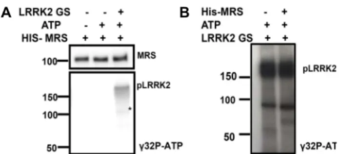
LRRK2와 MRS의 상호 작용이 3가지 다른 종류의 반복적인 실험에서 확인되었기 때문에 LRRK2가 MRS를 인산화시키는 가를 관찰하였다. 인산화효소는 세포 내 신호전달회로의 중요 한 조절인자이다[6]. LRRK2 단백질은 serine/threonine 인산 화효소 활성을 가지고 있기 때문에 LRRK2에 의해 MRS가 인 산화되는지를 확인하기 위해 방사선 동위원소, [γ
32P]-ATP를 이용해 실험을 진행하였다. Fig. 2A에서 LRRK2의 자가인산화 가 관찰 되었으므로 인산화반응이 진행된 것을 확인하였다.
하지만 MRS위치에는 상대적으로 너무 약한 인산화 밴드가
보여서 인산화여부를 확신할 수 없었다. 그래서 다시 LRRK2
의 양을 증가시켜서 인산화반응을 진행하였다. 그 결과, auto-
radiography에서 MRS 밴드 위치에 아무런 밴드가 나타나지
않음을 반복적으로 확인하였다(Fig. 2B). 결론적으로 MRS는
LRRK2의 인산화 기질이 아님을 확인하였다.
A B
Fig. 2 MRS is not phosphorylated by LRRK2. (A) In vitro kinase assay was performed using recombinant His-MRS pro- tein and N-LRRK2 G2019S. His-MRS was incubated with or without LRRK2 G2019S and [γ-
32P] ATP. The reaction mixture was subjected to SDS-PAGE and the specific bands were detected by Western blotting (MRS) and au- toradiography (pLRRK2). (B) In vitro kinase assay was performed with LRRK2 G2019S protein and in the pres- ence or absence of His-MRS. * indicates non-specific band.
A
B
C
Fig. 3. Over-expression of MRS or LRRK2 does not increase sta- bility of the partner protein. (A) MRS over-expression does not increase LRRK2 stability. HEK293T cells were co-transfected with Myc-LRRK2 (LRRK2 WT) and in- creasing concentration (0. 0.5, 1, 2 μg) of Myc-MRS (MRS).
The cell lysates were analyzed by Western blotting. A representative image of more than 6 experiments was shown. (B) Down-regulation of MRS expression by siMRS RNA transfection showed no effect on LRRK2 stability in NIH3T3 cells. (C) LRRK2 over-expression does not in- crease MRS stability. NIH3T3 cells were transfected with Myc-LRRK2-WT, -G2019S or vector in a dose-dependent manner (0, 1, 3 μg). The cell lysates were analyzed by Western blotting. A representative image of 3 experi- ments was shown.
MRS와 LRRK2 단백질의 안정성에 대한 상대 단백질의 영향
파킨슨병은 단백질의 응집에 의해 초래되는 대표적 질병의 하나이며, LRRK2는 autophagy 조절자로서 단백질 분해에 영 향을 준다는 사실이 잘 알려져있다[13]. 그러므로 LRRK2와 MRS의 상호작용이 상대단백질의 안정성에 서로 영향을 주는 가를 각 단백질의 과발현을 통하여 관찰하였다.
먼저 MRS 유전자의 형질전환과 siMRS RNA를 이용한 넉 다운(Knock-down)으로 MRS 단백질의 양을 각각 증가, 또는 감소시킨 HEK293T 세포에서 LRRK2 단백질의 변화를 확인해 보았다. Fig. 3A에서 보인 것과 같이 MRS의 발현 양이 뚜렷하 게 증가했음에도 불구하고 LRRK2의 양적인 변화는 관찰할 수 없었다. 동일한 실험을 NIH3T3 세포에서도 수행하였으나 유의미한 변화는 없었다(data not shown). 이 결과를 확인하 기 위하여, MRS siRNA를 이용하여 MRS의 양을 감소시켜서 LRRK2의 양을 관찰하였다. a-tubulin의 양을 control로 조정 하여 LRRK2 양을 비교했을 때에 MRS의 양은 감소되었으나 LRRK2의 양적인 변화를 확인 할 수 없었다(Fig. 3B). 이와 반 대로 LRRK2 야생종이나 G2019S 돌연변이 단백질을 과발현시 켜 LRRK2의 증가에 따른 MRS 단백질 양의 변화를 조사하였 으나, LRRK2 단백질의 양이 증가함에 따른 MRS의 단백질 양의 변화는 관찰할 수 없었다(Fig. 3C). 이 결과는 LRRK2와 MRS의 상호작용이 각 단백질의 분해나 안정성에는 직접적인 영향이 없다는 것을 의미한다.
고 찰
인산화효소 활성이 증가된 LRRK2 G2019S 돌연변이가 파 킨슨병 환자에서 발견됨으로 인해 LRRK2에 의해 인산화되는
기질의 세포 내 기능변화가 파킨슨병 발병에 중요한 요인이 될 것이라는 추론하에 LRRK2에 의해 인산화되는 기질 단백질 의 규명과 인산화 후에 나타나는 기능변화에 LRRK2 연구의 초점이 모아지고 있다[8, 15, 23]. MRS는 과발현하지 않은, 세 포에 내재하는 단백질이 LRRK2와 결합한다는 것이 확인되었 으나(Fig. 1) LRRK2에 의해 인산화되지는 않는 것으로 관찰되 었다(Fig. 2). 그래서 우리는 LRRK2, MRS 상호작용이 MRS의 다른 기능에 변화를 나타내는 지를 관찰하였으나 한 단백질의 과발현은 다른 상대단백질의 양을 변화시키지 못하였다(Fig.
3). 이는 LRRK2가 MRS와 결합함으로 인해 생기는 기능변화 가 단백질의 분해에 관련한 것은 아니라는 것을 제시한다.
MRS는 산화스트레스가 있을 때 ERK에 의해 인산화되어 산화
스트레스에 의한 세포독성을 저하시킨다고 보고되었다[9]. 산
화스트레스는 파킨슨병의 발병원인중의 하나이므로[9] 우리
는 LRRK2의 발현이 MRS의 산화스트레스에 대한 방어 기작 을 변화시키는가를 조사하였으나 뚜렷한 결과를 관찰할 수 없었다. 또한 LRRK2, MRS의 두 단백질을 NIH3T3 세포에서 과발현 하였을 때에도, 과발현으로 인한 세포 독성의 유의미 한 차이를 관찰할 수 없었다.
본 연구에서 MRS는 LRRK2와 상호작용은 하면서 인산화 효소 기질이 아닌 것으로 확인되었다. LRRK2는 LRR, WD40 이라는 두 개의 단백질 상호작용 도메인을 가지고 있기 때문 에 많은 단백질과 상호작용하며 이들 단백질은 인산화효소의 기질인 단백질도 있으나, 아닌 것도 있음이 보고되었다[15].
후자의 경우, LRRK2와 상호작용한다고 보고된 AP3B1와 Tau 는 LRRK2와의 상호작용의 결과로 인산화되지는 않았더라도 자신의 고유의 기능이 변화하였다[5].
본 연구는 NIH3T3나 HEK293T 세포에서 진행되었다. 이는 LRRK2의 내재적인 발현이 상대적으로 높은 NIH3T3 세포에 서 단백질 상호작용이 관찰되었고, MRS의 산화스트레스에 관 한 선행 연구결과[9]도 HEK293T 세포에서 진행되었기 때문이 다. 하지만, 파킨슨병 발병이 도파민성 신경세포의 사멸로 나 타난다는 점을 고려할 때, LRRK2와 MRS의 상호작용이 도파 민성 신경세포에서도 일어나고, 그 작용이 이들 세포에서만 파킨슨병과 관련되는 기능의 유의미한 변화를 초래할 가능성 을 배제할 수는 없다. 또한 LRRK2는 여러 종류의 리보솜 단백 질과 결합하며, 그 중 특히 s15를 인산화하여 단백질 합성을 증가시킨다고 보고되었다[15]. MRS가 단백질 합성의 첫 번째 아미노산인 메티오닌을 tRNA에 붙이는 효소라는 걸 감안할 때, LRRK의 MRS에 대한 작용이 단백질 합성조절에 영향을 미칠 수도 있을 것이다. 앞으로 이 부분의 연구도 필요할 것으 로 보인다.
감사의 글
MRS 관련 발현 벡터는 서울대 약대 김성훈교수에게서 제 공받았으며, 본 연구는 원광대학교 산본병원 인암뇌신경연구 센터, 한국연구재단 신진연구 지원사업(2015R1C1A2A01051 755)에 의해 수행되었음.
References