Effects of Glutamine Deprivation and Serum Starvation on the Growth of Human Umbilical Vein Endothelial Cells
Jin-Woo Jeong
1, Hye Hyeon Lee
2, Cheol Park
3, Wun-Jae Kim
4and Yung Hyun Choi
5,6*
1
Center for Core Research Facilities, Daegu Gyeongbuk Institute of Science & Technology, Daegu 711-873, Korea
2
Department of Biotechnology, College of Natural Resources and Life Science, Dong-A University, Busan 604-714, Korea
3
Department of Molecular Biology, College of Natural Sciences, Dongeui University, Busan 614-714, Korea
4
Department of Urology, College of Medicine, Chungbuk National University, Chungbuk 361-763, Korea
5
Department of Biochemistry, Dongeui University College of Oriental Medicine, Busan 614-052, Korea
6
Anti-Aging Research Center & Blue-Bio Industry RIC, Dongeui University, Busan 614-714, Korea Received May 1, 2013 /Revised July 6, 2013 /Accepted July 8, 2013
Glutamine and serum are essential for cell survival and proliferation in vitro, yet the signaling path- ways that sense glutamine and serum levels in endothelial cells remain uninvestigated. In this study, we examined the effects of glutamine deprivation and serum starvation on the fate of endothelial cells using a human umbilical vein endothelial cell (HUVEC) model. Our data indicated that glutamine deprivation and serum starvation trigger a progressive reduction in cell viability through apoptosis induction in HUVECs as determined by DAPI staining and flow cytometry analysis. Although the apoptotic effects were more predominant in the glutamine deprivation condition, both apoptotic ac- tions were associated with an increase in the Bax/Bcl-2 (or Bcl-xL) ratio, down-regulation of the in- hibitor of apoptosis protein (IAP) family proteins, activation of caspase activities, and concomitant degradation of poly (ADP-ribose) polymerases. Moreover, down-regulation of the expression of Bid or up-regulation of truncated Bid (tBid) were observed in cells grown under the same conditions, in- dicating that glutamine deprivation and serum starvation induce the apoptosis of HUVECs through a signaling cascade involving death-receptor-mediated extrinsic pathways, as well as mitochon- dria-mediated intrinsic caspase pathways. However, apoptosis was not induced in cells grown in glu- tamine- and serum-free media when compared with cells exposed to glutamine deprivation or serum starvation alone. Taken together, our data indicate that glutamine deprivation and serum starvation suppress cell viability without apoptosis induction in HUVECs.
Key words : HUVECs, glutamine deprivation, serum starvation, apoptosis
*Corresponding author
*Tel:+82-51-850-7413, Fax:+82-51-853-4036
*E-mail : [email protected]
This is an Open-Access article distributed under the terms of the Creative Commons Attribution Non-Commercial License (http://creativecommons.org/licenses/by-nc/3.0) which permits unrestricted non-commercial use, distribution, and reproduction in any medium, provided the original work is properly cited.
Journal of Life Science 2013 Vol. 23. No. 7. 926~932 DOI : http://dx.doi.org/10.5352/JLS.2013.23.7.926
서 론
내피세포(endothelial cell)는 염증, 혈관재생 및 혈관신생에 서 중요한 역할을 하는 세포이다. 내피세포의 항상성은 내피 세포의 증식과 사멸의 균형에 의존적이며, 혈관 벽의 보존을 위하여 내피세포는 일반적으로 세포의 사멸에 대하여 내성을 지니고 있다. 이러한 측면에서 apoptosis는 내피세포에 의해 형성되는 조직의 항상성뿐만 아니라 다양한 질환의 원인과 진행에 중요한 역할을 하는 것으로 생각되어지고 있다[10, 20].
특히 apoptosis에 의한 내피 손상은 아테롬성 동맥 경화증, 동종 이식 맥관 장애, 고혈압, 폐혈증 등과 많은 염증성 장애 과정에서 중요한 역할을 하는 것으로 알려져 있다[4, 15]. 현재
내피세포의 대표적인 apoptosis 유도 인자로서 lip- opolysaccharide [11], tumor necrosis factor (TNF)-α [26], in- terferon-γ [27], 산화적 스트레스 [29] 및 transforming growth factor β1 [31], amyloid-β [8] 등이 알려진 반면, fibroblast growth factor (FGF) [16], vascular endothelial growth factor (VEGF) [9] 및 endothelin [28] 등은 내피세포의 apoptosis 억 제인자로 알려져 있다. 이러한 측면에서 in vitro 조건에서 혈 청(serum) 공급의 차단은 내피세포의 apoptosis를 촉진하는 환경을 제공해 줄 수 있다[3, 17, 25, 27]. 또한 에너지 대사 측면에서 글루타민의 공급제한 역시 내피세포의 apoptosis를 유도하는 인자로서 작용[24]할 수 있으나, 그에 대한 구체적인 기전은 알려진 바 없다.
일반적으로 내피세포의 apoptosis는 Fas 및 TNF re-
ceptor-1과 같은 세포막 죽음의 수용체 경로(extrinsic path-
way)와 미토콘드리아 의존적 경로(intrinsic pathway)에 의
하여 유도될 수 있다[2, 13]. 특히 심혈관계 질환과 연계된
내피세포의 미토콘드리아 경로를 통한 apoptosis 조절에는
Bcl-2 family 단백질들의 발현 변화와 caspase 단백질의 활
성이 주요 역할을 하는 것으로 알려져 있으며[21], 미토콘드 리아에서의 활성산소종(reactive oxygen species, ROS) 생성 이 결정적 인자로 작용할 수 있음이 밝혀졌다[18, 23]. 그러 나 최근 연구에 의하면 내피세포에서의 apoptosis에서 ROS- 의존적 caspase-8의 활성화가 세포막 수용체 경로와 미토콘 드리아 경로를 연결 및 증폭할 수 있는 것[8, 19, 22, 32]으로 알려져 아마도 내피세포의 apoptosis 유발은 내피세포가 처 한 환경적 스트레스의 종류에 매우 의존적일 수 있음을 의 미한다. 따라서 본 연구에서는 내피세포의 기본적인 증식에 필요한 글루타민과 혈청의 유무에 따른 내피세포의 반응을 조사하였다. 이를 위하여 인체 재대정맥 내피세포(human umbilical vein endothelial cells, HUVECs)가 글루타민 또 는 혈청이 단독 또는 동시에 결핍된 조건에서 apoptosis 유 발에 어떤 반응을 보이는지를 조사하였다.
재료 및 방법
세포배양
HUVECs은 American Type Culture Collection (ATCC, Rockville, MD, USA)에서 구입하였으며, 10% fetal bovine se- rum (FBS) (Invitrogen, Carlsbad, CA, USA) 및 100 μg/ml penicillin/streptomycin이 함유된 정상 RPMI 1640 (글루타민 농도: 2.05 mM) 및 글루타민 결핍 RPMI 1640 (Invitrogen) 배지를 이용하여 배양하였다.
Hemocytometer를 이용한 세포 생존율 및 MTT assy에 의한 세포 증식율 측정
글루타민 또는 혈청 결핍이 HUVECs의 증식에 미치는 영 향을 조사하기 위하여 6-well plate에 세포를 1 X 10
5개/ml로 분주하고 24시간 동안 안정화시킨 다음, 글루타민 또는 혈청 이 결핍된 RPMI-1640 배지로 교체하였다. 적정 시간 경과 후 상층액을 제거하고 0.05% trypsin-EDTA를 처리하여 세포를 부유시킨 다음 phosphate-buffered saline (PBS)와 0.5% try- pan blue solution을 각 well 당 첨가하였다. 약 2분 후 sample 을 hemocytometer에 적용한 후 도립 현미경을 이용하여 살아 있는 세포를 계수하여 상대적 수치로 비교하였다. MTT assay 를 위하여 적정 시간 후 배지를 제거하고 tetrazolium bromide salt (MTT, Sigma-Aldrich Chemical Co., St. Louis, MO, USA) 를 0.5 mg/ml 농도가 되게 배지로 희석하여 분주하고 3시간 동안 CO
2incubator에서 배양시킨 다음 MTT 시약을 깨끗하게 제거하고 dimethylsulfoxide (DMSO)를 적정량 분주하여 well 에 생성된 formazin을 모두 녹인 후 96 well plate에 200 μl씩 옮겨서 ELISA reader (Molecular Devices, Sunnyvale, CA, USA)로 540 nm에서 흡광도를 측정하였다.
Apoptosis 유도의 확인
글루타민 또는 혈청 결핍에 의한 HUVECs의 증식억제가
apoptosis 유도와 연관성이 있는지의 여부를 조사하기 위하여 4',6-diamidino-2-phenylindole (DAPI, Sigma-Aldrich) 염색 을 이용한 핵의 형태적 변화를 관찰하였다. 이를 위하여 동일 조건에서 배양된 HUVECs를 37% formaldehyde 용액과 PBS 를 1 : 9의 비율로 섞은 fixing solution을 이용하여 상온에서 10분 동안 고정하고 DAPI 용액으로 염색시켰다. 15분 정도 염색시킨 후, mounting solution을 처리한 후 형광 현미경 (Carl Zeiss, Germany)을 이용하여 400배의 배율로 핵의 형태 변화를 관찰하였다. 또한 apoptosis 유도의 정량적인 비교 분 석을 위하여 준비된 세포를 모은 후 PBS로 충분히 씻고 CycleTEST Plus DNA Reagent Kit (Becton Dickison, San Jose, CA, USA)를 사용하여 고정 및 propidium iodide (PI)를 처리하여 4℃, 암조건에서 10분 이상 염색시켰다. 염색된 세포 를 25-mm mesh를 이용하여 단일세포로 분리한 후 FACSCallibur (Becton Dickison)를 이용하여 형광반응에 따 른 Cellular DNA content 및 histogram을 Cell Quest software 프로그램을 이용하여 sub-G1기에 해당되는 세포의 빈도를 apoptosis가 일어난 세포로 정량화하였다.
단백질 분리, 전기영동 및 Western blot analysis
준비된 세포들을 적당량의 lysis buffer [25 mM Tris-Cl (pH 7.5), 250 mM NaCl, 5mM EDTA, 1% NP-40, 1 mM phenyl- methylsulfonyl fluoride (PMSF), 5 mM dithiothreitol (DTT)]
를 첨가하여 4℃에서 1 시간 반응 시킨 후, 14,000 rpm으로 30분간 원심분리하여 상층액을 모았다. 상층액의 단백질을 정 량화한 후 동량의 단백질을 sodium dodecyl sulphate (SDS)-polyacrylamide gel을 이용하여 전기영동으로 분리하 였다. 분리된 단백질을 함유한 acrylamide gel을 nitro- cellulose membrane (Schleicher and Schuell, Keene, NH, USA)에 전이시킨 후, 비특이적인 단백질에 대해 blocking을 실시하였다. 준비된 membrane을 적정 항체로 반응시킨 후 enhanced chemiluminescence (ECL) 용액(Amersham Life Science Corp., Arlignton Heights, IL, USA)을 처리하여 특정 단백질의 발현 정도를 비교하였다.
In vitro caspase activity 측정
동일한 조건에서 준비된 세포들의 단백질을 96-well plate 에 동량 분주하고 2X reaction buffer를 각각 50 μl씩 넣어 주어 총 100 μl가 되게 한 후 각각의 caspase 기질을 첨가하였다.
준비된 plate를 37℃ 암상태에서 3시간 동안 incubation 시킨
후 ELISA reader를 이용하여 405 nm의 파장에서 흡광도를
측정하였다. Caspase 활성 측정을 위한 colormetric assay kit
는 R&D Systems (Minneapolis, MN, USA)에서 구입하였고,
실험에 사용된 기질은 caspase-3의 경우에는 Asp-Glu-Val-
Asp (DEVD)-p-nitroaniline (pNA)이었고, caspase-8의 경우
에는 Ile-Glu-Thr-Asp (IETD)-pNA이었으며, caspase-9은
Leu-Glu-His-Asp (LEHD)-pNA였다.
통계 처리
모든 실험결과는 평균±표준편차로 표시하였고 SigmaPlot 을 이용하여 Student t-test를 이용하여 통계적 유의성을 얻었다.
결과 및 고찰
HUVECs의 생존 및 증식에 미치는 글루타민 및 혈청 결핍 의 영향
HUVECs의 생존 및 증식에 미치는 글루타민의 영향을 조 사하기 위하여 정상배지에서 배양된 HUVECs를 글루타민이 없는 조건에서 24 시간 및 48 시간 배양한 후 hemocytometer counting을 이용하여 생존한 세포의 수 및 MTT assay를 이용 한 증식율의 변화를 측정하였다. Fig. 1에 나타낸 바와 같이 글루타민이 결핍된 조건에서 HUVECs는 시간의 경과에 따라 세포의 생존율 및 증식율이 글루타민 결핍 시간의 증가에 따 라 모두 감소되었다. HUVECs의 증식에 혈청이 필수적으로 요구되며, 혈청이 결핍되었을 경우 apoptosis가 일어난다는 것은 잘 알려진 바 있다[12, 14]. 또한 HUVECs에서는 정확한 비교 선행 연구가 없지만, in vitro 조건에서 글루타민의 결핍 도 세포의 증식을 억제하며 apoptosis를 유발할 수 있음이 Drogat et al. [5]에 의하여 보고된 바 있다. 그러나 동일 조건에 서 혈청 결핍보다는 글루타민의 결핍에서 HUVECs의 증식율 은 현저히 낮게 나타났으며, 혈청과 글루타민가 동시에 결핍 된 조건에서 배양된 HUVECs의 증식율은 더욱 더 낮게 나타 났다(Fig. 2A 및 2B).
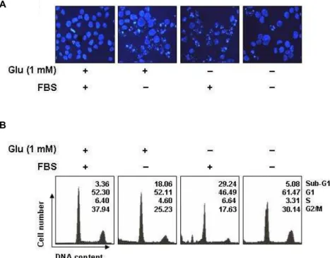
이러한 글루타민 및 혈청의 단독 및 동시 결핍에 의한 HUVECs의 증식율의 저하 현상이 apoptosis 유발과 연관성이 있는지를 조사하기 위하여 DAPI staining을 한 후 핵의 형태 변화 및 flow cytometry 분석에 의한 sub-G1기에 속하는 세포 의 빈도를 비교하였다. Fig. 3의 결과에서 알 수 있듯이 글루타 민 또는 혈청이 결핍된 조건에서 배양된 HUVECs는 전형적인 apoptosis가 일어났을 경우 관찰되는 염색질의 응축에 의한 apoptotic body 현상이 뚜렷하게 증가되었으며, 글루타민 및 혈청이 모두 결핍된 조건에서 배양된 HUVECs에서는 이러한 현상을 오히려 관찰할 수 없었다. 아울러 sub-G1기에 속하는 세포의 빈도 역시 글루타민 또는 혈청이 결핍된 조건에서 배 양된 HUVECs에서는 각각 18% 및 29% 정도로 관찰되어 각각 의 단독 결핍에 의한 생존율 저하 현상과 연관성이 있었으나, 글루타민과 혈청이 동시에 결핍된 조건에서 배양된 HUVECs 에서는 5% 정도로 정상배지에서 배양된 세포의 자연적 apop- tosis 유발 빈도인 3%에 유사한 빈도를 보여주었다. 즉 글루타 민과 혈청이 동시에 결핍된 조건에서 HUVECs의 증식 및 생 존율이 저하되었지만, 각각의 결핍에 의하여 나타나는 apop- tosis 유발은 오히려 차단되었음을 알 수 있었다. 또한 글루타
A
B
Fig. 1. Inhibition of cell viability and proliferation by glutamine deprivation in HUVECs. HUVECs were seeded into 6-well plate at 1×10
5cells/ml and incubated in normal medium (glutamine, 2.05 mM). After 24 hr incubation, the media was changed to glutamine-free media and the cells were collected at the indicated times. (A) Cell via- bility was measured using a metabolic-dye-based MTT assay. (B) The cells were trypsinized and the viable cells were scored by hemocytometer counts of trypan blue-ex- cluding cells. The data shown are means±SD of three independent experiments. Significance was determined using a Student’s t-test (*, p <0.05 vs. untreated control).
민과 혈청이 동시에 결핍된 조건에서 배양된 세포의 경우 G1 기에 해당되는 세포의 빈도가 다소 증가되었으며, 이러한 현 상 세포주기 교란과 연관이 있는지의 여부에 때해서는 추후 연구가 필요할 것으로 생각된다.
HUVECs의 caspase 활성에 미치는 글루타민 및 혈청 결 핍의 영향
이상에서 관찰된 혈청과 글루타민의 단독 및 동시 결핍에
의한 HUVECs에서 apoptosis 유발의 차이에 대한 기전의 해
석을 위하여 세포 내 apoptosis 유발에 중요한 역할을 하는
3가지 종류의 caspase (caspase-3, -8 및 -9)의 발현 및 활성
변화를 조사하였다. Fig. 4에서와 같이 Harfouche et al. [12]
A
B
Fig. 3. Induction of apoptosis by glutamine deprivation and FBS starvation in HUVECs. (A) The cells grown under same condition as Fig. 2 were collected, fixed, and stained nuclei with DAPI solution were photographed with a fluorescent microscope using a blue filter (Magnification, ×400). (B) The cells were stained with PI for flow cytometry analysis. The percentages of cells in the each phase are presented. The data represent the average of two independent experiments.
Fig. 2. Effects of glutamine starvation and FBS supplementation on the cell viability and cell morphology of HUVECs.
HUVECs were incubated in normal medium for 24 hr and then the media was changed to glutamine-free or/and FBS-free media and the cells were collected after 48 hr. (A) Cell viability was measured using a metabol- ic-dye-based MTT assay. The significance was de- termined by a Student’s t -test (
*p <0.05, compared with control). (B) Cells were visualized by an inverted light microscope (Magnification, ×200).
및 Date et al. [3]의 선행결과에서와 유사하게 혈청이 결핍된 조건에서 배양된 HUVECs에서 조사된 3가지 caspase의 활성 형 단백질의 발현을 모두 관찰할 수 있었으며, 글루타민이 결 핍된 조건에서는 배양된 세포에서는 각 caspase 활성형의 발 현이 더욱 증가하였다. 이를 정량적으로 비교하기 위하여 in vitro caspase 활성 정도를 해당 기질을 사용하여 비교한 결과, 글루타민이 결핍된 조건에서 각 caspase의 활성이 혈청 결핍 조건에서 보다 상대적으로 다소 높게 나타나 Western blotting 결과와 일치된 경향성을 보여 주었다(Fig. 4). 아울러 활성화된 caspase-3의 기질 단백질인 poly(ADP-ribose) polymerase (PARP)의 단편화 현상[30]도 글루타민 결핍 조건에서 다소 강 하게 나타났다. 그러나 혈청과 글루타민이 동시에 결핍된 조 건에서 배양된 HUVECs에서는 3가지 caspase의 활성 증가 및 PARP의 단편화는 전혀 관찰되지 않았다. 따라서 혈청과 글루 타민이 동시에 결핍된 조건에서 배양된 HUVECs의 증식율 저하 현상이 현저하게 증가하였음에도 apoptosis가 유발되지 않았음은 Fig. 4의 결과에서와 같이 caspase의 활성 변화와 밀접한 연관성이 있음을 알 수 있었다.
HUVECs에서 Bcl-2 및 IAP family 단백질의 발현에 미 치는 글루타민 및 혈청 결핍의 영향
이상의 결과에서 비록 혈청과 글루타민이 동시에 결핍된 조건에서 배양된 HUVECs의 증식율이 상대적으로 더 억제되 A
B
A
B
Fig. 4. Effects of glutamine deprivation and FBS supplementa- tion on the activation of caspases in HUVECs. (A) Cells were incubated in glutamine-free or/and FBS-free media for 48 hr. The cells were lysed and then cellular proteins were separated by SDS-polyacrylamide gels. Separated proteins in gels transferred to nitrocellulose and probed with the indicated antibodies. Actin was used as an in- ternal control. (B) The cells grown under same condition as (A) were collected and then lysed. The equal amounts of cell lysates were assayed for caspase-3, -8 and -9 activ- ity using DEVD-pNA, IETD-pNA and LEHD-pNA as substrates. The results are expressed as the mean±SD of three independent experiments. Significance was de- termined using a Student’s t-test (*, p <0.05 vs. untreated control).
었으나 apoptosis가 유발되지 않았음이 caspase의 활성 여부 와 연관성이 있음을 확인하였기 caspase의 활성을 조절하는 다양한 인자들의 발현에 미치는 혈청과 글루타민의 영향을 조사하였다. 이를 위하여 먼저 미토콘드리아를 중심으로 각 caspase의 활성을 조절하는 Bcl-2 family 인자들[7]의 변화 여 부를 조사하였는데, 혈청이 결핍된 조건에서 배양된 HUVECs 에서 anti-apoptotic Bcl-2 인자에 속하는 Bcl-2 및 Bcl-xL의 발 현이 다소 감소한 반면, 대표적인 pro-apoptotic 인자인 Bax의
Fig. 5. Effects of glutamine deprivation and FBS supplementa- tion on the levels of Bcl-2 and IAP family proteins in HUVECs. The cells grown under same condition as Fig.
3 were collected and then lysed. The cellular proteins were then separated by electrophoresis on SDS-poly- acrylamide gels and transferred onto nitrocellulose membranes. Next, the membranes were probed with the indicated antibodies and the proteins were visualized using an ECL detection system. Actin was used as an internal control.
발현은 약간 증가되었음을 알 수 있었다(Fig. 4). 그러나 이러 한 단백질들의 발현 변화는 글루타민 결핍 조건에서는 더욱 더 크게 나타났으며, 특히 caspase-8의 활성에 의한 Bid 단백질 의 truncation된 형태인 tBid의 발현[1]이 글루타민 결핍 조건 에서 매우 증가되었다. 한편 caspase 단백질들과 직-간접적인 결합을 통하여 그들의 활성을 억제함으로서 apoptosis를 억제 하는 역할을 하는 IAP family 단백질들[6]의 발현 역시 혈청 결핍 조건보다 글루타민 결핍 조건에서 배양된 HUVECs에서 더 감소되었다. 그러나 혈청과 글루타민이 동시에 결핍된 조 건에서 배양된 HUVECs에서는 이러한 변화 현상이 관찰되지 않았다.
이상의 결과를 종합하여 보면, HUVECs의 배양 조건에서
글루타민이 결핍될 경우 세포막 수용체 경로의 활성에 의하여
caspase-8의 활성이 증가되고, 증가된 caspase-8에 의하여 Bid
가 tBid로 전환되면서 미토콘드리아 경로 활성을 촉진하였을
것으로 생각된다. 이로 인하여 caspase-9의 활성화에 따른 cas-
pase-3의 활성 및 기질 단백질들의 분해로 연결되는 caspase
cascade 경로의 활성이 촉진됨으로 apoptosis가 유발되었을
것으로 추정된다. 그러나 이러한 경로가 혈청이 결핍된 조건
에서는 글루타민 결핍에 비하여 다소 약하게 일어나는 것으로
추정된다. 그러나 혈청과 글루타민이 동시에 결핍되었을 경우 에는 오히려 이러한 경로가 차단되어 apoptosis가 억제되는 현상이 관찰되었는데, 이에 대한 정확한 이유를 현재의 결과 로서는 추정할 수 없는 상태이며 이에 관한 추가적인 연구가 이루어져야 할 것으로 생각된다.
감사의 글
이 논문은 정부(교육과학기술부)의 재원으로 한국연구재단 의 지원을 받아 수행된 기초연구사업임(2010-0004415 및 2012-0000476).
References