Vol.24 No.2 p197-205, Dec. 2007
The Difference of Left Atrial Volume Index : Can It Predict the Occurrence of Atrial Fibrillation after
Radiofrequency Ablation of Atrial Flutter?
Ung Kim, Young Jo Kim, Sang Wook Kang, In Wook Song, Jung Hwan Jo, Sang Hee Lee, Geu Ru Hong, Jong Seon Park, Dong Gu Shin
Department of Internal Medicine,
College of Medicine, Yeungnam University, Daegu, Korea
-Abstract-
Background:The occurrence of atrial fibrillation after ablation of atrial flutter is clinically important. We investigated variables predicting this evolution in ablated patients without a previous atrial fibrillation history.
Materials and Methods:Thirty-six patients (Male=28) who were diagnosed as atrial flutter without previous atrial fibrillation history were enrolled in this study. Group 1 (n=11) was defined as those who developed atrial fibrillation after atrial flutter ablation during 1 year follow-up. Group 2 (n=25) was defined as those who has not occurred atrial fibrillation during same follow-up term. Echocardiogram was performed to all patients. We measured left atrial size, left ventricle end diastolic and systolic dimension, ejection fraction and left atrial volume index before and after ablation of atrial flutter. The differences of each variables were compared and analyzed between two groups.
1)Results:The preablation left ventricular ejection fraction (preLVEF) and postablation left ventricular ejection fraction (postLVEF) are 54±14%, 56±13% in group 1 and 47±16%, 52±13%
in group 2. The differences between each two groups are statistically insignificant (2.2±1.5 in group 1 vs 5.4±9.8 in group 2, p=0.53). The preablation left atrial size (preLA) and postablation left atrial size (postLA) are 40±4 mm, 41±4 mm in group1 and 44±8 mm, 41±4 mm in group 2. The atrial sizes of both groups were increased but, the differences of left atrial size between two groups before and after flutter ablation were statistically insignificant (0.6±0.9
책임저자:신동구, 대구광역시 남구 대명5동 317-1, 영남대학교 의과대학 내과학 교실 Tel: (053) 620-3313, Fax: (053) 621-3310, E-mail: [email protected]
mm in group 1 vs -3.8±7.4 mm in group 2, p=0.149). The left atrial volume index before flutter ablation was significantly reduced in group 1 than group 2 (32±10 mm
3/m
2, 35±10 mm
3/m
2in group 1 and 32±10 mm
3/m
2, 29±8 mm
3/m
2in group 2, p<0.05).
Conclusion:The difference between left atrial volume index before and after atrial flutter ablation is the robust predictor of occurrence of atrial fibrillation after atrial flutter ablation without previous atrial fibrillation.
Key Words: Atrial flutter, Ablation, Atrial fibrillation, Left atrium, Volume
Introduction
Radiofrequency catheter ablation targeting the isthmus between the tricuspid annulus and the inferior vena cava is an established therapy for typical atrial flutter (AFL). It is successful in more than 90% of patients.
1-7)But, in the clinical setting, AFL and atrial fibrillation (AF) often coexist, and the follow up of patients successfully treated with transisthmic ablation is complicated by the occurrence of AF in 10~47% of patients.
8-17)We already have known that the presence of preablation AF is the most significant predictor of postablation AF.
18-20)However, clinical and procedural predictors of postablation AF occurrence has always been evaluated in the mixed group of AFL patients with AF
18-20)and rarely in the group of patients without history of AF. +This study aimed that better identification of patients who are at risk for the development of AF may help to optimize the antiarrhythmic stratery during or after AFL ablation. So, we investigated variables predicting this evolution with
echocardiography in ablated patients without a previous AF History.
Materials and methods
1. Study population
The study group consisted of 36 consecutive patients who were diagnosed as AFL without previous history of paroxysmal AF and underwent radiofrequency catheter ablation for recurrent typical AFL from January 2000 to June 2005 at Yeungnam university hospital.
Typical AFL was diagnosed when the
surface ECG showed flutter waves that were
predominantly negative in leads II, III, aVF
and positive in lead V1 and defined as a
macroreentrant atrial tachycardia that exhibited
either counterclockwise or clockwise activation
around the tricuspid annulus and atypical
AFL was defined as an atrial flutter other
than typical AFL.Previous episodes of AF
were all excluded from this study. Postablation
AF development during 1 year follow-up
was defined as the documentation of AF
during ECG or Holter ECG monitoring of at
least one episode of AF lasting more than one minute.
2. Electrophysiological study and radiofrequency catheter ablation
Written informed consent was obtained before transcatheter ablation from all patients.
Four multipolar catheters were inserted from right femoral vein and left subclavian vein:
One quadripolar catheter (Boston scientific, USA) positioned at the His bundle and RV;
One decapolar catheter (Daig, USA) positioned in the coronary sinus with the proximal electrode pair positioned at the ostium; a 20 electrode Halo catheter (Cordis, USA) positioned around the tricuspid valve to assess annular activation; and the ablation catheter. All measurements were performed with the Cardiolab system (Prucka engineering, USA).
The ablation was anatomically guided. The end-point of the procedure was the achievement of a complete bi-directional isthmus block according to the method reported in detail in the land mark study by Poty et al
21)using activation mapping.
3. Echocardiographic measurements
Transthoracic echocardiography was performed before and after 24 hour of the radiofrequency ablation procedure by one observer blinded to the patient’s electrophy- siological status. Ultrasound studies were performed with Acuson Sequoia C256 (SIEMENS). M-mode measurements were
made according to the recommendations of the American Society of Echocardiography.
22)Left ventricular systolic function was evaluated on two-dimensional echocardiographic imaging of the left ventricle. Left ventricular volumes and ejection fraction were calculated by planimetry in the apical two and four-chamber views with the modified Simpson rule. Left atrial long axis, short axis and area were obtained by planimetry of the atrial inner borders with maximized atrial chamber size at end-systole in the four and two-chamber views. All two dimensional echocardiographic measurements were averaged over five cardiac cycles and the differences of each variable before and after AFL ablation were compared and analyzed between two groups.
4. Statistical analysis
Continuous variables are presented as mean±SD. Discrete variables are presented as percentages(%). Variables were compared by Fisher’s exact test for categorical variables and independent sample t test for continuous variables. A probability value of p<0.05 was considered significant.
Results
Thirty-six patients (36, Male=28) were enrolled in this study. Among all patients, AFL ablation succeeded in 35 patients (35/
36, 97%) and AFL recurred in four patients
(4/36, 11%). During 1 year follow-up, eleven
AF procedure success AFL recur AF occurrence Total N=36
N=11 (31%) N=4 (11%)
N=35 (97%)
AF procedure success AFL recur AF occurrence Total N=36
N=11 (31%) N=4 (11%)
N=35 (97%)
Fig. 1. One Year Follow-up Results of Patients After Ablation of Atrial Flutter.
347911131417192233
Case Number 25
30 35 40 45 50 55
LA s ize
PreAbl-Lasize Post-LA size
1 2 5 6 8 10 12 15 16 18 20 21 23 24 25 26 27 28 29 30 31 32 34 35 36 Case Number 36
38 40 42 44 46 48 50
LA s ize
PreAbl-Lasize Post-LA size
LA SizeLA Size
LA Size
Pre LAsize PostLAsize
Pre LAsize PostLAsize
347911131417192233
Case Number 25
30 35 40 45 50 55
LA s ize
PreAbl-Lasize Post-LA size
1 2 5 6 8 10 12 15 16 18 20 21 23 24 25 26 27 28 29 30 31 32 34 35 36 Case Number 36
38 40 42 44 46 48 50
LA s ize
PreAbl-Lasize Post-LA size
LA SizeLA Size
LA Size
Pre LAsize PostLAsize
Pre LAsize PostLAsize
Group 1(n=11) Group 2(n=25)
Group 1(n=11) Group 2(n=25)
Fig. 2. Comparison of LA size between two groups.
Group 1 (n=11) Groups 2 (n=25) P
Age(yr) Male
Organic Ht Ds Valvular Ischemic CHF HCM DM HTN
57±13 10(90%)
0 0 0 0 2(18%)
1(9%)
58±11 18(72%)
3 1 2 1 3(12%) 5(20%)
NS NS NS
NS NS
Ht: Heart, Ds: Disease, CHF: Congestive heart failure, HCM: hypertrophic cardiomyopathy
Table 1. Baseline characteristics between two groups
Group 1 (n=11) Group 2 (n=25) P
LVEDD LVESD LVEF LA size LA Vol Index
50±3 34±3 57±10 41±7 33±14
49±8 35±8 49±14
43±7 38±30
NS NS NS NS NS
LVEDD: Left ventricular end-diastrolic dimension, LVESD: Left ventricular end-systolic dimension, LVEF: Left ventricular ejection fraction, LA: Left atrium, Vol: Volume, NS: not significant
Table 2. Echocardiogrphic findings between two groups patients developed AF (Group 1, n=11) but, eighteen patients has not occurred AF (Group
2, n=25) (Fig 1). Baseline characteristics between two groups are summarized in Table 1. No differences between two groups are existed in age, sex, DM, hypertension
and organic heart disease. Table 2 showed
echocardiographic differences between two
groups and the result was statistically
insignificant. The findings between two
groups during electrophysiologic study are
Group 1 (n=11)
Group 2
(n=25) P
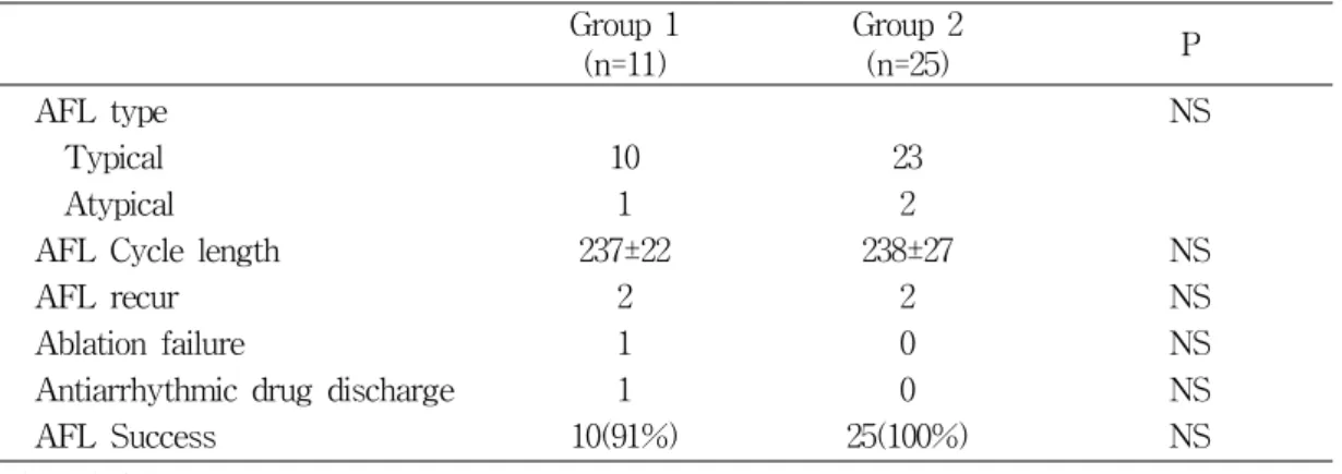
AFL type Typical Atypical AFL Cycle length AFL recur Ablation failure
Antiarrhythmic drug discharge AFL Success
10 1 237±22
2 1 1 10(91%)
23 2 238±27
2 0 0 25(100%)
NS
NS NS NS NS NS
AFL: Atrial flutter
Table 3. Electrophisiologic findings between two groups
Group 1 (n=11)
Group 2
(n=25) P
PreAbl-LVEF PostAbl-LVEF Differences
PreAbl-LA size PostAbl-LA size Differences
PreAbl-LA Vol index PostAbl-LA Vol index Differences
57±10 59±8 2±9
41±7 42±7 0.8±1
33±14 35±14 2.1±2
49±14 54±11 5±8
43±7 41±4 -2.8±6
38±30 29±7 -9±28
0.138 0.354 0.613
0.745 0.745 0.009
0.846 0.303 0.000
PreAbl: preablation, PostAbl: postablation, LVEF : Left ventricular ejection fraction, LA: Left atrium, Vol:
volume
Table 4. Echocardiographic Comparison Between two group
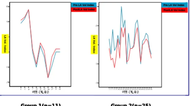
represented in Table 3. Statistical significance was not found in this examination. Table 4 showed echocardiographic differences between two groups. Differences of Left ventricular ejection fraction (LVEF) before and after AFL ablation were not statistically different between two group after 1 year follow-up (p=0.613). The atrial sizes of both groups were increased but, pre-ablational left atrium
(LA) size were significantly increased in
group 1 but, decreased in group 2 after AFL
ablation (p=0.009) and pre-ablational LA
volume index were significantly increased in
group 1 but, decreased in group 2 (p=0.000)
during same follow up period. In summary,
Differences of LA size and LA volume index
were increased or not decreased in case of
patients who had development of AF after
347911131417192233 Case Number 10
20 30 40 50 60
LA V ol In de x
Pre-LA vol index Post-LA vol index
1 2 5 6 8 10 12 15 16 18 20 21 23 24 25 26 27 28 29 30 31 32 34 35 36 Case Number 10
20 30 40 50
LA Vo l In de x
Pre-LA vol index Post-LA vol index
Group 1(n=11) Group 2(n=25)
LA Vol. Index LA Vol. Index
PostLA Vol Index Pre LA Vol Index
PostLA Vol Index Pre LA Vol Index
347911131417192233
Case Number 10
20 30 40 50 60
LA V ol In de x
Pre-LA vol index Post-LA vol index
1 2 5 6 8 10 12 15 16 18 20 21 23 24 25 26 27 28 29 30 31 32 34 35 36 Case Number 10
20 30 40 50
LA Vo l In de x
Pre-LA vol index Post-LA vol index
Group 1(n=11) Group 2(n=25)
LA Vol. Index LA Vol. Index
PostLA Vol Index Pre LA Vol Index
PostLA Vol Index Pre LA Vol Index
Fig. 3. Comparison of LA Vol. Index between two groups.
AFL ablation and differences of LA size and LA volume index were decreased in those who had not occurred AF after AFL ablation (Fig 2, Fig 3).
Discussion
Predictors of postablation AF occurrence have been recognized as history of preablation AF, left atrial size, inducibility of AF at the end of the ablation, the presence of structural heart disease, and a reduced left ventricular ejection fraction. Among these, history of preablation AF has emerged as the most significant predictor of postablation AF.
8, 10, 11,13, 18-20)