Vol.25 No.2 p165-170, Dec. 2008
폐쇄성 황달로 내원한 담관내 증식을 동반한 간세포암 1예
김승범⋅김태년⋅이호찬⋅박재현⋅은종렬⋅장병익⋅이헌주⋅윤성수*⋅배영경
†영남대학교 의과대학 내과학교실, 외과학교실*, 병리학교실†
A Case of Hepatocellular Carcinoma with intradural growth Presenting as Obstructive Jaundice
Sung-Bum Kim, Tae-Nyeun Kim, Sung-Jun Kim, Ho-Chan Lee, Jae-Hyun Park, Jong-Ryul Eun, Byung-Ik Jang, Heon-Ju Lee, Sung-Su Yun*, Young-Kyung Bae
†Department of Internal Medicine, *Department of Surgery and
†Department of Pathology, College of Medicine, Yeungnam University, Daegu, Korea
1)
-Abstract-
The incidence of hepatocellular carcinoma presenting as obstructive jaundice is 0.7∼9%.
The mechanisms of obstructive jaundice include bile duct invasion by tumor, tumor thrombi, blood clots, direct bile duct compression by tumor, and intraductal tumor growth. We report a rare case of hepatocellular carcinoma with intraductal growth. A 46-year-old woman was admitted due to colicky right upper abdominal pain and jaundice for 4 days. Computed tomography showed dilatation of the left intrahepatic duct, and endoscopic retrograde cholangiography showed a filling defect in the left main intrahepatic duct. We performed a left lobectomy with a Roux-en-Y hepaticojejunostomy. The tumor was diagnosed as a hepatocellular carcinoma with intraductal growth.
Key Words: Hepatocellular carcinoma, Intraductal, Obstructive jaundice
서 론
간세포암 환자에서 황달은 대부분 간경변증 이나 종양의 미만성 침윤에 의한 간기능 부전
에 의해 발생하는 경우가 대부분이며, 담관 침 범에 의한 폐쇄성 황달로 발현하는 경우는 드 믈어 그 빈도가 전체 간세포암의 0.7∼9% 정 도로 알려져 있다.
1-3)책임저자:김태년, 대구광역시 남구 대명동 317-1번지, 영남대학교 의과대학 내과학교실 Tel: 053) 620-3830, Fax: (053) 654-8386, E-mail: [email protected]
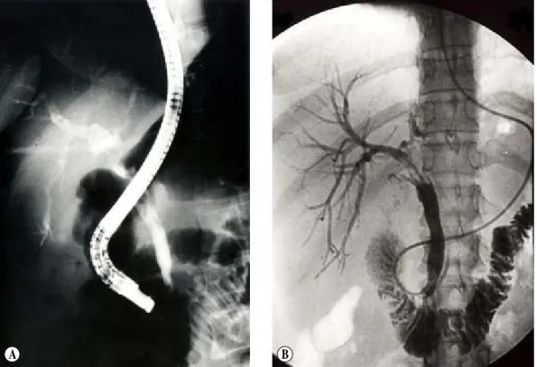
Fig. 1. Computed tomography. Dilatation of left intrahepatic duct and air-biliary gram of right intrahepatic duct is seen.
Fig. 2. A; Endoscopic retrograde cholangiogram. multiple filling defects in common hepatic duct and dilatation of right intrahepatic duct is seen. Contrast is not filled in left intrahepatic duct. B;
Follow-up cholangiogram after one week. Previously noted multiple filling defects are not seen, but stricture of common hepatic duct and left intrahepatic duct is seen.
저자들은 황달로 내원하여 내시경역행담췌 관조영술(Endoscopic retrograde cholangiopa- ncreatography, ERCP) 및 수술을 시행하여 담 관내 증식한 간세포암이 진단된 증례를 경험하 였기에 문헌고찰과 함께 보고하는 바이다.
증 례
46세 여자가 내원 4일전부터 시작된 찢어질 듯한 우상복부 통증과 황달이 있어 타병원을 방문하여 시행한 검사에서 담관결석을 의심하 여 ERCP를 이용한 경비담도배액술을 시행하 였으나 잘 배액되지 않고 혈성담즙이 배출되어 본원으로 전원되었다. 과거력에서 간염의 병력
은 없었고, 내원당시 활력징후는 혈압 120/80 mmHg, 맥박 74회/분, 체온 37.4℃였다. 진찰
A B
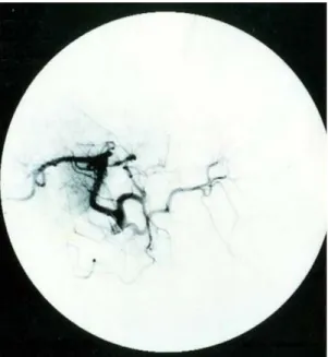
Fig. 3. Angiogram. typical tumor stain is not seen.
Fig. 4. Gross pathology. 2.5×1.7cm sized mass in segment 2 and 9.5×6.0cm sized intraductal mass in segment 3 are seen.
소견에서 급성병색이었고 공막은 노란색을 띄 었으며 우상복부 압통을 호소하였다. 검사실 소견은 백혈구 13,400 /uL, 혈색소 11.6 g/dL, 혈소판 159,000 /uL였고, 총빌리루빈 9 mg/dL, 직접빌리루빈 6.2 mg/dL, AST/ALT 106/297 IU/L, 총단백 7.4 g/dL, 알부민 4.1 g/dL, 프로 트롬빈 시간 12.0초였다. 바이러스 표지자는 HBsAg 음성, HBsAb 음성, HBcAb (IgG) 양 성, anti-HCV 음성이었고, 종양표지자는 AFP 277.2 ng/mL, CA19-9 6.33 U/mL였다.
복부 전산화 단층촬영에서 좌측 간내담관의 확장과 우측 간내담관내 공기음영이 관찰되었 다(Fig. 1). 입원 1일째 ERCP를 시행하였는데 총담관과 좌측 주간관에 충만결손이 있어 경비 담도배액관을 삽입하였다(Fig. 2A). 배액도관 을 통해 생리식염수로 담관을 매일 세척하였고 1주일 후 도관을 통하여 담도조영술을 다시 시 행하였는데, 이전 담도조영사진에서 보였던 다 발성 충만결손은 사라졌지만 주간관의 협착은
계속 관찰되었다(Fig. 2B). 혈관조영술에서 종 양은 염색되지 않았고, 좌측 간문맥이 보이지 않았다(Fig. 3). 입원 16일째 간좌엽절제술 및 Reux-en-Y 간공장문합술을 시행하였고, 조직 검사에서 간세포암이 진단되었다(Fig. 3, 4). 환 자는 14개월 후 간내에 간세포암이 재발하여 간동맥 화학색전술 및 경피적 에탄올 주입술을 시행하였으나 종양의 진행으로 수술한지 28개 월 후 사망하였다.
고 찰
간세포암에 의해 폐쇄성 황달이 발생하는 기전으로는 간외담관 외부로부터의 압박과 담 관내강의 폐쇄에 의한 경우로 구분할 수 있다.
전자의 경우는 비교적 흔하게 발생할 수 있으 나 후자의 경우는 드물다.
4)Lin 등
5)은 332명 의 간세포암 환자들 가운데 4명에서 담관 침윤 이 있었다고 보고하면서 이처럼 폐쇄성 황달이 동반된 간세포암을 “icteric type hepatoma”로 명명하였다.
담관 내강 폐쇄의 기전으로는 담관을 침윤
한 종괴가 담관내강으로 성장하여 폐쇄된 경
Fig. 5. Microscopic findings. A; Hepatocellular carcinoma. Note that the cancer cell nests in intraductal area. B; AFP stain, which stain hepatocellular carcinoma. C; Hepatocellular carcinoma. Cancer cell nests in liver were noted. D; Cytokeratin-7 stain, which revealed intact bile duct epithelium.
우, 담관 내강으로 성장한 종양이 떨어져서 하 부 담관으로 이동하여 담관 폐쇄를 초래하는 경우 그리고 종괴의 출혈에 의해 형성된 혈괴 가 담관을 폐쇄하는 경우 등이 있다.
4-7)드물게 는 간 실질의 원발 병소는 없으면서 간외 담관 내에 원발성으로 간세포암이 발생하였다는 보 고도 있으나 아직까지 그 기전에 대해서는 논 란이 많다.
8, 9)간세포암에 의한 폐쇄성 황달의 진단은 매 우 어려워 대부분 사후에 부검을 통하여 확인 되거나 생존시에 진단되는 경우에도 수술 전에 는 의심하지 못하였다가 절제조직의 병리검사 를 통해 진단되는 경우가 많은데, 이와 같이 진단이 어려운 경우는 담관내 간세포암의 발생
이 매우 드물기 때문에 의심하지 못하는 이유 가 있으며, 전신상태 불량으로 조직검사 등의 관혈적 검사를 시행하지 못하기 때문이다.
2)진단을 위해서는 초음파 검사, 컴퓨터 단층 촬영 등을 시행한 후 경피경간 담관조영술 혹 은 ERCP 등이 필요하다.
10)영상학적으로 담관 내 간세포암과 담관암을 구별하려는 노력이 있 어왔는데, 김 등
11)은 간세포암이 담관암보다 초음파 음영이 낮고 낭성확장을 보이며, 만성간 질환이 있고 경계가 부드러워 임상검사와 초음 파 소견을 종합하면 담관내 간세포암과 담관암 을 구별하는데 도움이 될 수 있을 것이라 하였 다. 그러나 실제 초음파와 컴퓨터 단층촬영으로 술전에 진단을 하기는 쉽지 않다. Murakami
A B
C D
등
12)은 ERCP를 통한 조직검사로 술전에 간세 포암을 진단한 증례를 보고하였다.
수술전 진단을 위해서는 담관조영술상 담관 암을 의심하는 충만결손이 보일때 간세포암의 가능성에 대해서도 고려해야 하며, 바이러스 간염, 간경변증의 존재 및 알파태아단백의 증 가가 있을 때는 담관내 간세포암의 가능성을 생각하여 ERCP를 통한 조직검사 등으로 진단 에 적극적으로 임해야 할 것이다.
그러나 안타갑게도 현재까지 간세포암의 담 관 침범에 대한 치료 지침은 확립되어 있지 않 다. 최근 Esaki 등
13)은 담관 침범된 간세포암 환자의 수술 후 평균 생존기간이 31개월로 간 내 전이가 없는 경우 적극적으로 수술을 권유 하고 있다. 수술 후 재발한 경우의 치료방법으 로는 간동맥 화학색전술, 경피적 에탄올 주입 술, 고주파 열치료 등이 알려져 있다.
본 증례는 담관을 침범한 원발성 간세포암 으로 수술 후 재발하여 간동맥 화학색전술, 경 피적 에탄올 주입술 등을 시행하였으나 암종의 진행으로 사망한 경우이다. 앞으로 담관 침범 된 간세포암의 치료방법과 수술 후 재발을 줄 일 수 있는 방법에 대한 연구가 더 필요하다.
참 고 문 헌
1. Nonomura A, Ohta G, Kanai M, Kobayashi K.
Hepatocellular carcinoma presenting extrahepatic hepatocellular biliary obstruction. Acta Patho Jap 1983 Jul;33(4):789-806.
2. Park SW, Song SY, Chung JB, Kang JK, Park IS, Lee WJ, et al. Obstructive jaundice in patients with hepatocellular carcinoma due to extrahepatic bile duct obstruction - focused on cholangiographic characteristics -. Korean
J Gastroenterol 1995 Jan;27(1):83-95.
3. Qin LX, Tang XY. Heatocellular carcinoma with obstructive jaundice: diagnosis, treatment and prognosis. World J Gastroenterol 2003 Mar;9(3):385-91.
4. Song IH, Koh MS, Choi HS, Lee SK, Chung YH, Kim MH, et al. Cholangiographic findings in hepatocellular carcinoma patients with obstructive jaundice. Korean J Gastroenterol 1996 Jan;28(1):101-10.
5. Lin TY, Chen KM, Chen YR, Lin WS, Wang TH, Sung JL. Icteric type hepatoma. Med Chir Dig 1975;4(5-6):267-70.
6. Choi SY. Hepatocellular carcinoma presenting as obstructive jaundice. Korean J Gastroenterol 1993;25(1):123-8.
7. Lee KC, Sakai K, Kinoshita H, Hirohashi K, Tsuji Y, Kubo S, et al. Resection of hepatocellular carcinoma with obstructive jaundice caused by compression of common hepatic duct. J Surg Oncol 1988 Nov;39(3):
201-5.
8. Badve SS, Saxena R, Wagholikar UL.
Intraductal hepatocellular carcinoma with normal liver-case report. Indian J Cancer 1991 Sep;28(3):165-7.
9. Tsushimi T, Enoki T, Harada E, Orita M, Noshima S, Masuda M, et al. Ectopic hepatocellular carcinoma arising in the bile duct. J Hepatobiliary Pancreat Surg 2005;12 (3):266-8.
10. Wu CS, Wu SS, Chen PC, Chiu CT, Lin SM, Jan YY, et al. Cholangiography of icteric type hapatoma. Am J Gastroenterolol 1994 May;89 (5):774-7.
11. Kim NR, Kim SH, Lee JM, Lee KH, Kim YJ, An SK, et al. Sonographic features of an intraductal polypoid mass: differentiation between hepatocellular carcinoma and intraductal cholangiocarcinoma. J Ultrasound Med 2004
Oct;23(10):1283-91.
12. Murakami Y, Yokoyama T, Kanehiro T, Uemura K, Sasaki M, Morifuji M, et al.
Successful diagnosis and resection of icteric type hepatocellular carcinoma. Hepatogast- roenterology 2003 Sep-Oct;50(53):1634-6.
13. Shiomi M, Kamiya J, Nagino M, Uesaka K, Sano T, Hayakawa N, et al. Hepatocellular carcinoma with biliary tumor thrombi: aggressive operative approach after appropriate preoperative management. Surgery 2001 Jun;129:692-8.