Ⅰ. 서 론
점액성 유상피암종은 타액선에서 흔하게 발생하는 악성종 양이며 전체 타액선 종양의 약 8%를 차지한다. 이하선에서 약 50%, 구개에서 약 20%의 빈도로 발견되지만 드물게 중 심성으로 하악골에서 발생하기도 하는데 이는 전체 점액성 유상피암종의 2~3%에 이른다1).
Browand은 하악골내 점액성 유상피암종이 상악보다 3~4배 더 빈번히 발생한다고 보고하였다2). Brookstone은 이의 기원으로 치성낭종을 이장하는 상피를 지목하였는데, 이는 악골내 점액성 유상피암종이 치성낭종과 연관되어 발 생하는 경우가 많고(34~48%) 이러한 낭종에서 점액분비 세포가 40%의 빈도로 발견되는데 이의 신생성 변형(neo- plastic transformation)에 의해 병소가 발생될 수 있다는 것이다1).
본 발표는 제3대구치 발거와 연관된 하악골내 점액성 유 상피암종의 증례보고로서 진단과 처치에 대한 일련의 과정 을 소개하고 양성질환으로 간과하기 쉬운 악골내 점액성 유 상피암종을 보고하고자 한다.
Ⅱ. 증례보고
2002년 5월 21일 25세의 남자가 하악 좌측 우각부의 종 창을 주소로 내원하였다. 증상으로는 안면부 종창, 좌측 구 치부 발치창의 열개, 개구제한, 하순의 감각이상 등을 호소 하였으며 농양 배출은 나타나지 않았다.
환자는 1999년 여름 군대에서 하악 좌측 제3대구치를 발 거한 뒤 발치창이 낫지 않아 여러 군데서 치료를 받다가 2000년 2월 2일 모 대학병원 치주과에 내원하여 발치창의 소파술을 시행받았다. 이후 연고지 관계로 자택근처 중소병 Jong-Ko Bae, Myung-Rae Kim, Nara Kang, Jae-Hwa Kim
Department of Oral and Maxillofacial Surgery, Ewha Womans University College of Medicine
Mucoepidermoid carcinoma is a common salivary gland tumor. It comprised 8% of all salivary gland tumor and originated mainly in parotid gland.
Central mucoepidermoid carcinoma is rare. It comprised 2~3% of all mucoepidermoid carcinoma, but it occurs in the mandible two or three times more frequently than in the maxilla.
Central Mucoepidermoid carcinoma are frequently associated with an odontogenic cyst, such as dentiger- ous cyst, in which mucous goblet cell would have neoplastic transformation.
In May 2002, a 25 year-old male visits in our clinic, presented with a progressive facial swelling after surgical tooth extraction of left mandibular third molar at 1999 in the army. After incisional biopsy, the lesion was confirmed as mucoepidermoid carcinoma so we performed tumor resection and reconstruction surgery of mandible.
Key words : Central mucoepidermoid carcinoma, Third molar extraction
원으로 전원되어 2000년 2월 18일 골수염으로 가진단 하 고 #37 발치 및 saucerization을 시행받았으나 여전히 불 편하여 2001년 11월 모 대학병원 구강외과로 재의뢰, 절개 생검을 시행받았다. 조직검사 결과 골수염진단을 받고 통원 가료 받다가 2002년 5월 6일 재시행된 조직생검에서 점액 성 유상피암종이라는 진단을 받고 본원으로 의뢰되었다.
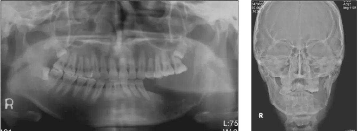
파노라마 검사상 경계가 불명확한 골파괴성 병소가 관찰 되었으며 컴퓨터 단층사진에서는 협,설측 피질골이 광범위 하게 파괴되는 소견을 보였다(Fig. 1, 2).
좌측 경부곽청술 및 하악골을 절반을 포함한 종물의 절제 와 동시에 광배근 근-피부 피판을 이용한 연조직 재건을 계 획하였다. 광배근 근-피부 피판은 넓은 결손부위 재건에 유 용하며 혈액공급이 풍부하여 오염된 창상일지라고 결손부 위의 혈액공급을 강화시키며 공여부의 반흔이 작다는 장점 이 있다.
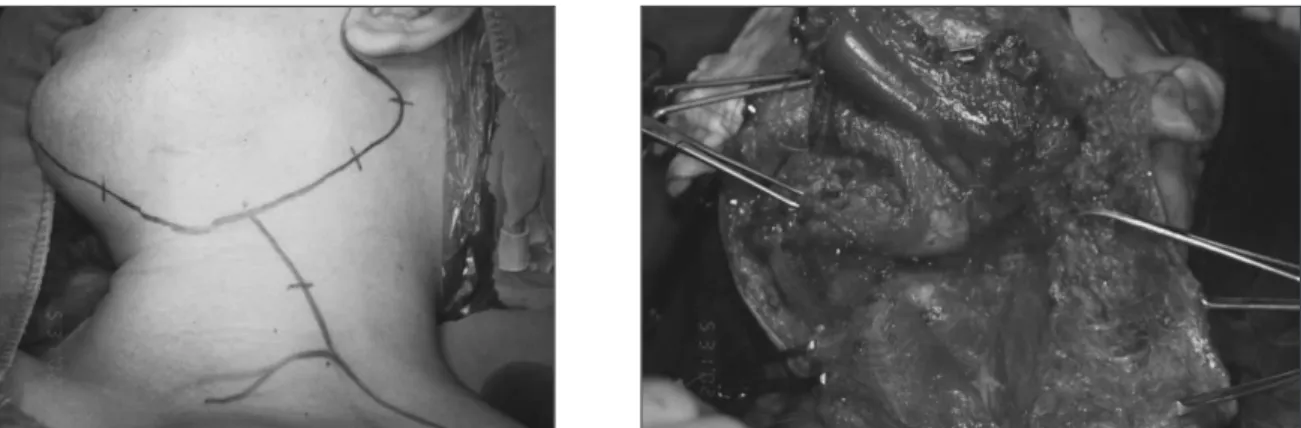
2002년 5월 31일 전신마취하에서 수술이 시행되었으며 종물절제 및 근치적 경부곽청술 후 우측 facial a.&v.과 광 배근 피판의 주 혈관인 thoracodorsal a.&v.의 미세혈관 접합이 이루어 졌다(Fig. 3, 4). 조직검사상 고등도의 점액 성 유상피암종으로 판명났으며 림프절로의 전이도 발견되 었다. 술 후 환자는 중환자실에서 집중 가료를 받았으며 약 한 달 이후에 퇴원하여 방사선치료를 6주간 시행 받았다.
방사선 치료가 끝난지 6개월째에 장골이식을 이용한 경조 직 재건을 계획하였고 2003년 2월 12일 수술이 시행되었 다. 술후 촬영된 컴퓨터 단층사진을 보면 우측 악궁과 비교 하였을 때 형태와 폭에서 유사하게 재건되었음을 알 수 있 었다(Fig. 5).
장골 이식을 시행한지 2개월 후에 좌안면 누공이 발생하 였다(Fig. 6). 전형적인 감염소견을 나타내었으며 항생재 감수성 검사 결과 MRSA 균이 검출되어 감염내과 협진하에 Fig. 1.Preoperative panoramic view and skull PA showing the radiolucent lesion at left mandible body.
Fig. 2.Axial CT image shows bony destruction of buccolingual cortex of left mandible and coronal CT image shows destruction of lingual cortex.
Fig. 3.Clinical photo shows incision line for radicular neck dissection and elevation of lymph nodes of level II, III, IV, V.
Fig. 4.Clinical photo shows the latissimus dorsi myocutaneous flap was positioned and anastomosis was made.
Fig. 5. Clinical photo shows the reconstruction of mandible defect using iliac block bone and postop- erative panoramic view and axial CT view shows the continuity of mandible.
Fig. 6. Clinical photo shows incision and drainage at the site of bone graft due to postoperative infection in postoperative 2 months.
Fig. 7.Clinical photo shows the pedicled Deltopectoral flap and after 3 weeks, flap revision was made.
Fig. 8.Panoramic view shows the implant restoration in postoperative 2 years.
임플란트를 3개 식립하였으며 연조직의 두께가 두껍고 장 골 이식부의 높이가 낮아서 임플란트 상부 나사가 완전히 골내로 식립되지 못하였다(Fig. 8). 술 후 2주째 확인하였 을 때 임플란트 주변의 연조직에 치유가 불완전 하였으며 음식물 저류가 관찰되었다. 특별한 처치는 하지 않고 생리 식염수 주수와 클로르헥시딘 양치교육을 시행하였으며 이 후 약 2개월에 follow up 되었을 땐 좋은 연조직 치유를 나 타내었다.
Ⅲ. 고 찰
악골에 발생하는 점액성 유상피암종의 기원을 밝히기 위 한 여러 가설이 있다. 악골내에 존재하는 이소성 타액선조 직으로부터 발생된다는 가설, 다분화 능력을 가지는 배아의 구강 외배엽 세포가 악골에 매몰되어 발생한다는 가설이 있 고 또 하나는 치성낭종의 이장상피에서 발견되는 점액분비 세포가 신생성 변형(neoplastic transformation)을 일으켜 악골내 점액성 유상피암종이 발생된다는 가설이다. 그러나 조직학적 발생빈도는 후자가 우세한 것으로 알려져 있다1,3,4).
Brookstone은 악골에 발생한 타액선 종양환자 11증례 중 8명이 조직학적으로 치성낭종과 연관되었거나 병소확진 전에 발치 기왕력이 있었음을 보고하였고1) Eversole은 하 악골 종양의 약 50%가 치성 낭종이나 매복치와 연관 있음 을 보고했다5,6).
본 증례의 경우는 군대에서 초진이 시행되었기에 어떠한 과정을 통해 진단 및 발치가 이루어 졌는지 알 수 없었다.
추측건대 제3대구치과 연관된 낭종이 있었고 주변부로 점 액성 유상피암종이 존재했으나 초기 진료자가 단순 치성낭 종으로 판단하고 소홀히 했을 가능성이 높아 보인다. 환자 의 제3대구치가 발거했을 당시 치아 주변의 연조직을 적절 히 생검했더라면 더 효과적인 치료가 진행되었으리라 사료 된다.
악골내 점액성 유상피암종의 방사선학적 소견은 경계가 분명한 낭종성 형태에서부터 피질골 팽창 및 천공에 이르는 형태에 이르기까지 다양하기 때문에 방사선 사진만으로 감
치료는 외과적 절제가 필요하며 단순 적출 및 소파술만 시 행했을 경우 재발율이 약 40%에 이르지만 광범위한 절제 를 시행했을 때는 약 9%에서 13%의 재발율이 보고되고 있 다1,7). 본 증례에서는 초기에 골수염으로 잘못 진단하여 소 파술을 시행하였다. 이는 결과적으로 종양세포를 더 파급시 키는 것에 기여했으리라 생각된다.
Ⅳ. 결 론
악골내 발생한 점액성 유상피암종으로 광범위한 하악골 절제술을 시행 받아 안면의 기능적 심미적 결손이 심한 환 자에게서 광배근 피판으로 연조직을 재건하고 장골이식으 로 하악골을 재건하여 양호한 결과를 얻었으며 임플란트 또 한 안정적인 osseointegration을 나타내었다. 환자는 술 후 약 3년 6개월이 지난 현재까지 재발의 소견 없이 사회생활 을 잘 영위하고 있다.
점액성 유상피암종은 치성 낭종 및 기타 악골내 양성질환 과 감별해야 하며 매우 드물게 발생하기에 놓치기 쉬운 질 환이지만 세밀한 조직검사와 확실한 절제, 그리고 안정적인 재건이 이루어진다면 충분히 극복될 수 있으리라 사료된다.
참고문헌
1. Brookstone MS, Huvos AG : Central salivary gland tumors of the maxilla and mandible: a clinicopathologic study of 11 cases with analysis of the literature. J Oral Maxillofac Surg 50(3) : 229, 1992.
2. Browand BC, Waldron CA : Central mucoepidermoid tumors of the jaws: report of nine cases and review of the literature. Oral Surg Oral Med Oral Pathol 40(5) : 631, 1975.
3. Winkle MR, Harrington PC, Maronian N : Central Mucoepidermoid Carcinoma of the mandible. Am J Otolaryngol 20(3) : 169, 1999.
4. Inagaki M, Yuasa K, Nakayama E, et al.:
Mucoepidermoid carcinoma in the mandible: findings of panoramic radiography and computed tomography. Oral Surg Oral Med Oral Pathol Oral Radiol Endod 85(5) : 613,1998.
5. Eversole LR, Sabes WR, Rovin S : Aggressive growth and neoplastic potential of odontogenic cyst: With special refer- ence to central mucoepidermoid carcinomas. Cancer 35(1) : 270, 1975.
6. Eversole LR : Mucoepidermoid carcinoma: Review of 815 reported cases. J Oral Surg 28(7) : 490, 1970.
7. Waldron CA, Koh ML : Central mucoepidermoid carcino- ma of the jaws: report of four cases with analysis of the literature and discussion of the relationship to mucoepider- moid, sialodontogenic, and glandular odontogenic cysts. J Oral Maxillofac Surg 48(8) : 871, 1990.
저자 연락처
우편번호 158-710
서울특별시 양천구 목동 911-1
이화여자대학교 의과대학 목동병원 구강악안면외과 김 명 래
원고 접수일 2006년 1월 2일 게재 확정일 2006년 3월 7일
Reprint Requests Myung-Rae Kim
Dept. of OMFS, Ewha Womans University College of Medicine 911-1 Mok 6-dong Yangcheon-gu, Seoul, 158-710, Korea Tel: +82-2-2650-5196
E-mail: [email protected] Paper received 2 January 2006 Paper accepted 7 March 2006