© The Korean Society of Plant Pathology
http://dx.doi.org/10.5423/RPD.2013.19.4.300Leaf Rot and Leaf Ring Spot Caused by Rhizoctonia solani in Chinese Cabbage
Chang-Ki Shim, Min-Jeong Kim*, Yong-Ki Kim, Hyeong-Jin Jee, Sung-Jun Hong, Jong-Ho Park, Eun-Jung Han and Jong-Chul Yun
Organic Agriculture Division, National Academy of Agricultural Science, Rural Development Administration, Suwon 441-707, Korea
(Received on November 15, 2013; Revised on November 25, 2013; Accepted on November 26, 2013)
This study was conducted to determine the occurrence of leaf rot and leaf ring spot, caused by Rhizoctonia solani in Chinese cabbage under seedling nursery and cultivation greenhouses. Symptoms of leaf rot and leaf ring spot were found in three Chinese cabbage cultivars, Brassica campestris subsp. pekinensis, ‘Ryeokgwang’,
‘Daetong’, and ‘CR mat’. In Hwacheon, the disease incidence was 73.8% in the seedling stage of the Chinese cabbage. In Icheon, the symptoms were observed on the upper leaves of the Chinese cabbage cultivar,
‘Norangmini’ with 20.5% of disease incidence. The symptoms appeared as primary lesions consisting of small, circular necrotic ring spots with gray color, 1.4−3.0 mm in diameter, accompanied by secondary rot lesions with large irregular borders of leaves. The color of mycelial mat of 20 isolates was dark brown and light brown. The average hyphal diameter of all the isolates was within 5.01−11.12 µm. Among the 20 strains isolated from Chinese cabbage, 16 isolates and four isolates anastomosed with the AG-1 (IB) and AG-1 (IC), respectively. Twenty isolates tested were only virulent on foliage parts of Chinese cabbage leaves but were avirulent on stem parts of the plants. Based on the mycological characteristics and pathogenicity test on host plants, the fungus was identified as Rhizoctonia solani.
Keywords : Anastomosis group, Bottom rot, Brassica campestris subsp. pekinensis, Rhizoctonia solani
Introduction
Chinese cabbage (Brassica rapa subsp. pekinensis) is one of the economically important vegetable crops in eastern Asia including Korea. It is usually harvested after completing the leaf heading period. Chinese cabbages as the major component in the preparation of Kimchi, a traditional fermented food and contain a majority of the components essential for human nutrition in Korea (Hong and Kim, 2011).
Bottom rot is a common disease caused by Rhizoctonia solani Kühn [telomorph: Thanatephorus cucumeris (Frank) Donk] in Chinese cabbage cultivation areas (Carling, 1996). Kim et al. (1995) reported that R. solani is one of the 65 diseases causing have to a wide range of plants including rice, potato, pepper, pine tree, etc.
in Korea. Kang and Kim (1986) first reported 30% of overall bottom rot infection rate in Chinese cabbage under plastic film house cultivation in Gyeongnam province was caused by R. solani. The disease severely
occurred in poly vinyl mulching fields during the wet and cold seasons. The symptoms were bottom rot of leaves, root rot and damping off of seedlings during cultivation. R. solani is one of the potent soilborne pathogens of economically important crops which develop both in cultured and non-cultured soils, causing diseases in different crops such as rice, bean and tomato, among others (Sneh and Akira, 1991). Numerous Rhizoctonia species have been reported to occur on many hosts in Korea (Kim et al., 1993; Kim, 1996).
The pathogen shows considerable diversity in morphology, geographic location, host specificity and pathogenicity (Ogoshi, 1987). Isolates of R. slolani have been classified into fourteen anastomosis group (AG), AG-1 through AG-13 and AG-BI (bridging isolates) by different in pathogenicity (Carling, 1996; Ogoshi, 1987). Among the fourteen anastomosis group, AG-1 is subgrouped as AG-1 (1A), AG-1 (1B), and AG-1 (1C) and AG-2-2 (III B). The best known subsets of AG-2 are AG-2-1, AG-2-2 (IIIB), AG-2-2 (IV), AG-2-2 (LP), and AG-2-3 (Hyakumachi and Sumino, 1984; Watanabe and Matsuda, 1966).
R. solani species and anastomosis groups are reported to differ in sensitivity to common fungicides (Campion
*Corresponding author
Phone)+82-31-290-0545, Fax) +82-31-290-0507 Email) [email protected]
Research Article Open Access
et al., 2003). Anastomosis occurs between fungal isolates of the same AG but not between isolates of different AG’s. The concept of anastomosis group is a widely accepted principle for identifying intraspecific groups in the R. solani complex and providing genetic relations for the breeding of new varieties (Carling et al., 1988).
AG-1 isolates have been subdivided into four subgroups, AG-1 (IA), AG-1 (IB), AG-1 (IC) and AG-1 (ID) based on pathogenicity and culture morphology (Priyatojo et al., 2001). Currently, two intraspecific groups of R.
solani AG-1 comprise the RFB (Rhizoctonia foliar blight) complex: intraspecific group A (IA), the causal agent of sheath blight of rice which causes aerial blight, intraspecific group B (IB), which causes web blight of soybean, and intraspecific group C (IC), which causes damping off (Joy et al., 1990; Sneh et al., 1991; Yang et al., 1990).
However, anastomosis group concept is not an ideal method for classification of R. solani as misidentifi- cation is caused from the varied frequency of hypal fusion in some AG (Liu and Sinclair, 1992). Recently, molecular biological techniques have been used in combination with morphological and physiological markers for the analysis of population (Guleria et al., 2007;
Jeon et al., 2010) and the detection of genetic variability among the isolates of R. solani (Hong et al., 1998;
Lee et al., 1998; Sharma et al., 2005)
This paper will discuss the incidence and the causal organism of leaf rot and leaf ring spot diseases of Chinese cabbage under the organic Chinese cabbage seedling nursery and cultivation greenhouses in Hwacheon and Icheon.
Materials and Methods
Disease incidence. The study was conducted in 2011 at the three seedling nursery greenhouses in Hwacheon and ten cultivation greenhouses in Icheon.
Chinese cabbage with leaf rot and leaf ring spot symptoms were visually determined and recorded based on the percentage of diseased plants in 20 replicates of each greenhouse one week before transplanting and harvesting
time, respectively, in the seedling nursery and cultivation greenhouses.
Isolation and purification. R. solani isolates were taken from symptomatic plants of Chinese cabbage collected from Icheon and Hwacheon farms in Korea.
Eight pieces (1 cm
2) were excised from the margin of healthy and diseased tissues of Chinese cabbage that showed symptoms of R. solani. The excised species were surface-sterilized in 1.5% NaOCl for two minutes and extensively rinsed in sterile distilled water. Four pieces per plant were placed on potato dextrose agar (PDA, Difco), all plates were incubated at 25ºC. Several isolates of R. solani were obtained by single hyphal tip isolation. The isolated R. solani were stored at 5ºC in PDA slants.
Mycological characterization. The isolates were identified to genus level, based on microscopic and cultural characteristics. From the colony morphology of 20 isolates, color of mycelial mat was recorded after 10 days incubation on PDA. Three weeks after inoculation on PDA, sclerotia production was evaluated at 20
oC.
Vegetable growth of the 20 isolates of R. solani was observed at seven temperatures levels, 5, 10, 15, 20, 25, 30 and 35
oC. Mycelial growth was determined in petri dish containing 15 ml of PDA inoculated with a 6 mm diameter of mycelial disk cut from a 5-day-old culture of 20 isolates on PDA. Mycelial growth in four replicated cultures at each temperature was measured at 24 hr intervals.
Determination of anastomosis groups. The standard isolates of the eight anastomosis groups, AG-1 to AG- 4, and unidentified two strains, R. cerealis, and R.
edophytica were obtained from the Korean Agricultural Culture Collection (KACC) (Table 1). The determination of the anastomosis group of the isolates was determined by using the methodology of Kim et al. (1993). Twenty isolates of R. solani were paired with the ten standard isolates of anastomosis groups of R. solani. Tester strains of multinucleate Rhizoctonia including AG-1 (IA), AG- 1 (IB), AG-1 (IC), AG-2-I, AG-2-2 (IIIB), AG-2-2 (IV), AG-3, AG-4 and unidentified two strains, R. cerealis, and R. edophytica were used. Single 5-mm-diameter Table 1. Standard isolates of anastomosis groups (AG), Rhizoctonia solani AG-1 to AG-4, R. cerealis, and R. edophytica obtained from the Korean Agricultural Culture Collection (KACC)
KACC No. Species Host KACC No. Species Host
40101 R. solani AG-1 (IA) Oryza sativa 40131 R. solani AG-2-2 (IV) Daucus carota
40108 R. solani AG-1 (IB) Lactuca sativa 40136 R. solani AG-3 Solanum tubersoum
40113 R. solani AG-1 (IC) Brassica campestris spp. pekinensis 40139 R. solani AG-4 Raphanus sativus
40119 R. solani AG-2-1 Brassica campestris spp. pekinensis 40154 R. cerealis Agrostis palustris
40127 R. solani AG-2-2 (III-B) Citrullus lanatus 40713 R. endophytica Orchid symbiont
agar disc was cut from the perimeter of a 2- to 3-day- old colony of each isolate on PDA. Tester isolates were placed 3 to 4 cm away from each tested isolate and incubated at 25
oC for 24 to 48 h in the dark. When the hyphae from the two disks were overlapping, they were stained with safranin O and 3% KOH and examined microscopically to determine anastomosis reaction (Carling, 1996).
Pathogenicity tests. Pathogenicity testing and leaf or root damage assessments of the isolates were carried out based on Kang and Kim (1986). The only modification was that the testing was conducted at 28
oC instead of 21
oC. Agar-disk assay was used in the pathogenicity tests. From stored cultures, isolates were transferred to the PDA plates and incubated at 25
oC in the dark for seven days. Inoculations were made by placing a mycelial-agar disk (6 mm in diameter, taken from a 3-day-old culture on PDA) in the foliage part and stem of each leaf. Twenty isolates of R. solani were tested for pathogenic potential on Chinese cabbage,
‘Daetong’ with 15 seedlings for each isolate. After inoculations, leaves were covered with a plastic bag for five days. Leaves were similarly prepared, but only PDA disks served as an untreated control. All experiments were replicated twice. Five days after inoculation, the leaf or root damage assessment scale was 0 = no damage, 1 = minor discoloration of leaves, 2 = discoloration plus small necrotic lesions (< 1 mm in diameter) on leaves, 3 = discoloration with large necrotic lesions (1 mm or larger in diameter) on leaves, and 4 = death of the seedling.
Results
Disease incidence. In 2011, the occurrence of bottom rot diseases of Chinese cabbage caused by R. solani were investigated at the Chinese cabbage nursery green- houses in Hwacheon and the cultivation greenhouses in Icheon. In Hwacheon Chinese cabbage nursery farm, only leaf ring spot symptom was observed on the upper leaves of three Chinese cabbage cultivars after 25 days of sowing with disease incidence of 22.6% for
‘Ryeokgwang’, 22.6% for ‘Daetong’ and 20.8% for
‘CR mat’, respectively (Table 2). At 35 days of seedlings, leaf ring spot and leaf rot symptoms occurred on leaves of three cultivars of Chinese cabbage with disease incidence of 60.1% both for ‘Ryeokgwang’ and ‘Daetong’
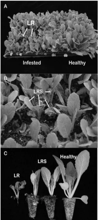
and 58.9% for ‘CR mat’ (Table 2). One week before transplanting, leaf rot and leaf ring spot symptoms of three Chinese cabbage cultivars were investigated having 73.8% of disease incidence (Fig. 1).
In Icheon organic Chinese cabbage farm, the leaf
Table 2. Occurrence of leaf rot and leaf ring spot caused by Rhizoctonia solani on three cultivars of Chinese cabbage seedlings grown in spring season of 2011
Cultivar Disease incidence (Mean ± SD, %)
25 days
a35 days 45 days
Ryeokgwang 22.6 ± 0.05 60.1 ± 0.06 73.8 ± 0.13 Daetong 19.6 ± 0.03 60.1 ± 0.07 75.4 ± 0.13 CR mat 20.8 ± 0.08 58.9 ± 0.10 74.2 ± 0.11
aDays after sowing of Chinese cabbage seed.
Fig. 1. Symptoms of leaf rot (LR; A, C) and leaf ring spot (LRS;
B, C) caused by Rhizoctonia solani in Chinese cabbage seedlings
in Hwacheon in 2011.
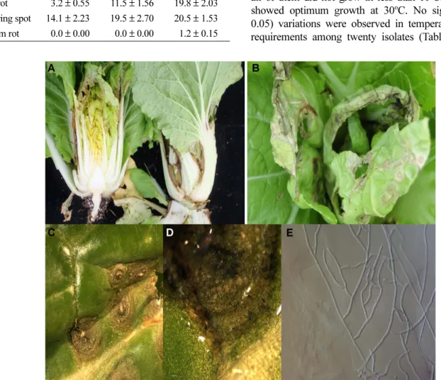
ring spot incidence of 14.1% was observed 15 days after transplanting and leaf rot incidence 3.2% was observed on the upper leaves of ‘Norangmini’ Chinese cabbage cultivar. At 25 days after transplanting, disease incidence of the leaf ring spot was 19.5% and the leaf rot was 11.5% observed only on the upper leaves. At 35 days after transplanting, the bottom rot symptoms were observed on the stem of the Chinese cabbage with 1.2% disease incidence, counted as 20.5% for leaf ring spot and 19.8% for leaf rot symptoms (Table 3, Fig. 2).
Fungal isolates and characterization. In spring season of 2011, Rhizoctonia samples we collected from the main organic Chinese cabbage cultivation areas of
Hwacheon and Icheon. Twenty isolates collected and were grown on PDA for 6−7 days.
The colony morphology of these isolates was recorded after 10 days incubation on PDA. The color of mycelial mat of 20 isolates was dark brown except for four isolates, ICC15, ICC16, ICC17 and ICC18 collected from Icheon which was light brown. The average hyphal diameter of all the isolates was within 5.01−11.12 µm which concurs with the previous reported data for R.
solani.
After four days of growth on PDA, white sclerotial initials were visible and after at eight days, all sclerotia of 20 isolates were dark brown and showed small (0.38−1.28 mm) and uniform of sclerotia formation on PDA. Formation stopped after 9 days, even if the colony had reached profusely at the edge of the petri dish.
The temperature for the twenty tested isolates ranged from 5 to 35
oC, with optimum of 25 to 30
oC. All of the twenty isolates grew relatively well at 15
oC, where all of them did not grow in less than 10
oC. Most isolates showed optimum growth at 30
oC. No significant (p = 0.05) variations were observed in temperature optimum requirements among twenty isolates (Table 5).
Fig. 2. Symptoms of leaf rot (A) and leaf ring spot (B, C) caused by Rhizoctonia solani on Chinese cabbage in Icheon in 2011.
Microscopic observation shows the mycelia stained with 0.02% cotton blue staining solution on the surface of lesions (D, ×60; E, ×200).
Table 3. Occurrence of leaf rot and leaf ring spot caused by Rhizoctonia solani on Chinese cabbage cultivar ‘Norangmini’
grown in Icheon in 2011
Symptom Disease incidence (Mean ± SD, %)
Feb. 25 Mar. 7 Mar. 17
Leaf rot 3.2 ± 0.55 11.5 ± 1.56 19.8 ± 2.03
Leaf ring spot 14.1 ± 2.23 19.5 ± 2.70 20.5 ± 1.53
Bottom rot 0.0 ± 0.00 0.0 ± 0.00 1.2 ± 0.15
Anastomosis Grouping and Pathogenicity Tests.
Twenty isolates of R. solani obtained from Chinese cabbage leaves found in Hwacheon and Icheon belonged to two anastomosis groups, AG-1 (IB) and AG-1 (IC) (Fig. 3). Ten isolates collected from Hwacheon anas- tomosed with AG-1 (IB). From Icheon, four isolates were anastomosed with AG-1 (IB) and six isolates anastomosed with AG-1 (IC) (Table 4, Fig. 3).
The pathogenicity of twenty isolates of R. solani was
also investigated on 35-day-old Chinese cabbage seedlings through the biological assay method. All isolates tested were only highly virulent on foliage parts of Chinese cabbage leaves but were avirulent on stem parts. None of the control seedlings developed symptoms. R. solani was reisolated from infected seedlings and compared with the original isolate by morphological characteristics.
All the isolates had characteristics similar to the original fungus.
Fig. 3. Determination of anastomosis group (AG) of Hwacheon isolates (A, ICC01) and Icheon (B, ICC11) isolates of Rhizoctonia solani from lesions of leaf rot and leaf ring spot symptoms of Chinese cabbage.
Table 4. Anastomosis group of 20 isolates of Rhizoctonia slolani isolated from spring season Chinese cabbage in Icheon in 2011
Isolates Local region
Anastomosis group AG-1 (IA) AG-1
(IB) AG-1
(IC) AG-2-1 AG-2-2 (IIIB) AG-2-2
(IV) AG-3 AG-4 R.
cerealis R.
edophytica
ICC01 Hwacheon − − + − − − − − − −
ICC02 Hwacheon − − + − − − − − − −
ICC03 Hwacheon − − + − − − − − − −
ICC04 Hwacheon − − + − − − − − − −
ICC05 Hwacheon − − + − − − − − − −
ICC06 Hwacheon − − + − − − − − − −
ICC07 Hwacheon − − + − − − − − − −
ICC08 Hwacheon − − + − − − − − − −
ICC09 Hwacheon − − + − − − − − − −
ICC10 Hwacheon − − + − − − − − − −
ICC11 Icheon − − + − − − − − − −
ICC12 Icheon − − + − − − − − − −
ICC13 Icheon − − + − − − − − − −
ICC14 Icheon − − + − − − − − − −
ICC15 Icheon − + − − − − − − − −
ICC16 Icheon − + − − − − − − − −
ICC17 Icheon − + − − − − − − − −
ICC18 Icheon − + − − − − − − − −
ICC19 Icheon − − + − − − − − − −
ICC20 Icheon − − + − − − − − − −
Discussion
This study identified twenty isolates of R. solani obtained from organic Chinese cabbage fields by mor- phology and anastomosis grouping. R. solani Kühn [teleomoph: T. cucumeris (Frank) Donk] is a destructive and widely spread fungal pathogen with wide host range including rice, turfgrass, Chinese cabbage, and others vegetable crops. Bottom rot results from infection by R. solani, which lives in garden soil. When bottom rot occurs, dark brown, possibly soft and watery, lesions are found at the cabbage base (Kang and Kim, 1986;
Keinath, 1995).
The results indicated that only leaf rot and leaf ring spot symptoms were observed on the foliage part of Chinese cabbage of seedlings and plants in Hwacheon seedling nursery and Icheon cultivation greenhouse.
Earlier researches showed that most bottom rot pathogen, R. solani causal agent is known to infect moist soil.
Infection in young plants into the environment due to improper plant growth will severely slow its progress (Kang and Kim 1986; Kim et al., 1993). The other
hand, the twenty tested isolates showed leaf rot and leaf ring spot symptoms and did not infect stem of Chinese cabbage except for bottom rot symptoms at Icheon Chinese cabbage cultivation field with 1.2%
disease incidence one week before harvesting.
Unlike many other sclerotia forming plant pathogens, R. solani sclerotia only undergo direct myceliogenic germination, whereby vegetative hyphae capable of infecting the host grow directly out of the sclerotium.
Mycelia and sclerotia can grow and develop on plant debris, allowing inoculum to survive in the soil as well as on seed from season to season (Dijst, 1988). Genetic variability among the isolates of R. solani was reported by Nelson et al. (1996). Kaminski and Verma (1985) found a variable response of different R. solani isolates at different levels of temperatures. It was found that there were uniforms among the isolates regarding colony diameter and sclerotia formation on PDA with same temperature. It also suggested that there was an existence of homogenous among the 20 isolates of R. solani.
Anastomosis groups appear to be fairly host plant specific. The classification of R. solani isolates into Table 5. Morphological characteristics and optimum temperature for mycelia growth of 20 isolates of Rhizoctonia slolani isolated from Chinese cabbage in Icheon in 2011
Isolates Colony diameter (mm) of Rhizoctonia solani on potato dextrose agar
a5
oC 10
oC 15
oC 20
oC 25
oC 30
oC 35
oC Mycelial color
ICC01 0 0 4.3 16.9 28.5 24.4 12.4 dark
ICC02 0 0 5.1 18 28.9 24.3 13.1 dark
ICC03 0 0 4.5 16.9 28.8 25 13.1 dark
ICC04 0 0 4.6 16.9 28.9 24.3 12.9 dark
ICC05 0 0 4.6 16.9 28.9 25.2 13.1 dark
ICC06 0 0 5.0 18.2 29.0 25.2 13.3 dark
ICC07 0 0 4.6 16.9 29.0 25.0 12.4 dark
ICC08 0 0 4.9 18.1 29.1 24.7 12.4 dark
ICC09 0 0 4.9 18 28.9 25.1 11.9 dark
ICC10 0 0 4.9 18.2 28.3 24.6 13.4 dark
ICC11 0 0 4.8 16.9 28.5 23 12.9 dark
ICC12 0 0 4.8 17.1 28.2 24.3 12.8 dark
ICC13 0 0 5.0 18.1 30.0 26.4 14.2 dark
ICC14 0 0 4.7 16.9 28.5 24.8 12.5 dark
ICC15 0 0 4.7 16.4 28.5 25 12.5 light
ICC16 0 0 4.8 18.2 28.4 24.2 13.3 light
ICC17 0 0 4.5 16.8 28.3 24.4 12.5 light
ICC18 0 0 4.6 16.5 28.3 25.1 12.5 light
ICC19 0 0 5.2 17.8 30.1 25.7 13.4 dark
ICC20 0 0 4.5 16.4 28.7 24.6 12.0 dark
aValues are the mean mycelial growth of four replicates measured at 7 days.