Vol. 29, No. 3, September, 2016 http://dx.doi.org/10.20408/jti.2016.29.3.82
� Address for Correspondence : Han Ho Do, M.D.
Department of Emergency Medicine, Dongguk University Ilsan Medical Center, 27, Dongguk-ro, Ilsandong-gu, Goyang-si, Gyeonggi-do, Korea
Tel : 82-31-961-7777, Fax : 82-31-961-7529, E-mail : erdohh@naver.com
Submitted : June 16, 2016 Revised : June 17, 2016 Accepted : September 25, 2016
The Effectiveness of Extended Focused Assessment with Sonography for Trauma Education Conducted
on the Medical College Students
Kyu Ho Oh, M.D., Han Ho Do, M.D., Hee Young Kim, M.D., Jun Seok Seo, M.D.
Department of Emergency Medicine, Dongguk University Ilsan Medical Center, Gyeonggi-do, Korea
Purpose: Sonongraphic examinations such as extended Focused Assessment with Sonography in Trauma (eFAST) are widely used in Emergency Departments. This study is designed to determine student achievement by teaching medical college students through short training.
Methods: 38 participants in their 3rd year of medical school were enrolled in this study. An Emergency Medicine physician trained the students to 2 hours of theoretical training followed by 2 hours of hands on training.
Results: The average age of students was 28.1±3.4, with 21 male students. The average of pre-educational test results were 60.4±8.9 and post-educational exam results were 80.1±14.5 (p<0.001). The average success rate of eFAST was 87.5%. But success rate of each items were lowest in checking the hepatorenal recess and the splenorenal recess, each success rate, 65.8% and 68.4%, consecutively. The questionnaires filled out after the study showed that the students were highly interested in this education and that they found the education easy to understand. They also answered that eFAST education is necessary in the medical college curriculum.
Conclusion: This study shows that eFAST can be effectively taught to students through short training. [ J Trauma Inj 2016; 29: 82-88 ]
Key Words: Ultrasonography, eFAST, Medical students, Education
I. Introduction
Ultrasonography is a non-invasive, bedside study that can be used quickly and efficiently so it is widely used in trauma patients. Extended Focused Assessment with Sonography in Trauma: eFAST is a sonography protocol designed to check the extent of injury in trauma patients by checking for injuries like pneumothorax, hemothorax, intra-abdominal hemorrhage and pericardial effusion.(1) Few reports show that the skill to carry out eFAST is relatively easy to acquire, letting sonographic novices carry out eFAST after a training of a few hours.(2)
Unfortunately, the ability to perform and read ultrasonography is not included in current medical school education. This study was designed to find out the possibility of whether sonographic training such as eFAST would be a feasible plan at medical school.
II. Method
1. Study Population
From April 2014 to June, a prospective study was carried out in an emergency medicine department.
Students in their 5th year of a Medical school, who have had no training in ultrasonography, were included as subjects. Students were grouped in 6 to 7 students. A preliminary study was done with the first group of seven students, and a total of 38 stu- dents were included as subjects. A written consent was received from the students and the study was approved by the Institutional Ethics review board (IRB) (Approval number: 2014-12).
2. Sonographic Education
The aim of the curriculum was to train personnel capable of preforming eFAST. We consulted sono- graphic specialists by using questionnaires in devis- ing the curriculum, training materials and evalua- tion guidelines. International and national papers and data were collected and a curriculum and evalu- ation guideline was developed by one month of con- ferences and adjustments.
Training was conducted for four hours per team. 1 hour was used for training on the basic theory and the usage of the ultrasound machine. The next 1 hour of training was for the anatomy, clinical appli- cation and normal and abnormal findings related to eFAST. The training was done through slides and videos. Since then, an emergency medicine physician demonstrated the eFAST with standard patient for 1 hour, and the students did direct hands-on training for 1 hour. The order of the items is followed by the order proposed by Kirkpatrick et al.(3) as follows:
First the students start the procedure at both ante- rior chest walls. The student scans the anterior chest wall at the midclavicular line of third to fourth rib space. The inspector checks for the pleura at between rib spaces by scanning the probe vertically.
Once the inspector checks the lung sliding sign caused by breathing, the probe is moved to the where the posterior axillary line meets the nipple line. With these four point exam, the inspector can check for pneumothorax and pleural effusion. The fifth item to check is pericardial effusion. The transducer is placed in the subxiphoid region and the beam is projected upwards, showing the heart transmitted through the liver. The sixth view is the hepatorenal recess, the seventh, splenorenal recess,
eighth is the transverse and horizontal cross section of bladder. All images were saved as a still image and lung sliding was saved as a video.
The instructor was an Emergency physician who has more than 10 years of experience in training stu- dents and in carrying out examinations, who is the director of ultrasound education in the Emergency room. The sonography machine used in this study was SONOACE X8� (Samsung Medison, Co., Ltd.
KOREA) and the probe was a curved 2-8 MHz (C2-8, Samsung Medison, Co., Ltd. KOREA) probe.
3. Evaluating the effectiveness of training
To check the student’s prior knowledge of materi- al before training, a pre-test was performed. The sonographic interpretation skills and the knowledge of ultrasound imaging were checked after the lessons by post-training examinations. Both pre- test and post-test were performed by 20 questions with the slides. Each test asked for the same ques- tions with different images. Each item consisted of 2 questions of basic theory of ultrasound, 7 questions of anatomy of eFAST, and 11 questions of interpre- tation of eFAST. The success rate of the eight test locations was recorded and the performance of eFAST was assessed by the overall adequacy of the obtained images and the appropriateness of the technique. The appropriateness of the technique was assessed by an emergency medicine resident in his fourth year. The adequacy of the image was deter- mined by an emergency physician who was in charge of the ultrasound education. The assessment guide- lines were made through the ratification of six emergency medicine physicians who specialize in emergency ultrasound. Evaluate for the usage of ultrasound technique, appropriateness of acquired image, and the ability to interpretation of the image. When the student pass for every sub-items of each region considered as acceptance (appendix 1). After the lessons a questionnaire was drawn about the interest the students felt, the degree of understanding and the need the students felt about eFAST. The survey was on a point five scale. Score one was highly disagree, two, disagree, three, aver- age, four, agree and score five strongly agree.
4. Statistical analysis
Categorical variables were expressed by a fre- quency and percentage. The continuous variables were checked for the normal distribution using Komogorov-Smirnov test. If the variables followed the normal distribution, the results were shown as standard deviations. If they did not follow the nor- mal distribution, the results were shown by median and quartile numbers. In order to confirm the improved level of knowledge before and after the training, the test results underwent a paired sample t-test. The statistical analysis was performed using SPSS version 18 (IBM Inc., Chicago, USA) and p value under 0.05 was considered statistically significant.
III. Results
A total of 38 students completed the ultrasound education and all students consented to the before
and after training tests and the surveys. The aver- age age of students was 28.1±3.4. 21 students were male. All students had theory education of sonogra- phy but none had hands-on training. 20 students replied that they knew about sonographic screening of trauma patients (Table 1).
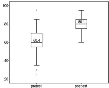
The average of pre-test results were 60.4±8.9 and the post-results 80.1±14.5, showing a statistically significant increase in knowledge gain (p<0.001) (Fig. 1).
The total success rate of eFAST was 87.5%. The location-specific performance rate was lowest in hepatorenal recess and splenorenal recess. The suc- cess percentage rate was 65.8% and 68.4% respec- tively. Checking for pleural effusion through a sub-
Table 1. Baseline characteristics of the students
Characteristics Value
Age (Yrs) 28.1±3.4
Gender
Male 21 (55.2%)
Female 17 (44.8%)
Previous knowledge of eFAST
Yes 20 (52.6%)
No 18 (47.4%)
Fig. 1. Comparison of pretest score and posttest score.
Fig. 2. Success rate of each categories of eFAST.
costal approach was 89.5%. In contrast, the success rate for bilateral anterior and lateral chest wall ultrasonography was all over 90% (Fig. 2).
The students replied that the curriculum was very satisfactory and easy to understand and they answered that eFAST education is needed in the medical college curriculum (Table 2).
IV. Discussion
As sonography is more widely used clinically, reports showing the effectiveness of sonography education in medical college are increasing.(4) This study is significant in confirming that ultrasonogra- phy education is effective in domestic medical schools. Also in this study we checked the perfor- mance rate of each item, differentiating the easy- to-learn items from the not-so-easy items which are a different point from other studies.
Our research showed significant increase in knowledge concerning the principles of the eFAST and the image interpretation skills. The pre-test results were 60.4±8.9 and post-test results were 80.1±14.5 showing a statistically significant increase. This is a similar result to Arger et al.(5).
Bentley et al.(6) reported the test results before and after FAST training to be 58.5 and 78.1, which is similar to ours.
The success rate of eFAST was 87.5% but each showed a big difference with each locations. The success rate of lung sonography was over 90% but the success rate of hepatorenal recess and the splenorenal recess was 65.8% and 68.4%. The suc- cess rate of checking for pericardial effusion using subcostal view was 89.5% and the success rate of perivesicular space was 94.7%.
Gogalniceanu et al.(7) reported a total of 86.0%
success rate in teaching 25 medical college students and Heegaard et al.(8) taught FAST to 104 emer-
gency medical technicians and showed an overall success rate of 92.3%. No report has shown the suc- cess rate of each item. We think that the success rate of each item will be an important data for future curriculum development.
To judge as a successful examination, we required the sonographer to check the whole length of the kidney. This requires delicate controlling of the probe such as moving and rotating the probe to avoid the ribs. It would have been a difficult skill for students to acquire in a short education time. So we think that more time needs to be distributed for the students to acquire these skills.
Students showed high success rate in the lung sonography. The success rate was higher, showing that the students found lung sonography easier than the checking for free fluid in the abdominal cavity.
Lung sonography is helpful in checking for pneu- mothorax or hemothorax and this is recommended in early examinations of trauma patients. This study along with other reports shows that medical college student, after completing a short course in sonogra- phy to complete this examination with ease.(9,10)
The post-training tests show that a short session of eFAST training was effectively delivered to stu- dents and the students understood the material eas- ily and were highly satisfied with the lesson. Also they replied that tests to screen trauma patients effectively like eFAST was essential in the medical college curriculum. We think that this fact could play a key role in developing future medical college curriculum.
This study has a few limitations. First, only 38 students from a medical college participated in this study, so it is difficult to generalize the findings.
Second, the aim of the study was for the students to check and obtain a normal anatomical structure in a standard patient so the students did not observe a pathologic lesion. All training was done on a healthy standard patient but the students were taught abnormal findings using slides and video lectures and the post test results show that the image inter- pretation skills were improved.
Recent studies have brought up this limitation and propose a simulator to solve it. Sara et al.(11) reports that the confidence of using sonography and image Table 2. Student questionnaires about eFAST education
Categories Median (IQR)
Learning interest 5 (4, 5)
Degree of understanding 4 (3, 4)
Need for medical college curriculum 5 (4, 5)
interpretation skills show no difference in groups who have been trained using standard patient or those who have been trained using simulators.
Lastly, as the standard patient was an average build male, who had a 4 hour of fasting time before the hands-on sessions. This would have made acquiring the images easier than if it would have been a real patient. A subsequent study is necessary to supple- ment these limitations.
V. Conclusion
This study shows that the short-term training of eFAST targeted at medical college students were effectively delivered to students. We suggest that there is need to include eFAST education in medical school curriculum.
REFERENCES
01) Korner M, Krotz MM, Degenhart C, Pfeifer KJ, Reiser MF, Linsenmaier U. Current Role of Emergency US in Patients with Major Trauma. Radiographics: a review publication of the Radiological Society of North America, Inc 2008; 28: 225- 42.
02) Counselman FL, Sanders A, Slovis CM, Danzl D, Binder LS, Perina DG. The status of bedside ultrasonography training in emergency medicine residency programs. Academic emer- gency medicine: official journal of the Society for Academic Emergency Medicine 2003; 10: 37-42.
03) Kirkpatrick AW, Sirois M, Laupland KB, Liu D, Rowan K,
Ball CG, et al. Hand-held thoracic sonography for detecting post-traumatic pneumothoraces: the Extended Focused Assessment with Sonography for Trauma (EFAST). The Journal of trauma 2004; 57: 288-95.
04) Amini R, Stolz LA, Gross A, O’Brien K, Panchal AR, Reilly K, et al. Theme-based teaching of point-of-care ultrasound in undergraduate medical education. Internal and emergency medicine 2015; 10: 613-8.
05) Arger PH, Schultz SM, Sehgal CM, Cary TW, Aronchick J.
Teaching medical students diagnostic sonography. Journal of ultrasound in medicine: official journal of the American Institute of Ultrasound in Medicine 2005; 24: 1365-9.
06) Bentley S, Mudan G, Strother C, Wong N. Are Live Ultrasound Models Replaceable? Traditional versus Simulated Education Module for FAST Exam. The western journal of emergency medicine 2015; 16: 818-22.
07) Gogalniceanu P, Sheena Y, Kashef E, Purkayastha S, Darzi A, Paraskeva P. Is basic emergency ultrasound training feasible as part of standard undergraduate medical education? Journal of surgical education 2010; 67: 152-6.
08) Heegaard W, Hildebrandt D, Spear D, Chason K, Nelson B, Ho J. Prehospital ultrasound by paramedics: results of field trial. Academic emergency medicine: official journal of the Society for Academic Emergency Medicine 2010; 17: 624-30.
09) See KC, Ong V, Wong SH, Leanda R, Santos J, Taculod J, et al. Lung ultrasound training: curriculum implementation and learning trajectory among respiratory therapists. Intensive care medicine. 2015.
10) Soldati G, Sher S. Bedside lung ultrasound in critical care practice. Minerva anestesiologica 2009; 75: 509-17.
11) Damewood S, Jeanmonod D, Cadigan B. Comparison of a multimedia simulator to a human model for teaching FAST exam image interpretation and image acquisition. Academic emergency medicine: official journal of the Society for Academic Emergency Medicine 2011; 18: 413-9.
A
Appppeennddiixx 11.. UUllttrraassoouunndd cchheecckklliisstt ffoorr ssttuuddeenntt Continue
Students number: exam date: YES NO PASS FAIL
1) Right anterior lung 1. Probe manipulation
1) Transducer was placed at the midclavicular line of the right 3rd~4th intercostal space
2) Direction of the transducer was placed by the cephalad and longitudinally
2. The adequacy and interpretation of image
1) Check between the two vertical ribs to confirm the ribs, pleura, and movement of pleural.
2) explain structures of the acquired image 2) Left anterior lung 1. Probe manipulation
1) Transducer was placed at the midclavicular line of the Left 3rd~4th intercostal space
2) Direction of the transducer was placed by the cephalad and longitudinally
2. The adequacy and interpretation of image
1) Check between the two vertical ribs to confirm the ribs, pleura, and movement of pleural.
2) explain structures of the acquired image 3) Right posterolateral lung 1. Probe manipulation
1)Transducer was placed at the posterior axillary line of the right 5th~6th intercostal space
2) Direction of the transducer was placed by the cephalad and longitudinally
2. The adequacy and interpretation of image
1) Check between the two vertical ribs to confirm the ribs, pleura, and movement of pleural.
2) explain structures of the acquired image 4) Left posterolateral lun 1. Probe manipulation
1)Transducer was placed at the posterior axillary line of the left 5th~6th intercostal space
2) Direction of the transducer was placed by the cephalad and longitudinally
2. The adequacy and interpretation of image
1) Check between the two vertical ribs to confirm the ribs, pleura, and movement of pleural.
2) explain structures of the acquired image 5) Pericardium 1. Probe manipulation
1) Transducer was placed at the area of xiphisternum 2) Transducer was moved to slightly right to use the liver as the acoustic window
3) The indicator of transducer was directed at Rt. shoulder 2. The adequacy and interpretation of image
1) Check 4 cardiac chambers and pericardium 2) explain structures of the acquired image
Students number: exam date: YES NO PASS FAIL 6) Hepatorenal recess 1. Probe manipulation
1) Transducer was placed at the mid axillary line of the Rt. 8th~11th intercostal space
2) Direction of the transducer was placed by the cephalad and longitudinally
2. The adequacy and interpretation of image
1) Check more than two-thirds of the boundaries of the Liver and Kidney
2) Check the Rt. Hemidiaphragm and costophrenic angle.
3) explain structures of the acquired image 7) Splenoreal recess 1. Probe manipulation
1) Transducer was placed at the mid axillary line of the Lt. 6th~9th intercostal space
2) Direction of the transducer was placed by the cephalad and longitudinally
2. The adequacy and interpretation of image
1) Check more than two-thirds of the boundaries of the spleen and Kidney
2) Check the Lt. Hemidiaphragm and costophrenic angle.
3) Check the entire length of the spleen 4) explain structures of the acquired image 8) Perivesical view 1. Probe manipulation
1) Transducer was placed at the midline of the suprapubic area
2) Direction of the transducer was placed by the left sided and transversally
2. The adequacy and interpretation of image 1) Check the entire length of the bladder 2) explain structures of the acquired image