식이지방에 첨가한 Conjugated Linoleic Acid가 지방산 조성, delta-5 Desaturase(D5D)와 Fatty Acid Desaturase2(FADS2)
유전자 발현에 미치는 영향
황윤희․강금지† 덕성여자대학교 식품영양학과
Effects of Conjugated Linoleic Acid Supplement on the Composition of Fatty Acids, Expressions of delta ⁃ 5 Desaturase (D5D) and
Fatty Acid Desaturase2 (FADS2) Genes in Mice
Yun⁃Hee Hwang and Keum⁃Jee Kang†
Department of Food and Nutrition, College of Natural Science, Duksung Women's University, Seoul 132-714, Korea
Abstract
We investigated the effects of conjugated linoleic acid (CLA) on the fatty acid composition in the plasma and liver, and the expressions of delta⁃5 desaturase (D5D) and fatty acid desaturase2 (FADS2) genes in ICR male mice using two different sources of fats in the diets. The experimental groups were divided into four groups: beef tallow (BT) and fish oil (FO), BT with CLA supplementation (BTC), and FO with CLA supplementation (FOC) groups. Ten mice in each group were fed with the experimental diets for 4 weeks.
All mice were fed experimental diets containing 12% of total dietary fat (w/w) either with or without 0.5%CLA (w/w). Fatty acid compositions were analyzed in the plasma and liver using gas chromatography. The levels of D5D and FADS2 expression were analyzed by RT⁃PCR in the liver. The results showed that CLA participates competitively with C18:2 in the elongation and desaturation processes, leading to significant increase in the levels of C20:4 and C22:6 in BTC group (p<0.05). The expression levels of D5D and FADS2 were higher in BT and BTC group than those of FO and FOC group. In particular, the expression of D5D gene was greatly upregulated in BTC group. Furthermore, the conversion ratios from C18:2 to C20:4 in the liver were higher in BTC group than those in other groups. Thus our results suggest that increased expressions of D5D and FADS2 genes may be responsible for the enhanced CLA effects on the desaturation in the BT containing saturated fatty acids rather than the FO rich in n⁃3 PUFA.
Key words: CLA, beef tallow, fish oil, D5D, FADS, fatty acids composition
†Corresponding author. E-mail: [email protected]
†Phone: 82-2-901-8363, Fax: 82-2-901-8372
서 론
Conjugated linoleic acid(CLA)는 자연적으로 유제품이나 쇠고기에 존재하며, n-6계 필수지방산인 linoleic acid(cis-9, cis-12, octa-decadienoic acid)로부터 생성된 공여 이중결합 을 갖는 이성체를 통틀어 지칭한다(1). CLA는 항비만작용 (2), 항암작용(3), 항동맥경화작용(4), 면역반응에 영향을 주 는 등(5) 다양한 생리활성이 보고되고 있다.
지방산의 desaturation과 elongation에 관여하는 여러 효 소 중 중요한 두 효소는 delta(Δ)-5 desaturase(D5D)와 del- ta(Δ)-6-desaturase(FADS2, fatty acid desaturase)이다.
D5D는 eicosatrienoic acid(C20:3n-6)와 eicosatetraenoic acid(20:4n-3)를 각각 arachidonic acid(AA, C20:4n-6)와
eicosapentaeonic acid(EPA, C20:5n-3)로 elongation해주는 효소이다(6). C20:4는 저장형태인 인지질로 전환되는데 이 는 자극이나 세포손상 등이 있을 때 phospholipases에 의해 분비되는 일종의 eicosanoids가 된다(7). Eicosanoids는 면 역, 항상성, 소화관의 내막보호역할 등 다양한 역할을 수행 한다(6).
FADS2는 n-6계 linoleic acid(C18:2)혹은 n-3계 α-lino- lenic acid(C18:3)를 각각 C18:3과 C18:4로 불포화시키는 효 소이다(6). FADS2로 인해 생성된 high unsaturated fatty acid(HUFA)는 대부분 인지질로 되기 때문에 막의 유동성에 영향을 끼치며, eicosanoids의 신호전달, 세포의 흡수작용 (pynocytosis), 막의 이온채널조절과 유전자 발현에 영향을 준다(6).
Cook 등(8)은 인체 내에서 FADS2는 세포신호전달기전, 심혈관 조절, 신장기능, 혈액응고에도 중요한 역할을 한다고 보고하였다. D5D와 FADS2의 활성은 대게 함께 그 비율을 보아 C18:2, C18:3로부터 C20:4, C20:5 혹은 C22:6의 합성에 끼치는 영향을 보고 있으며 이로 인해 생성된 C20:4나 C22:6 은 막의 유동성에 영향을 미쳐 인슐린 등의 세포신호전달을 조절하거나, C20:4의 경우에는 eicosanoids로의 전환되는 중 요 지방산으로써 암이나 면역반응에 영향을 끼치게 된다(9).
이처럼 지방산의 불포화도에 관계하는 desaturase의 발현은 식이조성에 따라, 간에서의 다른 유전자의 활성에 따라 혹은 체내 지방산 조성에 따라 활성화되거나 억제되어 eicosa- noids 생성 뿐만 아니라 항상성을 유지하는 데에도 큰 역할 을 하고 있다.
그러므로 본 연구는 ICR 수컷 마우스에 포화지방산이 많 은 쇠기름과 n-3계 불포화지방산이 다량 함유된 어유식이에 0.5% CLA를 첨가하여 D5D와 FADS2 유전자 발현 및 지방 산 조성에 미치는 영향을 관찰하여 식이지방에 따른 효과를 살펴보고자 하였다.
재료 및 방법
시료 및 시약
실험동물은 평균 체중이 32~35 g인 4주령의 ICR 수컷 마우스(Orient, Seoul, Korea) 40마리를 사용하였다. 식이에 첨가한 CLA(Natural ASA., Norway)는 순도가 80%로 50%
의 c9t11, 50%의 t10c12를 포함하고 있다. 대부분의 시약은 덕산종합과학(Seoul, Korea)과 Sigma-Aldrich Co.(St. Louis, MO, USA)를 사용하였으며, 지방산 분석에 사용된 시약은 JT. Baker Inc.(Phillipsburg, NJ, USA) HPLC 등급을 사용 하였다. 기타 시약은 그 구입처를 별도로 표시하였다.
실험계획
4주령의 ICR 수컷 마우스 40마리를 1주 동안 고형사료로 적응시킨 후 체중에 따라 난괴법으로 식이지방의 종류와 CLA첨가의 유무에 따라 BT(beef tallow)군, BTC(BT with 0.5% CLA)군, FO(fish Oil)군, FOC(FO with 0.5% CLA)군 으로 나누어 4주동안 사육하였다.
실험식이
실험식이는 전체 열량 중 당질이 59.2%, 단백질이 19.7%, 지방이 21.9%(12% w/w)가 되게 구성하였으며, 다른 성분 은 AIN93 diet를 기초로 동일하게 첨가하였다(Table 1). 실 험식이에 사용된 지방급원으로는 포화지방산이 주로 함유 된 쇠기름(삼강유지)과 n-3계 불포화지방산이 주로 함유된 어유(동원산업 식품연구소)를 사용하였고, 부족한 필수지방 산을 제공하기 위해 n-6 linoleic acid(C18:2)가 포함된 대두 유(soybean oil)를 공통으로 사용하였다. CLA는 총 식이무 게의 0.5%(w/w)(10)수준으로 첨가하였다. 어유의 산화를
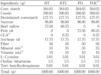
Table 1. Diet composition of experimental groups
Ingredients (g) BT BTC FO FOC1)
Corn starch Casein
Dextrinized cornstarch Sucrose
Beef tallow Fish oil CLA2) Soybean oil Fiber Mineral mix3) Vitamin mix4) L-Cysteine Choline bitartarate Tert-butylhydroquinone
384.63 200.05 127.75 96.80 72.50 0 0 17.75 50 35 10 3.01 2.5 0.01
384.63 200.05 127.75 96.80 66.25 0 6.25 17.75 50 35 10 3.01 2.5 0.01
384.63 200.05 127.75 96.80 0 72.50 0 17.75 50 35 10 3.01 2.5 0.01
384.63 200.05 127.75 96.80 0 66.25 6.25 17.75 50 35 10 3.01 2.5 0.01 Total (g) 1000.00 1000.00 1000.00 1000.00
1)BT: Beef tallow diet, BTC: Beef tallow diet+0.5% CLA, FO:
Fish oil diet, FOC: Fish oil diet+0.5% CLA.
2)CLA rich oil contained 80% of total conjugated linoleic acid (50% c9t11, 50% t10c12).
3)AIN 93 Mineral mixture.
4)AIN 93 Vitamin mixture.
방지하기 위해 dl-α-tocopherol (145.6 mg/100 g oil)을 어유 군에 첨가하였다. 각 식이지방의 지방산조성은 Table 2와 같다. 실험동물은 12시간 주기로 조절하였고 물은 증류수를 제공하였으며, 물과 식이는 자유롭게 섭취할 수 있도록 공급 해 주었다. 체중은 1주일에 한번 씩 같은 시간에 측정하였다.
시료 채취
각 실험군의 식이섭취 기간이 끝나는 날 12시간 공복시킨 마우스를 ethyl ether로 가볍게 마취시켜 단두로 희생시킨 후 헤파린 처리된 시험관에 혈액을 모았다. 혈액채취 직후 4oC, 800×g에서 15분간 원심분리하여 혈장을 취하고 -70oC 에서 보관하였다.
간은 0.9% NaCl로 씻은 후 물기를 제거하고 총 무게를 측정한 다음 반은 지방산조성을 측정하기 위해 -70oC에 보 관하고 나머지 반은 유전자 발현을 보기 위해 0.1% diethyl pyrocarbonate(DEPC) 처리된 phosphate buffer solution (PBS)에 씻어 물기를 제거한 다음 액체질소에 급속 냉동시 켜 -70oC에서 보관하였다.
Ribonucleic acid(RNA)추출에 이용될 조직은 RNase오염 을 막기 위해 해부에 이용되는 모든 기구는 0.1% DEPC에 24시간 담궈 두었다가 120oC, 30분간 autoclave하여 사용하 였다. 각 군마다 wash dish는 달리 사용하였으며 해부용 가 위, 핀셋은 그룹이 바뀔 때마다 70% ethanol에 소독한 후 30% H2O2에 30분 동안 담궈 사용하였다.
지방산 조성 분석
혈액의 지방산 조성: 혈액의 지방산 조성은 혈장 0.2 mL 를 취해 Lepage와 Roy(11)의 one-step methylation 방법에 따라 methylation 하였다. Fatty acid methyl ester는 gas
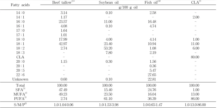
Table 2. Fatty acid composition of dietary oils
Fatty acids Beef tallow1)* Soybean oil Fish oil1)# CLA2)
g/100 g oil 14 : 0
14 : 1 16 : 0 16 : 1 17 : 0 17 : 1 18 : 0 18 : 1 18 : 2 18 : 3 CLA 20 : 0 20 : 1 20 : 5 22 : 6 Unknown
3.14 1.17 23.57 4.08 1.64 1.01 17.99 42.97 2.74 - - 1.15
- - - 0.60
0.10 - 11.00 0.10 - - 4.00 23.40 53.20 7.80 - 0.30
- - - 0.10
2.58 - 16.48 4.74 - - 4.14 10.94 1.08 2.19 - 1.56 0.36 5.47 27.65 22.81
- 2.00 - - - - 1.00 11.00 6.00 - 80.00
- - - - - Total
SFA3) MUFA4)
PUFA5)
100.00 47.49 49.23 2.74
100.00 15.40 23.50 61.10
100.00 24.76 16.04 36.39
100.00 1.00 13.00 86.00
S/M/P6) 1.0:1.04:0.06 1.0:1.53:3.98 1.0:0.65:1.47 1.0:13.0:86.00
*Supplemented 54.55 mg dl-α-tocopherol/100 g oil.
#Supplemented 145.46 mg dl-α-tocopherol/100 g oil, Tuna oil.
1)Supplemented with 4800 IU vitamin A per 15 g oil.
2)CLA rich oil contained 80% of total conjugated linoleic acid (50% c9t11, 50% t10c12).
3)SFA: Saturated fatty acids. 4)MUFA: Monoundaturated fatty acids. 5)PUFA: Polyundaturated fatty acids.
6)S/M/P: Saturated/ Monounsaturated/ Polyunsaturated fatty acid ratio.
chromatography(GC, Hewlett Packard, 6890Ⅱseries, USA) 를 이용하여 분석하였으며, column은 omegawax 250 (Supelco, Bellefonate, PA, USA)를 이용하여 3 point gra- dient temperature condition에서 분석하였다. 각 지방산의 retention time은 표준지방산 fatty acid methyl esters (FAME, Sigma Aldrich)와 omegawax column test mix (Sigma Aldrich, USA), PUFA animal source(Sigma Aldrich, USA)를 사용하여 비교 확인하였으며 그 양을 백분 율로 표시하였다.
간의 지방산 조성: 간에서의 지방산 조성을 보기 위해 0.2 g의 간을 0.2 M phosphate buffer(pH 7)에 넣어 tissue grinder로 갈아 균질화하여 이중 200 μL를 취해 혈장 지방산 분석법과 같이 methylation하여 GC로 분석하였다.
유전자 발현 분석
Ribonucleic acid(RNA) 분리: 간에서 mRNA를 분리 하기 위해 TRIzol reagent(Invitrogen, Life Technologies, Carlsbad, CA, USA)를 사용하였다. 간 조직 0.1 g을 1 mL의 TRIzol reagent에 넣고 glass homogenizer로 균질화한 후 Chomczynski와 Sacchi(12)에 의해 개발된 single step RNA isolation 방법으로 isolation하여 260/280 nm에서 O.D값을 이용해 정량하였다.
역전사반응: D5D와 FADS2의 유전자발현을 보기 위해
mRNA를 DNA로 역전사 시키는 과정을 수행하였다. RT (reverse transcription)과정은 1 μg의 RNA에 1 unit의 RQ1 DNase(Promega, Madison, WI, USA)와 25 mM의 MgCl2, 10 mM dNTP(Promega) 1 unit의 AMV RTase(Promega), 10X RT buffer, 20 unit의 RNasin(RNase inhibitor, Promega), oligo dT(Promega)를 넣고 42oC에서 1시간동안 polymerase chain reaction(PCR) thermometer(Applied Biosystems, GeneAmp PCR system 9600, CA, USA)로 수행하였다.
RT의 산물인 cDNA 2 μL에 D5D와 FADS2 유전자에 맞 게 제작된 primer(Cosmogenetech 제조, Seoul, Korea)와 2.5 mM dNTP, 25 mM MgCl2, 1 unit의 Taq DNA polymer- ase(JenanMed, Seoul, Korea), Taq buffer를 넣고 autoclave 한 증류수로 총 양을 50 μL로 맞춰 PCR thermometer로 각 primer의 최적조건에서 PCR을 수행하였다. PCR산물은 1.2% agarose gel(BMA, USA cat No. 50004)에서 전기영동 하여 image analyzer로 band를 관찰하였다. PCR산물의 발 현정도를 수치화하기 위해 TINA20 image program을 이용 하여 GAPDH(glyceraldehyde phosphate dehydrogenase) 발현에 대한 상대적인 표적유전자의 발현양의 비율을 통계 처리(p<0.05)하여 평균과 표준편차를 구하였다.
통계분석
실험결과는 SPSS(v12.0)프로그램을 이용하여 평균과 표
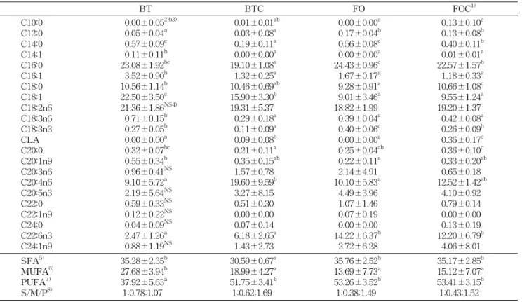
Table 4. The relative % of fatty acids in plasma
BT BTC FO FOC1)
C10:0 C12:0 C14:0 C14:1 C16:0 C16:1 C18:0 C18:1 C18:2n6 C18:3n6 C18:3n3 CLA C20:0 C20:1n9 C20:3n6 C20:4n6 C20:5n3 C22:0 C22:1n9 C24:0 C22:6n3 C24:1n9
0.00±0.052)b3) 0.05±0.04a 0.57±0.09c 0.11±0.11b 23.08±1.92bc 3.52±0.90b 10.56±1.14b 22.50±3.50c 21.36±1.86NS4) 0.71±0.15b 0.27±0.05b 0.00±0.00a 0.32±0.07bc 0.55±0.34b 0.96±0.41NS 9.10±5.72a 2.19±5.64NS 0.59±0.33NS 0.12±0.22NS 0.04±0.09NS 2.47±1.26a 0.88±1.19NS
0.01±0.01ab 0.03±0.08a 0.19±0.11a 0.00±0.00a 19.10±1.08a 1.32±0.25a 10.46±0.69ab 15.90±3.30b 19.31±5.37 0.29±0.18a 0.11±0.09a 0.09±0.08b 0.21±0.11a 0.35±0.15ab 1.57±0.78 19.60±9.59b 3.27±8.15 0.51±0.30 0.00±0.00 0.07±0.14 6.18±2.65a 1.43±2.73
0.00±0.00a 0.17±0.04b 0.56±0.08c 0.00±0.00a 24.43±0.96c 1.67±0.17a 9.28±0.91a 9.01±3.46a 18.82±1.99 0.39±0.04a 0.40±0.06c 0.00±0.00a 0.25±0.04ab 0.22±0.11a 2.14±4.91 10.10±5.83a 4.49±3.96 1.07±1.46 0.07±0.19 0.00±0.00 14.22±6.37b 2.72±6.28
0.13±0.10c 0.13±0.08b 0.40±0.11b 0.01±0.01a 22.57±1.57b 1.18±0.33a 10.66±1.08c 9.55±1.24a 19.20±1.37 0.42±0.08a 0.26±0.09b 0.36±0.17c 0.36±0.10c 0.33±0.20ab 0.65±0.18 12.52±1.42ab 4.10±0.92 0.79±0.14 0.00±0.00 0.13±0.19 12.20±6.79b 4.06±8.01 SFA5)
MUFA6) PUFA7) S/M/P8)
35.28±2.35b 27.68±3.94b 37.92±5.63a 1:0.78:1.07
30.59±0.67a 18.99±4.27a 51.75±3.41b 1:0.62:1.69
35.76±2.52b 13.69±7.73a 53.26±3.52b 1:0.38:1.49
35.17±2.85b 15.12±7.07a 53.41±3.15b 1:0.43:1.52 Fatty acid composition is expressed as percentage of total recovered fatty acids.
1)Groups are the same as in the Table 1. 2)Values are mean±SD.
3)Values sharing different superscripts in the same row are significantly different at p<0.05. 4)NS: not significant.
5-8)See the legend of Table 2.
준편차로 표시하였고 각 군 간의 평균치의 통계적 유의성 검정은 one-way ANOVA를 실시하였고 군 간의 차이는 p<0.05 수준에서 Duncan's multiple range test로 사후 검증 하였다.
결 과
체중증가량, 식이섭취량 및 식이효율
쇠기름과 어유에 0.5% CLA를 4주간 급여한 마우스의 식 이섭취량, 체중증가량 및 식이이용효율은 Table 3과 같다.
식이섭취량, 체중증가량 및 식이이용효율은 기름의 종류 또 는 CLA첨가에 따라 차이가 없었다.
혈장의 지방산 조성
혈장의 지방산 분석 결과는 Table 4와 같다. Palmitic acid(C16:0)가 BT와 FO군보다 CLA첨가군(BTC, FOC)에 서 낮은 수치를 보였으며, stearic acid(C18:0)은 FO군보다 FOC군에서 높은 수치를 보였다. Palmitoleic acid(C16:1), oleic acid(C18:1)의 함량은 BTC군이 BT군보다 유의적으로 낮았으며(p<0.05), FO군과 FOC군은 차이가 없었다. C18:2 수준은 CLA의 첨가유무나 식이지방에 따른 유의차이가 없 었다. CLA는 CLA를 섭취하지 않은 군에서는 전혀 검출되
Table 3. Effects of CLA on food intake, weight gain and food efficiency ratio in mice
Groups1) Food intake (g) Weight gain (g) FER2) BT
BTC FO FOC
5.2±0.53)NS4) 5.3±1.3 5.6±0.6 5.8±2.3
9.9±3.3 8.6±4.1 10.6±2.6 8.6±2.7
1.58±0.44 1.48±0.69 1.63±0.36 1.47±0.45
1)Groups are the same as in the Table 1.
2)FER: food efficiency ratio.
3)Values are mean±SD.
4)NS: not significant. N=10.
지 않았다. C20:4의 경우 BT군보다 BTC군에서 유의적으로 높은 함량을 보였다(p<0.05). n-3계 불포화지방산인 C20:5 는 기름의 종류나 CLA의 첨가유무에 따른 변화가 없었으나 C22:6의 경우 어유섭취군(FO, FOC)이 쇠기름섭취군(BT, BTC)보다 유의적으로 높은 수치를 보였다(p<0.05).
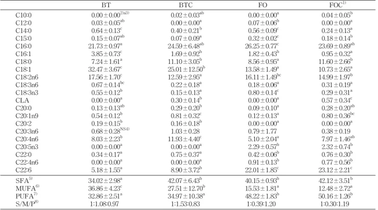
간의 지방산 조성
지방산의 총 합성에 대해 관찰할 수 있는 간의 지방산 조 성은 Table 5와 같다. C18:0이 BT와 FO군보다 CLA첨가군 (BTC, FOC)이 유의적으로 많이 검출되었고(p<0.05) C16:1, C18:1은 BT군보다 BTC군에서 유의적으로 낮았다. CLA를 섭취시킨 군에서만 소량의 CLA가 검출되었다. 필수지방산 인 C18:2의 경우 BT군에 비해 BTC군이 유의적으로 낮은
Table 5. The relative % of fatty acids in liver
BT BTC FO FOC1)
C10:0 C12:0 C14:0 C15:0 C16:0 C16:1 C18:0 C18:1 C18:2n6 C18:3n6 C18:3n3 CLA C20:0 C20:1n9 C20:2 C20:3n6 C20:4n6 C20:5n3 C22:0 C22:4n6 C22:6
0.00±0.002)a3) 0.03±0.05ab 0.64±0.13c 0.15±0.07ab 21.73±0.97a 3.85±0.73c 7.24±1.61a 32.47±3.67c 17.56±1.70c 0.67±0.14bc 0.55±0.12b 0.00±0.00a 0.13±0.13ab 0.54±0.12b 0.19±0.15b 0.68±0.28NS4) 8.03±2.23b 0.00±0.00a 0.34±0.17a 0.00±0.00a 5.18±1.55a
0.02±0.03ab 0.00±0.00a 0.40±0.21b 0.07±0.09a 24.59±6.48ab 1.69±0.92b 11.10±3.05b 25.01±12.50b 12.59±2.95a 0.22±0.18a 0.15±0.13a 0.30±0.14b 0.29±0.20b 0.81±0.32c 0.16±0.18b 1.03±0.28 11.93±4.40c 0.00±0.00a 0.75±0.37a 0.00±0.00a 8.90±3.72b
0.00±0.00a 0.07±0.06b 0.56±0.09c 0.32±0.02c 26.25±0.77c 1.82±0.43b 8.56±0.95a 13.58±1.49a 16.11±1.49bc 0.18±0.06a 0.80±0.14c 0.00±0.00a 0.09±0.10a 0.12±0.13a 0.00±0.00a 0.79±1.77 5.10±2.04a 2.29±0.57b 0.42±0.06b 0.91±0.13b 22.01±1.85c
0.04±0.05b 0.00±0.00a 0.24±0.13a 0.18±0.14b 23.69±0.89ab 0.95±0.32a 11.60±2.66b 10.73±2.65a 14.99±1.97b 0.31±0.19a 0.29±0.31a 0.57±0.34c 0.28±0.20ab 0.80±0.36bc 0.00±0.00a 0.38±0.19 7.97±1.46ab 2.32±0.74b 0.76±0.30b 0.77±0.56b 23.12±2.21c SFA5)
MUFA6) PUFA7) S/M/P8)
34.02±2.98a 36.86±4.23c 32.86±2.51a 1:1.08:0.97
42.07±6.43b 27.51±12.70b 34.97±10.38a 1:1.53:0.83
40.15±0.93b 15.53±1.81a 48.22±1.83b 1:0.39:1.20
42.12±3.51b 12.48±2.72a 50.16±1.26b 1:0.30:1.19 Fatty acid composition is expressed as percentage of total recovered fatty acids.
1)Groups are the same as in the Table 1. 2)Values are mean±SD.
3)Values sharing different superscripts in the same row are significantly different at p<0.05. 4)NS: not significant.
5-8)See the legend of Table 2.
A
BT BTC FO FOC FADS2D5D
G3PDH
Relative FADS2 expression
a
a
b b
0 0.2 0.4 0.6 0.8 1
FADS2/GAPDHmRNA ratio
BT BTC FO FOC Relative D5D expression
b
a
c c
0 0.2 0.4 0.6 0.8 1
D5D/GAPDHmRNA ratio
BT BTC FO FOC
B
C
Fig. 1. Relative ratios of FADS2 and D5D mRNA expressions.Three replicates of RT-PCR analysis were carried out to quanti- tate relative ratios of target genes over GAPDH as an endogenous gene. Relative mRNA ratios of FADS2 (B) and D5D genes (C) were quantitated as described in Materials and Methods section.
Values sharing different alphabet are significantly different at p<0.05.
수준을 보였다(p<0.05). C20:4의 경우 BT군보다 BTC군에 서 유의적으로 높은 수치를 보였다(p<0.05). C20:5는 쇠기름 섭취군(BT, BTC)에서는 발견되지 않았다. C22:6의 경우 어 유섭취군(FO, FOC)이 쇠기름섭취군(BT, BTC)보다 유의적 으로 높게 나타났으며(p<0.05), BTC군이 BT군보다 C22:6
수준이 유의적으로 높게 나타났다(p<0.05).
D5D와 FADS2의 유전자 발현
지방산의 desaturation과 elongation에 관여하는 유전자 인 D5D와 FADS2의 발현수준은 Fig. 1에 나타나있다.
NS
c a
bc b
0 5 10 15 20 25 30
Plasma Liver
Dietary group
Relative %
BT BTC FO FOC C18:2n6
b
b a
a c
c
a
b
0 0.2 0.4 0.6 0.8 1
Plasma Liver
Dietary group
Relative %
BT BTC
FO FOC
C18:3n3
a a
b
b
a a
c
c
0 0.2 0.4 0.6 0.8 1
Plasma Liver
Dietary group
Relative %
BT BTC
FO FOC
CLA
ab bc
a
b
a ab
c ab
0 0.2 0.4 0.6
Plasma Liver
Dietary group
Relative %
BT BTC
FO FOC
C20:0
b a
b
c
a a
ab ab
0 5 10 15 20 25 30 35
Plasma Liver
Dietary group
Relative %
BT BTC
FO FOC
C20:4n6
a a
a
b c b
c b
0 5 10 15 20 25 30 35
Plasma Liver
Dietary group
Relative %
BT BTC
FO FOC
C22:6n3
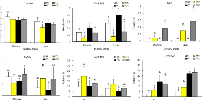
Fig. 2. Comparison of C18:2, C18:3, CLA, C20:0, C20:4 and C22:6 fatty acids in plasma and liver.
Groups are the same as in the Table 1.
Values sharing different superscripts in each tissue are significantly different at p<0.05.
FADS2의 발현은 BT군과 BTC군간의 발현의 차이를 보 이지 않았고, BT와FO, BTC와 FOC군 간의 발현은 유의적 인 차이를 나타내었다(p<0.05). D5D의 경우 어유섭취군 (FO, FOC)이 쇠기름섭취군(BT, BTC)보다 발현량이 유의 적으로 적었으며(p<0.05), BTC군은 BT군보다 발현량이 유 의적으로 많았다(p<0.05).
고 찰
CLA가 지방산 조성과 desaturase 유전자 발현에 미치 는 영향
혈장과 간의 지방산 조성: 혈장과 조직의 지방산 조성은 체내에서 합성되는 지방산과 식이로 섭취하는 지방산의 영 향을 받는다. 본 연구에서는 BT, BTC군이 FO, FOC군보다 식이 중 C16:0, C18:0, C18:1비율이 월등히 높으며, C20:5, C22:6의 함량이 낮았다(Table 2). 이들 지방산은 간에서 새 로이 합성이 되거나 혈장 등에 존재하게 되는데, C16:0은 BT를 제외한 다른 군에서 간에서의 함량 비율이 상대적으 로 많은 것으로 나타났다. C18:1의 경우 BT와 BTC군은 혈 장과 간에서의 비율이 FO와 FOC군보다 높았으며, 어유군 간(FO, FOC)의 혈장과 간에서의 수준은 비슷하였다(Table 4, 5). BT, BTC군의 혈장과 간에서 C18:1의 수준이 FO, FOC군의 수준보다 높은 것은 C18:1이 중성지방을 구성하는 주요 지방산이라는 보고에 의할 때(13), 혈장과 간의 중성지 방 수준에 영향을 받은 것으로 사료된다. C18:2도 혈장에서
더 높은 비율을 차지하였고, C18:3의 경우 혈장보다 간에서 의 수준이 더 낮게 나타났다(Fig. 2). CLA의 경우 혈장보다 간에서의 수준이 더 높았는데, 군별로 보았을 때 특히 BTC 군보다 FOC군의 수준이 높게 나타났다(Fig. 2). C20:0과 C20:4는 혈장에서 더 높은 수준으로 있으며 C22:6은 간에 더 많이 분포되어 있다(Fig. 2). CLA는 C18:2와 경쟁적으로 elongation과 desaturation에 참여한다는 보고가 있다(14,15).
본 실험 식이에서 쇠기름이 어유보다 C18:2함량이 2배 이상 많음에도 불구하고 혈장이나 간에서 C18:2 수준이 식이지방 에 따른 차이가 크지 않은 것과 CLA의 함량이 BTC군보다 FOC군이 많은 것으로 보아 C18:2는 비율 제한적으로 elongation과 desaturation에 참여하고 있으며 또 CLA와 경 쟁적으로 참여하고 있음을 알 수 있으며, 쇠기름에서 CLA의 참여가 더 높은 것으로 추정된다.
C20:4는 저장되기보다 세포막을 구성하고, C22:6 또한 주로 세포막을 구성하는 지방산이다(16,17). CLA가 C20:4 의 수준을 낮추어 준다는 보고와는 달리(18,19) 본 실험에 서는 BTC군과 FOC군에서 C20:4의 수준이 증가함을 보였 고, 간의 C22:6 또한 CLA의 첨가에 의해 BTC군에서 유의 적으로 증가함을 보임으로써 CLA의 첨가에 의한 C20:4와 C24:6의 합성은 인지질에 영향을 미쳐 막의 유동성을 조절 함으로써 세포의 신호전달에 영향을 끼칠 것이라 추정할 수 있다.
FADS2와 D5D 유전자 발현과 지방산 조성의 변화 지방산의 대사와 desaturase 효소의 활성은 밀접한 관련
Table 6. Effects of CLA on the conversion ratios of C18:3/C18:2 and C20:4/C18:2 in mice liver
BT BTC FO FOC1)
Liver 18:3/18:2
20:4/18:2
0.04±0.002)a3) 0.47±0.17ab
0.02±0.01b 0.94±0.30c
0.01±0.00c 0.32±0.13a
0.02±0.01bc 0.54±0.12b
1)Groups are the same as in the Table 1. 2)Values are mean±SD.
3)Values sharing different superscripts in the same row are significantly different at p<0.05.
이 있다. 간에서 지방산 합성시 불포화도를 높여주고 elon- gation해주는 데 직접적인 역할을 수행하기 때문이다. 본 실 험에서는 Δ6 desaturase의 한 종류인 FADS2와 Δ5 desatur- ase의 대표적인 D5D 두 유전자와 지방산 조성변화를 관찰 하였다.
Δ6 desaturase와 Δ5 desaturase의 활성은 불포화지방산 의 생성에 의거하여 두 효소의 활성을 동시에 보는데, 이는 eicosanoids 생성으로 인한 암, 면역체계와 불포화지방산 생 성으로 인한 혈관계 질환개선과 관계가 있다(18). 본 연구에 서는 n-3계 다가불포화지방산이 많이 함유된 어유군보다 쇠 기름군에서 FADS2와 D5D 유전자 발현이 유의적으로 높게 나타났다(Fig. 1). 각 desaturase에 의한 지방산의 전환비율 은 이들 유전자 발현과 일치하였다(Table 6). 식이지방에서 불포화지방산의 함량이 높을수록 Δ5, Δ6-desaturase의 활 성은 낮아지고, C20:4가 eicosanoids로 전환되는 비율이 커 지게 된다(6). CLA는 사람의 대동맥 내피세포에서 C18:2의 Δ6-desaturation을 감소시켜 C20:4 수준의 감소효과를 내며 이러한 감소효과는 특히 t10c12 CLA에서 두드러졌다 (18,19). 본 연구결과에서도 불포화지방산이 많이 들어있는 FO, FOC군은 BT, BTC군에 비해 FADS2와 D5D의 활성은 낮았고 BT와 FO에 비해 BTC와 FOC군은 C18:2에서 C18:3, C18:2에서 C20:4의 전환율이 낮았다(Table 6). FO와 FOC간 에는 FADS2와 D5D의 활성에는 차이가 없었으나 C18:2에 서 C20:4로의 전환은 FOC군이 FO군에 비해 유의적으로 많 았다(p<0.05). 반면에 BTC군은 BT군에 비해 FADS2와 D5D발현이 증가하였고, C20:4의 함량은 BT보다 BTC군이 더 많았다. 이들이 eicosanoids로 전환되었던지 또는 지방산 대사에서 degradation 되었을 것이라고 추정할 수 있으나 이에 관한 연구가 더 수반되어야 할 것이다.
반면에 ICR 마우스에 1.5% CLA첨가하여 사육하였을 때 Δ5 desaturase, Δ6 desaturase의 mRNA발현이 증가되었다 고 보고되었는데, 본 실험에서도 FADS2는 BT와 BTC간에 유의적인 차이는 아니지만 BTC군이 BT군에 비해 높은 경 향을 보였고, D5D의 발현은 BTC군에서 BT보다 유의적으 로 증가함을 보였다. 이는 peroxisome proliferator activated receptor α(PPARα)의 활성으로 CLA에 의한 지방산 산화의 증가 때문이라 가정하였는데, 본 실험에서도 CLA에 의한 지방산 산화증가로 FADS2, D5D 유전자 발현이 증가되었다 고 사료된다(20). C22:6의 생성은 C24:6으로부터 지방산의 β-산화에 의해 생성되는데 본 실험에서 BT군보다 BTC군
이 간의 C22:6함량이 유의적으로 높게 나타난(Fig. 2) 원인 도 CLA에 의한 β-산화 활성효과의 결과라 가정한다(21,22).
또 이로 인하여 세포막의 유동성에 변화를 가져와 세포신호 전달에 영향을 줄 수 있다고 본다.
Δ5, Δ6 desaturase의 활성과 지방산 대사와의 관계는 매 우 다양한 측면에서 해석할 수 있는데, 이 효소들이 n-3계 지방산과 n-6계 지방산들을 경쟁적으로 desaturation하기 때문이다. 또, 이들로 인해 생성된 불포화지방산들이 eico- sanoids로의 전환이 되는지의 여부에 따라 이들 유전자들은 지방산 조성의 항상성을 유지하기 위해 유전자 발현이 억제 되고 활성화되기도 한다(6). CLA와 desaturase 활성과의 상 관관계에 대한 연구는 아직 미비하며, CLA에 의한 desatur- ase의 활성 또는 억제효과와 관련하여 이들의 활성에 영향 을 미치는 다른 유전자[cyclooxygenase(COX), sterol regu- latory binding protein(SREBP), PPARα 등]와의 상관관계 도 앞으로 계속 수행되어야 할 과제이다.
요 약
본 연구에서 4주령 ICR 수컷 마우스에 쇠기름과 어유에 단독 또는 0.5% CLA를 첨가한 식이로 4주동안 사육하여 혈장과 간의 지방산 조성과 D5D와 FADS2의 유전자 발현을 관찰한 결과는 다음과 같다. CLA는 C18:2와 경쟁적으로 지 방산의 elongation과 desaturation에 참가하며, CLA의 첨가 는 BTC군에서 C20:4수준을 높여주는 효과가 있었다. 또, 지 방산의 β-산화에 의해 생성되기도 하는 C22:6은 BTC군에 서 간의 C22:6의 함량이 BT군보다 증가하였다. D5D와 FADS2는 쇠기름섭취군(BT, BTC)에서 발현이 유의적으로 증가하였는데, 특히 D5D의 발현은 BTC군이 다른 군보다 유의적으로 증가하였다. 이에 대한 지방산 전환 또한 BTC 군이 C18:2로부터 C20:4로 불포화하고 elongation하는 비율 이 혈장과 간 모두에서 가장 높았다. 결론적으로 BT에 CLA 를 첨가한 경우에 지방산 조성의 변화와 FADS2와 D5D의 발현을 증가시켜 지방산의 불포화도를 높여서 eicosanoids 합성과 항상성에 영향을 미치는 것으로 나타났으나 FO에 CLA첨가시에는 영향을 미치지 않는 것으로 나타났다.
감사의 글
본 연구는 한국과학재단 연구 지원(R04-2002-000-20039-
0)과 한국학술진흥재단의 지원(KRF-2005-005-J13002)에 의하여 수행되었으며 이에 감사드립니다.
문 헌
1. Kelly GS. 2001. Conjugated linoleic acid: a review. Altern
Med Rev 6: 367-382.
2. Park Y, Albright KJ, Liu W, Stroskon JM, Cook ME, Pariza MW. 1997. Effect of conjugated linoleic acid on body com- position in mice. Lipids 32: 853-858.
3. Liew C, Schut HA, Chin SF, Pariza MW, Dashwood RH.
1995. Protection of conjugated linoleic acids against 2-ami- no-3-methylimidazo [4,5-f]quinoline-induced colon carci- nogenesis in the F344 rat: a study of inhibitory mechanisms.
Carcinogenesis 16: 3037-3043.
4. Lee KN, Kritchevsky D, Pariza MW. 1994. Conjugated lino- leic acid and atherosclerosis in rabbits. Atherosclerosis 108:
19-25.
5. Cook ME, Miller CC, Park Y, Pariza M. 1993. Immune modulation by altered nutrient metabolism: nutritional control of immune-induced growth depression. Poult Sci 72: 1301-1305.
6. Nakamura MT, Nara TY. 2004. Structure, function, and di- etary regulation of Δ6, Δ5, and Δ9 desaturases. Annu Rev
Nutr 24: 345-376.
7. Funk CD. 2001. Prostaglandins and leukotrienes: advances in eicosanoid biology. Science 294: 1871-1875.
8. Cook HW, Byers DM, Palmer FB, Spence MW, Rokoff H, Duval SM, Emken EA. 1991. Alternate pathways in the de- saturation and chain elongation of linolenic acid 18:3(n-3), in cultured glioma cells. J Lipid Res 32: 1265-1273.
9. Brenner RR. 2003. Hormonal modulation of Δ6 and Δ5 desa- turases: case of diabetes. Prostagl Leukot Essent Fatty
Acids 68: 151-162.
10. DeLany JP, West DB. 2000. Changes in body composition with conjugated linoleic acid. J Am Coll Nutr 19: 487s- 493s.
11. Lepage G, Roy CC. 1986. Direct trans-esterification of all class of lipid in a one step reaction. J Lipid Res 27: 114-120.
12. Chomczynski P, Sacchi N. 1987. Single-step method of RNA isolation by acid guanidinium thiocyanate-phenol- chloroform extraction. Anal Biochem 162: 156-159.
13. Miyazaki M, Gomez FE, Ntambi JM. 2002. Lack of stearo- yl-CoA desaturase-1 function induces a palmitoyl-CoA delta 6 desaturase and represses the stearoyl-CoA desatur- ase-3 gene in the preputial glands of the mouse. J Lipid
Res 43: 2146-2154.
14. Belury MA, Kempa-Steczco A. 1997. Conjugated linoleic acid modulates hepatic lipid composition in mice. Lipids 32:
199-204.
15. Liu KL, Belury MA. 1998. Conjugated linoleic acid reduces arachidonic acid content and PGE2 synthesis in murine keratinocytes. Cancer Lett 127: 15-22.
16. Maritinez M. 1992. Tissue levels of polyunsaturated fatty acids during early human development. J Pediatr 120:
S129-S138.
17. Johnson DW, Beckman K, Fellenberg AJ, Robison BS, Poulosa A. 1992. Monoenoic fatty acids in human brain lip- ids: isomer identification and distribution. Lipids 27: 177- 180.
18. Eder K, Schleser S, Becker K, Körting R. 2003. Conjugated linoleic acids lower the release of eicosanoids and nitric ox- ide from human aortic endothelial cells. J Nutr 133: 4083- 4089.
19. Chuang LT, Thurmond JM, Liu JW, Kirchner SJ, Mukerji P, Bray TM, Huang YS. 2001. Effect of conjugated linoleic acid on fungal delta 6-desaturase activity in a transformed yeast system. Lipids 36: 139-143.
20. Takashi Y, Kushiro M, Shinohara K, Ide T. 2003. Activity and mRNA levels of enzymes involved in hepatic fatty acid synthesis and oxidation in mice fed conjugated linoleic acid.
Biochim Biophys Acta 1631: 265-273.
21. Cohen P, Friedman JM. 2004. Leptin and the control of me- tabolism: role for stearoyl-CoA desaturase-1 (SCD-1). J
Nutr 134: 2455s-2463s.
22. Cohen P, Miyazaki M, Socci ND, Hagge-Greenberg A, Liedtke W, Soukas AA, Sharma R, Hudgins LC, Ntambi JM, Friedman JM. 2002. Role for stearoyl-CoA desatur- ase-1 in leptin-mediated weight loss. Science 297: 240-243.
(2007년 7월 23일 접수; 2007년 10월 4일 채택)