Conjugated Linoleic Acid (CLA) Ameliorates Hydrogen Peroxide-Induced Oxidative Stress on Rat Cardiomyoblast H9c2 Cells
Jae Hong Park1, Yeon Gyu Moon2, Jung Min Kwon2, Yong Un Cho3, Jeong Ok Kim4 and Yeong Lae Ha2*
1Kangnam General Hospital, Yongin 446-907, Korea
2Division of Applied Life Sciences (BK21 Program), Graduate School, and Institute of Agriculture & Life Science, Gyeongsang National University, Jinju 660-701, Korea
3Gyeongnam National University of Science and Technology, Department of Pharmaceutical Engineering, Jinju 660-758, Korea
4HK Biotech. Co., Ltd., Jinju 660-778, Korea
Received October 20, 2012 /Revised December 22, 2012 /Accepted December 24, 2012
Conjugated linoleic acid (CLA) exhibits several beneficial biological activities including anticarcino- genesis and body-fat reduction. Now, we report that CLA ameliorated the oxidative stress in rat car- diomyoblast cells, H9c2, treated with hydrogen peroxide (H2O2). Cells were cultured in DMEM/F-12 media at 37℃ with humidified atmosphere of 5% CO2. The cells, cultured for 48 hrs, were seeded at a density 3.5×10³cell/well in a 24 well-plate and incubated for 24 hr. Using these cells, two experi- ments were performed: the cytotoxicity test of CLA (10, 20, 30, 40, and 50 μMs), and the oxidative stress amelioration test of CLA (20 and 50 μMs) against cells treated with H2O2(10 and 50 μMs) for 1 and 2 hrs. CLA enhanced the growth of H9c2 cells at any concentrations of CLA and at any in- cubation times (up to 6 days), indicating that CLA acts as a growth stimulant. No protective effect of CLA (20 and 50 μMs) was seen in cells treated 50 μM H2O2for 1 and 2 hr, but these CLA concen- trations ameliorated (p<0.05) the adverse effect of 10 μM H2O2 in cells treated for 1 hr. These CLA concentrations significantly (p<0.05) reduced the proportion of apoptotic cells, relative to control cells.
These results suggest that CLA protected H9c2 cells from the oxidative stress of H2O2 through the suppression of cell apoptosis and could be a useful compound for the prevention of cardiac diseases caused by oxidative stress.
Key words : Conjugated linoleic acid (CLA), hydrogen peroxide (H2O2), rat cardiomyoblast cell H9c2, cytotoxicity, apoptosis
*Corresponding author
*Tel:+82-55-772-1964, Fax:+82-55-772-1969
*E-mail : [email protected]
This is an Open-Access article distributed under the terms of the Creative Commons Attribution Non-Commercial License (http://creativecommons.org/licenses/by-nc/3.0) which permits unrestricted non-commercial use, distribution, and reproduction in any medium, provided the original work is properly cited.
Journal of Life Science 2012 Vol. 22. No. 12. 1658~1664 DOI : http://dx.doi.org/10.5352/JLS.2012.22.12.1658
서 론
심근경색(myocardial infarction)은 관상동맥의 폐쇄에 따 른 심근의 허혈로 심근세포의 괴사가 발생하는 질환이다 [16,20]. 관상동맥의 폐쇄는 관상동맥의 동맥경화가 주된 원인 이며, 혈전 등도 원인이다. 이는 당뇨, 고혈압, 비만, 지속적인 흡연, 스트레스 등에 의해 발생하지만 oxidative stress가 큰 원인이다[7,23,24,26,27]
최근까지 전 세계적으로 심근경색으로 인한 사망수가 증가 하고 있으며, 이는 65세 이상 노인의 80% 이상 발생한다고 보고되었다[3,5,6]. 한국에서도 최근 평균수명의 연장, 식생활 및 생활환경의 변화로 그 발생빈도가 증가하는 추세에 있다
[17,19]. 한국에서 심근경색으로 인한 사망률(인구 10만 명당) 은 남자 2.5명, 여자 5.8명으로, 남자보다 여자가 더 높은 사망 률을 보이고 있다[16,20,23,30,31]. 1960년대 이후 부정맥에 대 한 진단 및 치료, 심폐소생술의 발달과 울혈성 심부전에 대한 각종 약물의 개발로 심근경색 발생초기의 사망률이 감소되고 있으나[17,25], 천연물로부터 안전한 예방 및 치료물질의 개발 이 절실하다.
심근경색 치료제는 베타차단제, angiotensin converting enzyme (ACE) 억제제, 콜레스테롤 강하제, 칼슘 차단제 등의 약물이 개발되었다. 또한 심근경색은 섬유질이 많은 콩, 현미, 잡곡, 등푸른 생선, 딸기 등의 섭취로 예방할 수 있다[4,28,30].
최근 conjugated linoleic acid (CLA)는 1987년에 항암성 물질 로 개발[11]된 이후, 항당뇨성, 항관절염성 등에 관하여 많은 연구가 수행되었다[1,8,9,11-15,21,22]. Lee 등[21]과 David 등 [9]은 토끼에서 CLA의 항동맥경화성을 보고하였으나, 그 이 후 항동맥경화성에 관한 연구는 많이 보고되지 않았다. 따라 서 CLA의 심혈관 질환 특히 심근경색에 관한 연구를 할 필요 가 있다.
Clonal cell line인 H9c2 (2-1)는 grom embryonic BDIX rat ventricular heart tissue에서 유래한 rat cardiomyoblast cell (rat 배아심근 세포주)로 약간의 cardiac muscle 특성을 갖는다 [2,10]. 이 세포주는 electrophysiological 특성이 adult cardio- myocytes와 유사하여in vitro cardiac muscle로 많이 이용되 고 있다[2,32]. 따라서 본 연구에서는 CLA의 심근경색 예방효 과를 H9c2 세포를 이용하여 연구하였다. H9c2 세포에 hydro- gen peroxide (H2O2)를 처리하기 2일 전에 CLA를 처리하고 CLA의 H2O2유도 oxidative stress 완화효과를 세포 증식 및 apoptosis를 통하여 구명하였다.
재료 및 방법 재 료
H9c2 세포는 Korea Cell Line Bank (Seoul, Korea)에서 구 입하였다. Linoleic acid (99%; LA), H2O2(34.5%), dimethyl- sulfoxide (DMSO), penicillin (10,000 U/ml)과 streptomycin (10 mg/ml), 0.25% trypsin-EDTA는 Sigma-Aldrich (St.
Louis, MO), 3-(4,5-dime- thylthiazol-2yl)-2,5-diphenyltetra- zolium (MTT)과 bovine serum albumin (BAS)은 Amersco (Solon, OH)에서 구입하였다. Dulbecco's Modified Eagle's Medium/Ham's F-12 nutrient (DMEM/F-12)와 fetal bovine serum (FBS)은 Gibco BRL (Grand Island, NY)에서 구입하였 다. Cell culture plate (24 well plate와 96 well plate), culture dish (10 cm, i.d.)는 Nunc 사(NY, NY)로부터 구입하였다. 그 외 사용된 시약은 Sigma- Aldrich사로부터 구입하였다.
CLA와 BAS complex 제조
CLA는 LA를 KOH-propylene glycol 용액(280℃)에서 알카 리이성화하여 합성하였다[1]. CLA와 BAS complex는 Islam 등의 방법에 준하여 제조하였다[15]. 즉, CLA (11.28 mg)를 100 mM NaOH/ethanol 1.6 ml에 잘 혼합하고, 진공농축으로 ethanol을 제거한 후 2 mM BAS 5 ml를 가하여 용해하였다.
여기에 증류수 5 ml (60℃)를 가하여 BAS에 complex된 4 mM CLA 용액을 제조하였다.
세포 배양
H9c2 cell은 DMEM/F-12배지(10% FBS, 1% penicillin 10,000 U/ml, streptomycin 10 mg/ml 포함) 25 ml를 함유한 cell culture dish를 이용하여 CO2 incubator (5% CO2, 37℃) 에서 배양하였다. 세포가 80% 정도 증식하면 배지를 제거하 고, phosphate-buffered saline (PBS; 15 ml)로 세척(3회)한 후, 0.25% trypsin-EDTA (1 ml)를 처리(37℃, 5 min)하고 원 심분리(1,500 rpm, 5 min)하여 세포를 회수하였다. 세포 수
는 heamacytometer로 측정하였다.
H9c2 세포에 CLA 처리
H9c2 세포를 24 well plate (3.5×10⁴ cells/0.5 ml me- dia/well)에서 24시간 배양하였다. 배지를 갈아주면서 CLA를 처리(0, 10, 20, 30, 40, 50 μM)하고 2, 4 및 6일 간 배양하였다.
배지는 24시간마다 갈아주었다.
H9c2 세포에 H2O2 처리
H9c2 세포를 24 well plate (3.5×10⁴ cell/0.5 ml me- dia/well)에서 24시간 배양하고 배지를 갈아주면서 CLA를 처 리(0, 20, 50 μM)하였다. 배양 2일 후, 배지를 갈아주고 H2O2를 처리(0, 10, 50, 500 μM)하고 1, 2 및 4시간 배양하였다.
H9c2 세포 증식 측정
H9c2 세포 증식은 Silvanatham의 방법에 준하여 MTT 방법 으로 측정하였다[29]. 즉, 24 well plate에서 CLA를 처리한 세 포 및 CLA와 H2O2를 처리한 세포로부터 배지를 제거하고, PBS (200 μl)로 3번 세척하였다. 여기에 MTT 용액 200 μl (0.005 g/ml PBS, 0.2 μm filter로 filter)를 첨가하고 은박지를 덮고 3시간 동안 반응 시켰다. 반응 후 MTT 용액을 제거하고 PBS로 washing한 후, DMSO를 200 μl씩 첨가하여 생성된 for- mazin crystal을 용해하였다. 570 nm에서 흡광도를 micro- plate reader (Anthos Labtech Instruments, Wals., Austria)로 측정하여 세포증식을 측정하였다.
Apoptosis 측정
세포의 apoptosis 측정은 flow cytometer로 측정하였다 [1,15]. CLA 및 H2O2로 처리된 H9c2 세포를 PBS (200 μl)로 3번 세척한 후 0.25% trypsin-EDTA를 처리한 다음 원심분리 (1,500 rpm, 5 min)하여 pellet을 모았다. 여기에 다시 PBS (2 ml)로 세척한 다음 원심분리하였다. Pellet에 차가운 70%
ethanol (200~500 μl)을 첨가하여 4℃에서 1시간 동안 고정시 키고 PBS로 여러 번 씻어내고 원심분리하였다. Pellet에 1 mg/ml RNase와 50 μg/ml PI가 함유된 PBS 0.5 ml을 첨가시 켜 실온에서 30분 동안 염색했다. FACSCalibur flow cy- tometer (BD Bioscience, San Jose, CA)로 20,000개 세포의 DNA 함량을 CellQuestPro (BD Biosciences)를 이용하여 분석 하였다.
Gas chromatographic 분석
CLA는 Ha 등의[12] 방법에 준하여 4% sulfuric acid/meth- anol로 methylation하여 GC로 분석하였다[1]. GC는 FID와 Supelcowax-10 fused silica capillary column (60×0.32 μm, i.d.)이 부착된 Hewlett Packard 5890 (USA)을 사용하였다.
Oven temperature는 50℃에서 1분간 유지한 다음 250℃까지
2℃/min으로 program하였다. Detector와 injector의 온도는 각각 260℃와 280℃였고, carrier gas는 N2를 사용하였다.
통계분석
Data는 평균±표준오차(M±SD)로 표시하였다. 결과는 SAS (Statistical analysis system, Cary, NC.) 버전 8.1 프로그램을 이용하여 분석하고, 평균치 간의 유의성은 Duncan의 multi- ple rage test로 검증하였다.
결과 및 고찰 CLA의 순도
CLA는 다양한 이성체를 갖는다. 그 중 c9,t11-CLA와 t10,c12-CLA가 major 이성체이고, 나머지 c,c- 및 t,t-CLA는 minor 이성체이다[1,15]. 본 연구에서 사용된 CLA는 LA를 알 카리이성화하여 합성한 CLA로 Table 1에서 보는 바와 같이 c9,t11-CLA와 t10,c12-CLA 이성체가 각각 47.5%, 46.8%였고, t,t-CLA 이성체가 3.2%이고 나머지 CLA 이성체가 2.5%였다.
이와 같은 CLA의 이성체 조성은 다른 연구자들이 사용한 CLA의 이성체 조성과 유사하였다[15]
CLA의 H9c2 세포 증식 효과
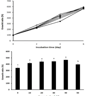
CLA는 인체유방암 MCF-7 세포, 인체전립선암 PC-3 세 포, 인체페암 A-549 세포 등의 암세포에 강한 독성을 나타낸 다[1,15]. 그러나 인체정상 세포나 rat cardiomyoblast H9c2 세포에 대한 세포독성 및 증식에 관한 연구는 많지 않다. 따 라서 본 연구에는 H9c2 세포에 대한 CLA의 세포증식을 MTT 방법을 이용하여 연구하였다. 세포배지에 CLA의 농도 를 0, 10, 20, 30, 40 및 50 μM로 처리한 후 0, 2, 4 및 6일 동 안 배양하면서 세포증식을 측정하였다(Fig. 1A). 세포증식은 CLA를 처리하고 2일 간 배양하였을 때 모든 CLA 처리 농도 에서 배양 시작보다 2배 이상 증가하였고, 대조처리와는 차 이가 없었다. 배양 4일에는 전 CLA 처리 농도에서의 세포증 식이 대조처리에 비해 증가하였다. 그러나 배양 6일에는 전 CLA처리 농도에서 세포증식은 증가하였지만 대조처리 세포
Table 1. Composition of CLA isomers used in this study CLA isomers1) Composition (%)2)
c9,t11-CLA 47.5
t10,c12-CLA 46.8
c,c-CLA 2.5
t,t-CLA 3.2
1)CLA isomers were identified by comparison of retention time in GC analysis with authentic CLA isomers.
2)Composition was calculated by area ratio of a given CLA to total CLA isomers in GC analysis.
증식과 차이가 없었다. 이와 같은 결과는 CLA가 H9c2에 독 성을 나타내는 것이 아니라 오히려 증식을 촉진하는 결과였 다. 다만, 6일 배양에서 CLA의 효과가 감소하는 것은 아마 도 H9c2 세포의 노화에 기인하는 것으로 추정되지만 이에 대한 연구는 더 수행되어야 할 것이다.
배양 4일에 CLA 처리 농도 간에 세포증식 차이가 있었다 (Fig. 1B). 세포증식은 CLA 10, 20, 30, 40 및 50 μM 농도에서 대조구 세포 342%에 비해 각각 418%, 441%, 445%, 468%, 373%로 유의성 있게 증가하였으나(p<0.05), 50 μM 농도에서 는 오히려 10-40 μM 농도보다 감소하였다(p<0.05). 따라서 CLA는 일정한 농도(10-40 μM)까지 농도 의존적으로 H9c2 세 포증식을 촉진하였다. 이와 같은 결과에 대한 기작에 관하여 서는 많은 연구를 수행하여야 하겠지만, CLA가 여러 암세포 에 대한 독성을 나타내는 것과는 달리 H2c9 세포에 대해서는 농도와 배양기간 의존적으로 증식효과가 있었다.
CLA의 H2O2 처리 H9c2 세포에 대한 oxidative stress 완화 효과
CLA가 실험동물, 인체 및 인체 세포주에서 체지방을 감소 하는 효과를 나타내고 있고, 발암 동물모델이나 인체 암세포
Fig. 1. Effects of CLA on the growth of rat cardiomyoblast H9c2 cells. Line identification (Top panel): control (●); 10 μM (■); 20 μM (▲); 30 μM (◯); 40 μM (△), 50 μM (□).
Bottom panel represents the growth rate of H9c2 cells treated with CLA for 4 days. Statistical comparisons of the results were done by Duncan's multiple range test (p<0.05).
Fig. 3. The protective effect of CLA on the growth of cardiomyoblast H9c2 cell treated with 50 μM H2O2. Cells, pre-incubated for 2 days with CLA, were treated with 50 μM H2O2for 1 hr (A panel) and 2 hr (B panel). Means with different small letters are significantly different at p<0.05 by Duncan's multiple range test.
주에서 암세포의 생장을 억제하는 보고는 많이 있지만, CLA 의 심근경색과 관련한 세포에 대한 연구는 아직 보고된 적이 없다. 따라서 심근경색의 원인이 되는 oxidative stress를 H2O2로 유발한 H9c2의 세포증식에 미치는 CLA의 영향을 연구하였다. 세포증식은 CLA 농도 의존적(Fig. 1A)이었기 때문에 H9c2 세포에 대한 H2O2유발 oxidative stress 연구에 서는 CLA 20 μM을 사용하여 증식이 양호하였던 2일간 배양 하였다
먼저, H9c2 세포에 대한 적정 H2O2농도를 연구하기 위하여 CLA (20, 50 μM)가 함유된 배지에서 2일간 세포를 배양하고 H2O2(10, 50, 500 μM)를 처리 한 후 1, 2 및 4시간 세포증식을 비교 검토하였다. Fig. 2는 H2O2를 500 μM 처리하고 H9c2 세 포증식과 CLA의 효과를 나타내고 있다. H2O2의 oxidative stress가 너무 강하여 세포의 생육이 급격히 억제 되었다.
Fig. 2. Effects of CLA on the growth of cardiomyoblast H9c2 cell treated with 500 μM H2O2. Cells, pre-incubated with CLA, were treated with 500 μM H2O2.for 1, 2 and 4 hrs. SD was less than 7% of mean values.
H2O2처리 1시간 후의 세포 생육은 CLA (0, 20, 50 μM) 처리 농도에서 각각 49%, 44%, 46%였다. 또한 H2O2 처리 2시간 후의 세포생육은 CLA (0, 20, 50 μM) 처리 농도에서 각각 50%, 47%, 53%였다. 그리고 H2O2 처리 4시간 후의 생육세포는 CLA (0, 20, 50 μM) 처리 농도에서 각각 56%, 52%, 55%였다.
따라서 CLA의 농도 및 처리 시간에 따른 효과를 볼 수 없었다.
H2O2저 농도 처리에 의한 CLA의 효과를 연구하였다. CLA (20, 50 μM)가 함유된 배지에서 2일간 세포를 배양하고 H2O2
(50 μM) 처리 1시간 및 2시간 경과 후에 세포증식을 비교 검토하였다(Fig. 3). H2O2처리 1시간 경과 후의 세포 생육에 대한 CLA 처리에 의해 다소 증가되었지만, 유의성은 없었다 (Fig. 3A). 대조처리의 생육은 73%였고, CLA 20, 50 μM 처리에 서는 각각 75%, 76%였다. 또한 H2O2 처리 2시간 경과 후의 세포생육은 CLA 처리로 세포의 생육은 다소 증가되었지만, 유의성은 없었다(Fig. 3B). 이들 결과 역시 H2O2농도가 높아 CLA의 효과를 볼 수 없음을 암시한다.
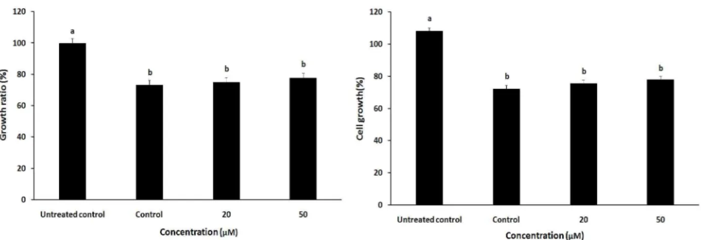
Fig. 4는 CLA (20, 50 μM)가 함유된 배지에서 2일간 세포를 배양하고 H2O2(10 μM) 처리 1 및 2시간 배양 후에 세포증식을 비교하였다. H2O2처리 1시간 후의 세포증식에 대한 CLA는 대조처리에 비해 유의성 있게 증가 시켰다(p<0.05) (Fig. 4A).
대조처리의 세포생육은 90%였지만, CLA 20 μM 처리로 108%
로 증가하였고, CLA 50 μM 처리로 110%로 증가 되었다. 즉, CLA 10 μM 처리는 대조처리에 비해 20% 증가하였고, CLA 50 μM 처리는 3% 증가하였다. 따라서 이 결과로부터 CLA는 H9c2 세포에서 H2O2의 oxidative stress를 제거하였음을 알 수 있었다. 또한 Fig. 4B에서는 H2O2처리 2시간 후의 세포증 식에 대한 CLA의 효과를 나타내고 있다. H2O2 처리 2시간 후의 세포증식에 대한 CLA의 효과는 H2O2처리 1시간 후의 처리 효과 보다 낮아서, 대조처리에 비해 세포증식은 증가하 였지만 유의성은 없었다. 즉, CLA 10 μM 처리는 대조처리에 비해 7% 증가하였고, CLA 50 μM 처리는 5% 증가하였다. 이
Fig. 4. The protective effect of CLA on the growth of cardiomyoblast H9c2 cell treated with H2O2. Cells, pre-incubated for 2 days with CLA, were treated with 10 μM H2O2.for 1 hr (Left panel) and 2 hr (Right panel). Means with different small letters are significantly different at p<0.05 by Duncan's multiple range test.
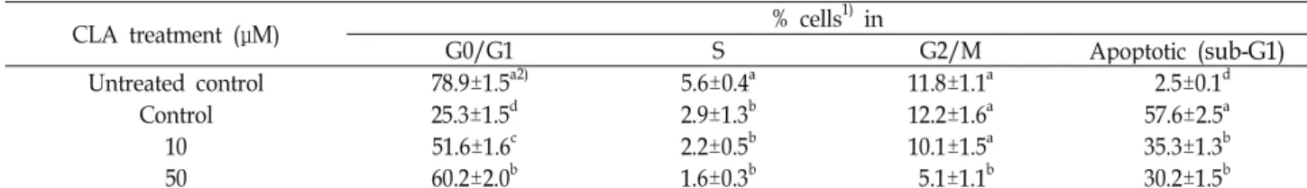
Table 2. Modulation of cell cycle progression and apoptosis in hydrogen peroxide-treated H9c2 cells by CLA
CLA treatment (μM) % cells1) in
G0/G1 S G2/M Apoptotic (sub-G1)
Untreated control Control
10 50
78.9±1.5a2) 25.3±1.5d 51.6±1.6c 60.2±2.0b
5.6±0.4a 2.9±1.3b 2.2±0.5b 1.6±0.3b
11.8±1.1a 12.2±1.6a 10.1±1.5a 5.1±1.1b
2.5±0.1d 57.6±2.5a 35.3±1.3b 30.2±1.5b
1)Cells, pretreated for 2 days with CLA, were incubated with 10 μM H2O2for 1 hr. Cell cycle distribution was assessed by flow cytometer. Results were averaged from three independent experiments.
2)Values with different small superscript letters in the same column were significantly different at p<0.01 by Duncan's multiple range test.
와 같은 결과는 CLA가 H2O2초기 반응시간(1시간 이내)에서 는 H2O2에 의한 oxidative stress를 제거하지만, 시간이 경과 할수록 그 효과는 낮아지고 있음을 암시하고 있다.
CLA는 H9c2 세포의 증식을 항진시켰으나(Fig. 1), H2O2에 의한 oxidative stress 완화효과는 CLA 농도(20, 50 μM)에는 영향을 받지 않았고, H2O2처리농도(10, 50, 500 μM)에 따라 차이가 있었다. 즉, CLA의 oxidative stress 완화효과는 H2O2
의 고농도(50, 500 μM)에서는 효과가 없었고, 저 농도(10 μM) 에서는 oxidative stress 완화효과가 있었다. 유리 상태의 CLA 는 항산화효과가 크지 않지만 CLA가 cell membrane 등에 in- corporation 되었을 경우 항산화효과가 있음이 많은 연구에 의해서 밝혀졌다[13,14]. 본 연구에서도 CLA가 첨가된 배지에 서 2일간 배양한 세포에서 oxidative stress 완화효과가 나타난 것은 CLA의 항산화 효과와 일치한다. 본 연구에서 간과하지 말아야 할 것은 CLA의 농도가 높은 경우에는 오히려 역효과 가 나타나다는 결과와 CLA에는 여러 가지 이성체가 존재한다 는 것이다. 이와 같은 결과에 대해서는 더 많은 연구가 수행되 어야 하겠지만, 본 연구는 Lee 등[21]과 David 등[9]의 rabbit에 서의 결과와 같이 CLA가 동맥경화에 긍정적인 역할을 한다는 보고와 유사한 결과이다.
CLA의 H2O2 처리 H9c2 세포의 apoptosis 억제 효과 Cardiac myocyte가 oxidative stress에 의해 apoptosis에 의 해 사멸되기 때문에[18], CLA의 H2O2로 처리된 H9c2 세포의 apoptosis 억제효과를 측정하였다. H2O2로 처리된 H9c2 세포 에서 CLA의 보호 효과에 대한 기전을 연구하기 위해 CLA (20, 50 μM)가 함유된 배지에 2일간 세포를 배양하고 H2O2
(10 μM) 처리 1시간 배양 후의 세포에 대한 cell cycle 및 apop- totic 세포의 DNA 분포를 flow cytometer로 측정하여 그 결과 를 Table 2에 나타내었다. H2O2와 CLA를 처리하지 않은 con- trol 세포 DNA는 apoptotic 세포 DNA (sub-G1 phase DNA) 가 2.5%로 거의 없으며, G0/G1 phase의 세포 DNA가 78.8%
로 가장 많았고, G2/M phase DNA가 11.8%, S phase DNA는 5.6%였다. 그러나 이들 세포에 H2O210 μM을 처리하였을 경 우 apoptotic 세포 DNA가 57.6%로 반 이상의 세포가 apopto- sis를 일으켜 사멸하였다. G0/G1, S, G2/M phase 세포 DNA 는 control 세포 DNA에 비해 상대적으로 감소되었다. 그러나 이와 같은 H2O2에 의한 apoptosis는 CLA처리로 유의성 있게 감소되었다(p<0.05). CLA를 20 및 50 μM 처리하였을 경우 apoptotic 세포 DNA의 분포는 각각 35.3%, 30.2%로 대조세포 DNA에 비해 유의성 있게 감소되었으나(p<0.05), CLA 처리
농도 간에는 큰 차이를 볼 수 없었다. 또한 G0/G1 phase 세포 DNA도 대조세포 DNA에 비해 상대적으로 유의성 있게 많았 다(p<0.05).
이와 같은 결과는 CLA가 H2O2에 의해 유발되는 reactive oxygen species (ROS)를 제거하거나 ROS와 관련되는 protein 의 발현과 밀접한 관련이 있다는 것을 의미한다. CLA의 인체 암세포에 대한 세포독성은 주로 mitochondrial dysfunction에 의한 apoptosis를 유도하기 때문이고 이와 같은 결과는 ROS 의 생성을 억제하기 때문이다[1,15]. 이들 결과와 유사하게 본 연구에서도 CLA는 H2O2로부터 유도되는 ROS의 활성을 억제 하여 H9c2 세포를 보호하여 세포증식을 촉진하였다. 본 연구 에서 사용한 CLA는 c9,t11-CLA와 t10,c12-CLA 이성체의 혼합 물이기에 이들의 기능성이 서로 다르기 때문에[8], 각각의 이 성체에 관한 H9c2 세포에서 ROS 제거 효과에 관한 연구도 하여야 할 것이다.
결론적으로, CLA는 rat의 배아심근 H9c2 세포에서 H2O2로 유발된 oxidative stress를 경감할 수 있는 능력이 있고, 이로 인해 H9c2 세포의 증식을 항진시켰다. 따라서 이와 같은 결과 는 CLA가 인체 또는 동물에서 심근경색이나 동맥경화를 예방 할 수 있다는 사실을 암시하고 있어 CLA의 이 분야에 관한 많은 연구를 수행하여야 할 것이다.
References
1. Abdur Rakib, M. D., Kim, Y. S., Jang, W. J., Jang, J. S., Kang, S. J. and Ha, Y. L. 2011. Preventive effect oft,t-conjugated linoleic acid on 12-O-tetradecanoylphorbol-13-acetate-in- duced inhibition of gap junctional intercellular communica- tion in human mammary epithelial MCF-10A cells.J. Agic.
Food Chem. 59, 4164-4170.
2. Aggeli, I. K. S., Beis, I. and Gaitanaki, C. 2008. Oxidative stress and calpain inhibition induce alpha B-crystalline phosphorylation via p38-MAPK and calcium signalling pathway in H9c2 cells. Cell Signal. 20, 292-1302.
3. Arias, E., Anderson, R. N., Kung, H. C., Murphy, S. L. and Kochanek, K. D. 2003. Deaths: final data for 2001.Natl. Vital Stat. Rep. 52, 111-115.
4. Arpita, B., Marci, W., Kavitha, P., Brandi, S., Nancy, M. B.
and Timothy, J. L. 2009. Freeze-dried strawberry powder improves lipid profile and lipid peroxidation in women with metabolic syndrome: baseline and post intervention effects. Nutrition J. 8, 43-50.
5. Bax, L., Ale, A., William, P. T. M., Michael, E., Jaap, J. B.
and Yolanda, G. 2008. Renal function as a risk indicator for cardiovascular events in 3216 patients with manifest arterial disease. Atherosclerosis 200, 184-190.
6. Camille, A. P., Curt, D. F., Ellen, S. O., Bruce, M. P., Lewis, K., Meil, R. P. and Teri, M. 2006. Characteristics and baseline clinical predictors of future fatal versus nonfatal coronary heart disease events in older adults: The cardiovascular
health study. Circulation 113, 2177-2185.
7. Canto, J. G., Shlipak, M. G., Rogers, W. J., Malmgren, J. A., Fredrick, P. D., Lambrew, C. T., Ornato, J. P., Barron, H.
V. and Kiefe, C. I. 2000. Prevalence, clinical characteristics, and mortality among patients with myocardial infarction presenting without chest pain. JAMA238, 3223-3229.
8. Daniel, E. B., Guangming, L. and Shane, M. H. 2001. The biologically active isomers of conjugated linoleic acid.Prog.
Lipid Res. 40, 283-289.
9. David, K., Shirley, A. T., Scott W., Patrick, T. and Susanne, K. C. 2000. Influence of conjugated linoleic acid (CLA) on establishment and progression of atherosclerosis in rabbits.
Am. Coll. Nutr. 19, 472S-477S.
10. Ellis, C. E., Naicker, D., Basson, K. M., Otha, C. J., Meintjes, R. A. and Schults, R. A. 2010. Cytotoxicity and ultra- structural changes in H9c2(2-1) cells treated with pavet- amine, a novel polyamine. Toxicon 55, 12-19.
11. Ha, Y. L., Grimm, N. K. and Pariza, M. W. 1987. Anticar- cinogens from fried ground beef: heat-altered derivatives of linoleic acid. Carcinogenesis 8, 1881-1887.
12. Ha, Y. L., Grimm, N. K. and Pariza, M. W. 1989. Newly recognized anticarcinogenic fatty acids: identification and quantification in natural and processed cheeses. J. Agric.
Food Chem. 37, 75-81.
13. Ha, Y. L., Strockson, J. and Pariza, M. W. 1990. Inhibition of benzo(a)pyrene-induced mouse forestomach neoplasia by conjugated dienoic derivatives of linoleic acid.Cancer Res.
50, 1097-1107.
14. Hur, S. J. and Park, Y. H. 2007. Effect of conjugated linoleic acid on bone formation and rheumatoid arthritis. Eur. J.
Pharma. 568, 16-24.
15. Islam, M. A., Kim, Y. S., Jang, W. J., Lee, S. M., Kim, H.
G., Kim, S. Y., Kim, J. O. and Ha, Y. L. 2008. A mixture oftrans, transconjugated linoleic acid induces apoptosis in MCF-7 human breast cancer cells with reciprocal expression of Bax and Bcl-2. J. Agric. Food Chem. 56, 5970-5976.
16. Kim, H. and Lee, S. W. 2005. Impacts of pain on clinical features and outcomes in patients presenting to the emer- gency department with acute myocardial infarction. Kor.
Soc. Emer. Med. 16, 511-518.
17. Kim, P. S., Cho, S. Y., Him, W. H., Chung, N. K., Jang, Y.
S., Ahn, J. B., Cho, J. Y. and Kim, S. S. 1993. Clinical ob- servation on acute myocardial infarction in korean adults.
Kor. Circ. J. 22, 498-509.
18. Kunapuli, S., Rosania, S. and Schwarz, E. R. 2006. How do cardiomyocytes die? apoptosis and autophagic cell death in cardiac myocytes. J. Card. Fail. 12, 381-391.
19. Kwon, O. H., Kim, Y. K., Kim, Y. D., Suh, B. K., Kim, Y.
J., Choi, Y. S., Seo, J. D. and Lee, Y. W. 1985. Clinical course of acute myocardial infarction. Kor. J. Med. 28, 441-452.
20. Lansky, A. J., Pietras, C., Costa, R. A., Tsuchiya, Y., Brodie, B. R. and Cox, D. A. 2005. Gender differences in outcomes after primary angioplasty versus primary stenting with and without abciximab for acute myocardial infarction: results of the controlled abciximab and device investigation to low- er late angioplasty complications (CADILLAC) trial.
초록:Hydrogen peroxide를 처리한 rat 배아심근 H9c2 세포에서 CLA의 oxidative stress 완화 효과
박재홍1․문연규2․권정민2․조용운3․김정옥4․하영래2*
(1강남병원, 2경상대학교 대학원 응용생명과학부,3경남과학기술대학 제약공학과, 4(주)HK바이오텍) Hydrogen peroxide (H2O2)를 처리한 rat cardiomyoblast H9c2 cell (rat 배아심근 세포)에서 conjugated lino- leic acid (CLA)의 oxidative stress 경감 효과를 연구하였다. H9c2 세포는 DMEM/F-12 배지에서 배양(37℃, 5%
CO2)하였다. CLA (10, 20, 30, 40, 50 μM)는 배양 6일간 H9c2 세포 증식을 거의 직선적으로 촉진하였다. 따라서 CLA (20 μM, 50 μM)를 H9c2 cell에 2일간 처리한 후 H2O2 (10 μM; 각각 1시간 및 2시간 처리)의 독성을 조사한 결과 CLA (20 μM, 50 μM)는 10 μM H2O2 1시간 처리에서 세포독성을 대조에 비해 유의성 있게 억제하였다 (p<0.05). 또한 이 CLA는 apoptotic DNA 분포를 대조 세포에 비해 유의성 있게 감소 시켰다(p<0.05). 따라서 이 결과는 CLA가 H2O2에 의한 apoptosis를 억제함으로서 oxidative stress로부터 H9c2 세포를 보호한다고 사료되 고, 이 CLA는 cardiac diseases를 보호할 수 있는 물질로 사용될 수 있을 것이다. 그러나 많은 연구가 수행되어야 할 것이다.
Circulation 111, 1611-1618.
21. Lee, K. N., Divid, K. and Pariza, M. W. 1994. Conjugated linoleic acid and atherosclerosis in rabbit.Atherosclerosis108, 19-25.
22. Martha, A. B., Annie, M. and Sebastiano, B. 2003. Conjugated linoleic acid (CLA) isomer, t10c12-CLA, is inversely asso- ciated with changes in body weight and serum leptin in subjects with type 2 diabetes Mellitus. J. Nutr. 133, 257S-260S.
23. Masami, K., Kazuo, K., Toshiyuki, I., Toshiaki, E., Kiyoshi, H., Kengo, T., Masahiko, K., Noriaki, I., Jyun, O., Naoki N., Hiroyuki, O., Hideto, Y., Tatsuya, N., Ikuyoshi, K. and Satoshi, U. 2006. Differences between men and women in terms of clinical features of ST-segment elevation acute my- ocardial infarction. Circ. J. 70, 222-226.
24. McGovern, P. G., Pankow, J. S., Shahar, E., Koliszny, K. M., Folsom, A. R., Blackburn, H. and Luepker, R. V. 1996.
Minnesota heart survey investigators. Recent trends in acute coronary heart disease: mortality, morbidity, medical care, and risk factors. N. Engl. J. Med. 334, 884-890.
25. Moss, A. J. and Benhorin, J. 1990. Prognosis and manage- ment after a first myocardial infarction.N. Engl. J. Med.322, 745-753.
26. Peter, W. F. W., Ralph, B. D., Daniel, L., Albert, M. B., Halit, S. and William, B. K. 1998. Prediction of coronary heart dis- ease using risk factor categories.Circulation97, 19837-19847.
27. Samarel, A. M. and Engelmann, G. L. 1991. Contractile activ- ity modulates myosin heavy-chain-β expression in neonatal rat heart cells. Am. Physiol. Soc. 261, H1067-H1077.
28. Sesso, H. D., Gaziano, J. M., Jenkins, D. J. and Buring, J.
E. 2007. Strawberry intake, lipids, C-reactive protein, and the risk of cardiovascular disease in women. J. Am. Coll.
Nutr. 26, 303-310.
29. Silvanatham, B., Kalimuthu, S., Govindaraj, S., Ramachan- dra, A., Marati, R. V. and Jagadeesan, A. 2009. Effect of zinc on regulation of insulin-like growth factor signaling in hu- man androgen-independent prostate cancer cell. Clinica Chimica Acta 411, 172-178.
30. Tamis-Holland, J. E., Palazzo, A., Stebbins, A. L., Slater, J.
N., Boland, J., Ellis, S. G. and Hochman, J. S. 2004. Benefits of direct angioplasty for women and men with acute myo- cardial infarction: Results of the global use of strategies to open occluded arteries in acute coronary syndromes angio- plasty (GUSTO Ⅱ-B) angioplasty substudy.Am. Heart J. 147, 133-139.
31. Vakili, B. A., Kaplan, R. C. and Brown, D. L. 2001. Sex-based differences in early mortality of patients undergoing pri- mary angioplasty for first acute myocardial infarction.
Circulation 104, 3034-3038.
32. Zorodocy, B. N. M. and El-Kadi, A. O. S. 2007. H9c2 cell line is a valuablein vitromodel to study the drug metaboliz- ing enzymes in the heart.J. Pharmacol. Toxicol. 56, 317-322.