Introduction
Anterior cruciate ligament (ACL) reconstruction has been widely accepted to be the standard of care for patients who sus- tain an ACL rupture to minimize the risk of further meniscal and chondral injuries, facilitate pre-injury level of activity, and to prevent posttraumatic osteoarthritis
1-3). Although ACL recon-
struction reduces the risk of secondary meniscal tears, a large percentage of ACL reconstructed patients have been reported to have radiographic evidence of osteoarthritis after surgery
4-8)and that only 66 to 76% of the patients return to their pre-injury level of activities
9,10). In addition, postoperative rotational instability, such as repeated episodes of giving-way in high-demand as well as daily living activities, has often been cited as a concern to ACL reconstructed patients
3,11-13). Widely practiced surgical techniques have yet to prove their efficacy in restoring normal knee joint function and preventing long term joint degeneration.
Sub-optimal performance of single-bundle ACL reconstruc- tion has sparked a renewed interest in anatomical reconstruction techniques and alterations to the conventional techniques, such as creating a more horizontal femoral tunnel
14-17). In an attempt to reproduce the native anatomical two-bundle structure of the ACL, double-bundle ACL reconstruction has been advocated by some investigators. Among the biomechanical studies, significant improvements in joint stability have been reported following
Kinematic Analysis of Five Different Anterior Cruciate Ligament Reconstruction Techniques
Hemanth R Gadikota, PhD, Ali Hosseini, PhD, Peter Asnis, MD, and Guoan Li, PhD
Bioengineering Laboratory, Department of Orthopaedic Surgery, Massachusetts General Hospital and Harvard Medical School, Boston, MA, USA
Several anatomical anterior cruciate ligament (ACL) reconstruction techniques have been proposed to restore normal joint kinematics. However, the relative superiorities of these techniques with one another and traditional single-bundle reconstructions are unclear. Kinematic responses of five previously reported reconstruction techniques (single-bundle reconstruction using a bone-patellar tendon-bone graft [SBR-BPTB], single-bundle reconstruction using a hamstring tendon graft [SBR-HST], single-tunnel double-bundle reconstruction using a hamstring tendon graft [STDBR- HST], anatomical single-tunnel reconstruction using a hamstring tendon graft [ASTR-HST], and a double-tunnel double-bundle reconstruction using a hamstring tendon graft [DBR-HST]) were systematically analyzed. The knee kinematics were determined under anterior tibial load (134 N) and simulated quadriceps load (400 N) at 0o, 15o, 30o, 60o, and 90o of flexion using a robotic testing system. Anterior joint stability under anterior tibial load was qualified as normal for ASTR-HST and DBR-HST and nearly normal for SBR-BPTB, SBR-HST, and STDBR-HST as per the International Knee Documentation Committee knee examination form categorization. The analysis of this study also demonstrated that SBR-BPTB, STDBR- HST, ASTR-HST, and DBR-HST restored the anterior joint stability to normal condition while the SBR-HST resulted in a nearly normal anterior joint stability under the action of simulated quadriceps load. The medial-lateral translations were restored to normal level by all the reconstructions.
The internal tibial rotations under the simulated muscle load were over-constrained by all the reconstruction techniques, and more so by the DBR- HST. All five ACL reconstruction techniques could provide either normal or nearly normal anterior joint stability; however, the techniques over- constrained internal tibial rotation under the simulated quadriceps load.
Keywords: Anterior cruciate ligament, Anatomical reconstruction, Single-bundle, Knee kinematics, Robotic testing system pISSN 2234-0726 · eISSN 2234-2451
Knee Surgery & Related Research
Received February 4, 2015; Revised March 5, 2015;
Accepted April 16, 2015
Correspondence to: Guoan Li, PhD
Bioengineering Laboratory, Department of Orthopaedic Surgery, Massa- chusetts General Hospital/Harvard Medical School, 55 Fruit Street - GRJ 1215, Boston, MA 02114, USA
Tel: +1-617-726-6472, Fax: +1-617-724-4392 E-mail: [email protected]
69
This is an Open Access article distributed under the terms of the Creative Commons Attribution Non-Commercial License (http://creativecommons.org/licenses/by-nc/4.0/) which permits unrestricted non-commercial use, distribution, and reproduction in any medium, provided the original work is properly cited.
Copyright © 2015 KOREAN KNEE SOCIETY
www.jksrr.org
double-bundle ACL reconstruction compared to single-bundle reconstruction
18-20). However, such improvements in patients’
outcomes are yet to be established and hence many surgeons re- main skeptical on practicing these technically challenging proce- dures
21). In an effort to minimize procedural complications while providing uncompromised joint stability, several authors have proposed innovative techniques to reproduce the two bundles of the ACL using the conventional single tibial and femoral tunnels familiar to all practicing sports medicine surgeons
14,22-26). While these various ACL reconstruction techniques have been shown to have different advantages in restoration of knee biomechanics, the relative superiorities of these techniques with one another is unclear.
As the ACL reconstruction techniques continue to evolve, our laboratory had the opportunity to conduct a series of in-vitro ro-
botic experiments to evaluate the efficacies of five different recon- structive techniques in restoring normal six-degrees-of-freedom (6DOF) kinematics of the knee. Among the five reconstructions evaluated, two were traditional single-tunnel single-bundle tech- niques using either a bone-patellar tendon-bone
27)or a quadruple hamstring tendon
18)autografts, and the three relatively new ana- tomical techniques that used quadruple hamstrang tendon auto- grafts were a single-tunnel double-bundle technique
14), double- tunnel double-bundle technique
18), and an anatomical single- tunnel technique
24). The data from these studies indicated that each ACL reconstruction may have a unique advantage in res- toration of normal knee biomechanics. Therefore, the objective of this study was to systematically compare the 6DOF knee joint kinematics of these five ACL reconstruction techniques. The hy- pothesis of this study was that anatomical ACL reconstructions
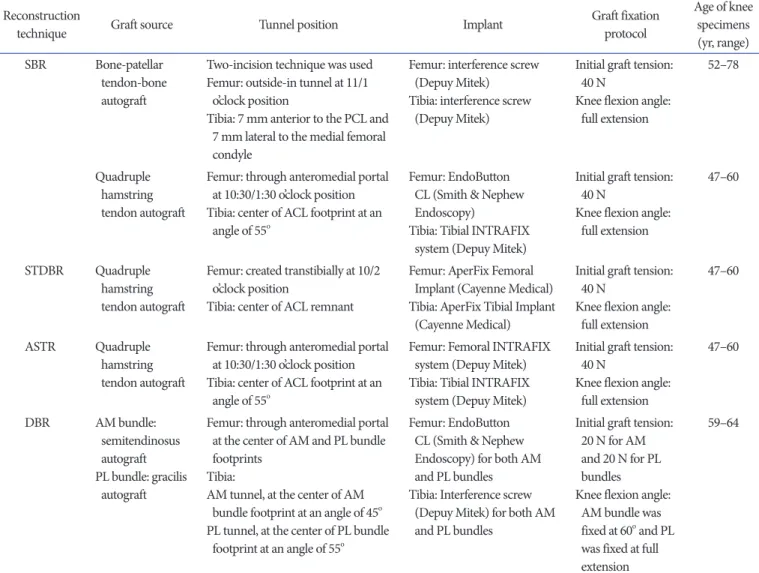
Table 1. A Summary of Four Anterior Cruciate Ligament (ACL) Reconstruction Techniques Reconstruction
technique Graft source Tunnel position Implant Graft fixation
protocol
Age of knee specimens (yr, range) SBR Bone-patellar
tendon-bone autograft
Two-incision technique was used Femur: outside-in tunnel at 11/1
o’clock position
Tibia: 7 mm anterior to the PCL and 7 mm lateral to the medial femoral condyle
Femur: interference screw (Depuy Mitek)
Tibia: interference screw (Depuy Mitek)
Initial graft tension:
40 N
Knee flexion angle:
full extension
52–78
Quadruple hamstring tendon autograft
Femur: through anteromedial portal at 10:30/1:30 o’clock position Tibia: center of ACL footprint at an
angle of 55o
Femur: EndoButton CL (Smith & Nephew Endoscopy)
Tibia: Tibial INTRAFIX system (Depuy Mitek)
Initial graft tension:
40 N
Knee flexion angle:
full extension
47–60
STDBR Quadruple hamstring tendon autograft
Femur: created transtibially at 10/2 o’clock position
Tibia: center of ACL remnant
Femur: AperFix Femoral Implant (Cayenne Medical) Tibia: AperFix Tibial Implant
(Cayenne Medical)
Initial graft tension:
40 N
Knee flexion angle:
full extension
47–60
ASTR Quadruple
hamstring tendon autograft
Femur: through anteromedial portal at 10:30/1:30 o’clock position Tibia: center of ACL footprint at an
angle of 55o
Femur: Femoral INTRAFIX system (Depuy Mitek) Tibia: Tibial INTRAFIX
system (Depuy Mitek)
Initial graft tension:
40 N
Knee flexion angle:
full extension
47–60
DBR AM bundle:
semitendinosus autograft PL bundle: gracilis
autograft
Femur: through anteromedial portal at the center of AM and PL bundle footprints
Tibia:
AM tunnel, at the center of AM bundle footprint at an angle of 45o PL tunnel, at the center of PL bundle
footprint at an angle of 55o
Femur: EndoButton CL (Smith & Nephew Endoscopy) for both AM and PL bundles
Tibia: Interference screw (Depuy Mitek) for both AM and PL bundles
Initial graft tension:
20 N for AM and 20 N for PL bundles
Knee flexion angle:
AM bundle was fixed at 60o and PL was fixed at full extension
59–64
SBR: single-bundle reconstruction, PCL: posterior cruciate ligament, STDBR: single-tunnel double-bundle reconstruction, ASTR: anatomical single-tunnel reconstruction, DBR: double-bundle reconstruction, AM: anteromedial, PL: posterolateral.
can more closely restore the intact knee kinematics than the tra- ditionally practiced single-bundle ACL reconstructions.
Materials and Methods
Kinematic responses of five reconstruction techniques (single- bundle reconstruction using a bone-patellar tendon-bone graft [SBR-BPTB], single-bundle reconstruction using a hamstring tendon graft [SBR-HST], single-tunnel double-bundle recon- struction using a hamstring tendon graft [STDBR-HST], ana- tomical single-tunnel reconstruction using a hamstring tendon graft [ASTR-HST], and double-tunnel double-bundle recon- struction using a hamstring tendon graft [DBR-HST]) were evaluated in eight human cadaveric knee specimens for each of these reconstructions
14,18,24,27). The kinematic responses of these specimens following all of the reconstructions have been previ- ously reported in the literature
14,18,24,27). All of the specimens were stored at –20
oC before they were thawed for 24 hours prior to the testing. Each specimen was prepared in a similar fashion as previously described in our studies to be tested using the robotic testing system. The operation of the robotic testing system to in- vestigate the biomechanics of the knee joint has been detailed in the literature
14,18,24,27).
After installation of the specimen on the robotic testing sys- tem, the passive flexion path of each specimen was determined from 0
oto 90
oof flexion for the specimens that underwent SBR- BPTB and STDBR-HST reconstruction procedures and from 0
oto 120
oof flexion for the specimens in which SBR-HST, ASTR- HST and DBR-HST reconstruction was performed. The passive flexion path is the combination of passive positions of the knee at 1
ointervals from 0
oto 90
oor 120
oof flexion. The passive position was recorded as the position of the tibia with respect to femur at which the forces and moments at the knee joint center were
<5 N and <0.5 N·m respectively. Following determination of the passive path, each specimen with an intact ACL was subjected to two external loading conditions (anterior tibia load of 134 N and simulated quadriceps load of 400 N) at 0
o, 15
o, 30
o, 60
oand 90
oof flexion, and the resulting tibiofemoral kinematics were re- corded. Thereafter, the ACL was transected at the mid-substance to simulate an ACL deficient condition. Responses of the ACL deficient knee were then evaluated under the same protocol that was used to test the intact knee. The ACL of each specimen was then reconstructed by one of the five reconstruction techniques and the kinematics was determined under the two external load- ing conditions and at the five selected flexion angles. A summary of the surgical techniques used are presented in Table 1.
1. Data Analysis
In this study, the kinematic responses of the cadaveric knee specimens before and after a certain reconstruction were evalu- ated in the same specimen, i.e., each reconstructed specimen had its own control group which is the intact ACL condition of the specimen. Since the kinematics of the ACL intact and recon- structed knee were obtained from the same specimen, paired student’s t-tests were used to determine if there were statistically significant differences between the two conditions at all flexion angles. The differences in the kinematics were considered statisti- cally significant when p<0.05.
Results
1. Kinematic Responses to 134 N of Anterior Tibial Load
Single-bundle ACL reconstruction using BPTB graft could not restore the normal anterior joint laxity at low flexion angles (≤30
o) (p<0.05). The residual laxity following SBR-BPTB ranged from 1.7±1.2 mm at full extension to 2.4±1.3 mm at 15
oof flexion (Fig. 1). Further, SBR-BPTB over-constrained the anteroposterior laxity beyond 60
oof flexion with a maximum over-constraint of –2.1±2.6 mm at 90
oof flexion (p>0.05). Significant residual ante-
0 15 30 60 90
6 5 4 3 2 1 0 1 2 3 4 5
Tibialtranslation(mm)
Flexion angle (degree) 6
SBR-BPTB SBR-HST
*
STDBR-HST
PosteriorAnterior
*
*
*
*
* * *
*
*
* ASTR-HST DBR-HST
*
Fig. 1. The difference in anterior-posterior tibial translation between the intact knee and five anterior cruciate ligament (ACL) reconstructions under an anterior tibial load (134 N). Error bars represent standard de- viations. SBR-BPTB: single-bundle reconstruction using a bone-patellar tendon-bone graft, SBR-HST: single-bundle reconstruction using a hamstring tendon graft, STDBR-HST: single-tunnel double-bundle reconstruction using a hamstring tendon graft, ASTR-HST: anatomical single-tunnel reconstruction using a hamstring tendon graft, DBR-HST:
double-tunnel double-bundle reconstruction using a hamstring tendon graft.
*p<0.05; significantly different compared to ACL intact knee.
rior laxities were observed at all selected flexion angles following SBR-HST (p<0.05). The maximum residual laxity of 3.6±1.8 mm occurred at 30
oflexion and the least residual laxity of 2.4±2.4 mm was observed at 90
oof flexion after SBR-HST. Anteroposterior joint laxity was significantly over-constrained by STDBR-HST between 15
oand 90
oof flexion (p<0.05). The amount of over- constraint observed due to STDBR-HST increased with knee flexion ranging from –0.8±1.1 mm at full extension to –2.7±2.0 mm at 90
oof flexion. No significant differences were observed between the anterior laxity of intact knee and ASTR-HST condi- tions at all selected flexion angles (p<0.05). Maximum residual anterior laxity of 1.8±3.1 mm in knee specimens reconstructed by the anatomical single-tunnel technique was observed at 90
oof flexion. DBR-HST closely restored the normal anterior joint lax- ity at all selected flexion angles (p<0.05). The maximum anterior residual laxity after DBR-HST was 1.3±2.3 mm, which occurred at 30
oof flexion.
2. Kinematic Responses to 400 N of Simulated Quadriceps Load
No significant differences were observed between the anterior laxities of intact knee and SBR-BPTB conditions at all selected flexion angles (p<0.05) (Fig. 2). However, SBR-BPTB over-con- strained the joint beyond 30
oof flexion with a maximum over-
constraint of –1.6±2.5 mm at 90
oof flexion (p<0.05). Significant residual laxities were observed following SBR-HST at 0
o, 15
o, and 30
oof flexion (p<0.05). Maximum anterior laxity of 2.5±2.0 mm was observed at 15
oof flexion after SBR-HST. Single-tunnel dou- ble-bundle reconstruction closely restored the normal anteropos- terior joint laxity at low flexion angles (≤30
o) (p<0.05). However, it significantly over-constrained the anteroposterior joint laxity at 60
oand 90
oof flexion (p<0.05). Both ASTR-HST and DBR-HST closely restored the normal anterior joint laxity at all selected flexion angles (p<0.05). The residual anteroposterior joint laxities following either ASTR-HST or DBR-HST were below 1 mm at all selected flexion angles.
The medial-lateral positions of the tibia with respect to the fe- mur of all the five reconstruction techniques were not significantly different compared to their respective intact knee conditions at all selected flexion angles (p<0.05) (Fig. 3). However, the tibiae of the SBR-BPTB and SBR-HST conditions were more medially lo- cated while the tibiae of STDBR-HST, ASTR-HST, and DBR-HST were more laterally located compared to their respective intact knee tibiae. A maximum medial tibial shift of 0.8±1.1 mm was observed at 30
oof flexion after SBR-HST and a maximum lateral tibial shift of –0.7±1.1 mm occurred at 60
oof flexion following STDBR-HST.
0 15 30 60 90
2.0
1.0
0
Tibialtranslation(mm) 1.0
Flexion angle (degree) 2.0
SBR-BPTB SBR-HST STDBR-HST
MedialLateral
ASTR-HST DBR-HST
Fig. 3. The difference in medial-lateral tibial translation between the intact knee and five anterior cruciate ligament (ACL) reconstructions under simulated quadriceps load (400 N). Error bars represent standard deviations. SBR-BPTB: single-bundle reconstruction using a bone- patellar tendon-bone graft, SBR-HST: single-bundle reconstruction us- ing a hamstring tendon graft, STDBR-HST: single-tunnel double-bundle reconstruction using a hamstring tendon graft, ASTR-HST: anatomical single-tunnel reconstruction using a hamstring tendon graft, DBR-HST:
double-tunnel double-bundle reconstruction using a hamstring tendon graft.
*p<0.05; significantly different compared to ACL intact knee.
0 15 30 60 90
5 4 3 2 1 0 1 2 3 4
Tibialtranslation(mm)
Flexion angle (degree) 5
SBR-BPTB SBR-HST STDBR-HST
AnteriorPosterior
*
* *
*
* ASTR-HST DBR-HST
Fig. 2. The difference in anterior-posterior tibial translation between the intact knee and five anterior cruciate ligament (ACL) reconstructions under simulated quadriceps load (400 N). Error bars represent standard deviations. SBR-BPTB: single-bundle reconstruction using a bone- patellar tendon-bone graft, SBR-HST: single-bundle reconstruction us- ing a hamstring tendon graft, STDBR-HST: single-tunnel double-bundle reconstruction using a hamstring tendon graft, ASTR-HST: anatomical single-tunnel reconstruction using a hamstring tendon graft, DBR-HST:
double-tunnel double-bundle reconstruction using a hamstring tendon graft.
*p<0.05; significantly different compared to ACL intact knee.
All the five reconstruction techniques induced an increase in external tibial rotation compared to the intact knee condition (Fig. 4). The tibial internal-external rotations were best restored to the normal condition by SBR-BPTB at all selected flexion angles compared to the other four reconstructions. Among the five reconstruction techniques, DBR-HST induced the largest ex- ternal tibial rotations compared to the intact knee at low flexion angles (≤30
o) (p<0.05). The maximum external tibial rotation (–4.0
o±2.4
o) compared to the intact knee condition was observed at 15
oof flexion following DBR-HST.
Discussion
As the efforts to further optimize the surgical techniques for a ruptured ACL continue in sports medicine, recent literature suggests an inclination of these efforts towards more anatomi- cal approaches. The purpose of this study was to systematically compare the two widely adopted traditional single-bundle re- construction techniques to three relatively new anatomical ap- proaches in restoring normal knee biomechanics. Our hypothesis that anatomical ACL reconstructions can more closely restore the normal knee kinematics than traditional SB ACL reconstructions
was partially supported by the findings of this analysis. More spe- cifically, the reconstructed knees were qualified as normal follow- ing ASTR-HST and DBR-HST and nearly normal following SBR- BPTB, SBR-HST and STDBR-HST as per the International Knee Documentation Committee knee examination form categoriza- tion based on the anterior stability under anterior tibial load. The internal tibial rotations under the simulated muscle load were over-constrained by all the reconstruction techniques, and more so by DBR-HST.
Single-bundle ACL reconstruction with either patellar tendon or hamstring tendon grafts is widely adopted to potentially re- store the normal joint laxity and to return the patients to their pre-injury level of activity. In this analysis, we found that both SBR-BPTB and SBR-HST were capable of restoring the anterior joint laxity to nearly normal. Further, the stability provided by SBR-BPTB was closer to the normal knee than by SBR-HST.
Similar observations have been reported in the literature
28). In general, single-bundle reconstruction has been reported to pro- vide good clinical outcomes
21,29). However, several biomechanical and clinical studies have often associated this technique with ro- tational instability and a prevalence of degenerative changes even after such a surgical intervention is commonly observed as early as within 15 month after surgery
4,5,11-13).
Over the years, several risk factors have been identified for the development of posttraumatic knee osteoarthritis
2,7). It remains obscure precisely what disrupts the homeostasis of healthy carti- lage, subsequently leading to cartilage degeneration. However, it has been hypothesized by some authors that the disease progres- sion may be accelerated due to abnormal loading of cartilage–
which is a manifestation of joint laxity–at locations that are otherwise unloaded or minimally loaded
30). With an objective to better control the rotational joint stability and to potentially miti- gate the incidence of osteoarthritis, several anatomical techniques have been proposed. Clinical evidence on the superiority of these relatively new techniques over the traditional single-bundle is sparse
21).
Among the three anatomical reconstructions evaluated in this study, DBR-HST and ASTR-HST were shown to provide normal joint stability while STDBR-HST restored the joint stability to a nearly normal condition. Similar to our observations, DBR-HST has been previously reported to provide normal joint stability
19,20). Quadriceps muscle action is known to induce anterior tibial translation and internal tibial rotation at low flexion angles. Yet, few studies have investigated the efficacy of ACL reconstructions under physiological loading conditions. The analysis of this study demonstrated that SBR-BPTB, STDBR-HST, ASTR-HST, and
Fig. 4. The difference in internal-external tibial rotations between theintact knee and five anterior cruciate ligament (ACL) reconstructions under simulated quadriceps load (400 N). Error bars represent standard deviations. SBR-BPTB: single-bundle reconstruction using a bone- patellar tendon-bone graft, SBR-HST: single-bundle reconstruction us- ing a hamstring tendon graft, STDBR-HST: single-tunnel double-bundle reconstruction using a hamstring tendon graft, ASTR-HST: anatomical single-tunnel reconstruction using a hamstring tendon graft, DBR-HST:
double-tunnel double-bundle reconstruction using a hamstring tendon graft.
*p<0.05; significantly different compared to ACL intact knee.
0 15 30 60 90
2 1 0 1 2 3 4 5 6 7 8 9 10 11 12
Tibialrotation(degree)
Flexion angle (degree)
SBR-BPTB SBR-HST STDBR-HST
InternalExternal
ASTR-HST DBR-HST
*
*
*
* *
*