115
자가 슬건과 슬개건을 이용한 관절경적 전방십자인대 재건술의 단기 추시 결과 비교
서재성․이동철․손욱진․장우혁․김세동
영남대학교 의과대학 정형외과학교실
목 적: 자가 슬건과 슬개건을 이용한 관절경적 전방십자인대 재건술의 술후 결과를 임상적, 방사선적으로 분석하여 비교하고자 하였다.
대상 및 방법: 1997년 9월부터 2003년 1월까지 전방십자인대 재건술을 시행받은 환자들 중 추시기간이 18개월 이상인 자가 슬건 군 22예와 자가 슬개건 군 30예를 대상으로 하였다. 임상적 평가는 IKDC 평가법, Lysholm 점수, Tegner 점수를 이용하였으며 방사선적 평가는 Telos기기를 이용하여 90도 굴곡위에서 전방 전위도를 정상측과 비교하였고 무릎을 꿇은 상태에서 나타나는 전방 슬부 동통의 빈도, 대퇴 사두근의 위축 정도 등을 비교하였다.
결 과: IKDC 평가법에 의하면 슬건 군은 A등급 2예, B등급 14예, C등급 6예였고 슬개건 군은 A등급 3예, B등급 20예, C등급 7예로 두 군간에 차이가 없었다. Lysholm 점수는 슬건 군이 술 전 평균 58.2점에서 최종 추시상 평균 88점으로, 슬개건 군은 평균 56.3점에서 평균 90.6점으로 호전되었고, Tegner 점수는 슬건 군이 술 전 평균 2.4점에서 최종 추시상 평균 5.8점으로, 슬개건 군은 평균 2.3점에서 평균 6.2점으로 활동도가 증가되었으나 두 군 간에 차이가 없었다. Telos기 기를 이용한 최종 추시 전방 전위도는 슬건 군이 정상측과 평균 2.6 mm의 차이를 보였고 슬개건 군이 평균 2.3 mm의 차이를 보여 두 군간에 차이가 없었다. 무릎을 꿇은 상태에서 나타나는 전방 슬부 동통은 슬건 군이 2예(13%), 슬개건 군이 11예(37%)로 통계적인 차이를 보였다(p<0.05).
결 론: 슬건을 이용한 관절경적 전방십자인대 재건술은 무릎을 꿇은 상태에서 나타나는 전방 슬부 동통의 빈도를 낮출 수 있으며 슬개건과 비슷한 정도의 만족할 만한 인대 안정성 및 임상적 결과를 얻을 수 있었다.
색인 단어: 전방 십자 인대, 재건술, 자가 슬건, 자가 슬개건
Comparison of Arthroscopic Anterior Cruciate Ligament Reconstruction between Autologous Hamstring Tendon and Bone-patellar
Tendon-bone Graft: Short Term Follow Up
Jae Sung Seo, M.D., Dong Chul Lee, M.D., Oog Jin Shon, M.D., Woo Hyuk Jang, M.D., and Se Dong Kim, M.D.
Department of Orthopaedic Surgery, Yeung Nam University Hospital, Daegu, Korea
P u rp o s e : To evaluate the stability, activity level and clinical results of an autologous hamstring and Bone-patellar tendon-bone (BPTB) graft in an Anterior cruciate ligam ent (ACL) reconstruction.
M a te ria ls a n d M e th o d s : Between September 1997 and January 2003, 22 patients with an ACL reconstruction with an autologous four strand ham string tendon, and 30 patients w ith a BPTB autograft were enrolled in this study. At the tim e of the final follow-up, the patients were evaluated by stress radiographs using a Telos instrum ent, the Lysholm score, Tegner activity score, IKDC activity grades, and anterior kneeling pain.
R e s u lts : An evaluation of the anterior laxity using the Telos instrument revealed no significant difference in the ham string and BPTB groups: side to side difference 2.6 m m in the ham string group
통신저자:김 세 동
대구광역시 남구 대명동 371-1 영남대학교병원 정형외과
TEL: 053-620-3642․FAX: 053-628-4020 E-mail: [email protected].
*본 논문의 요지는 2004년도 대한관절경학회 추계학술대회에서 발표되었음.
Address reprint requests to Se Dong Kim, M.D.
Department of Orthopaedic Surgery, Yeung Nam University Hospital, 317-1 Daemyeong-dong, Nam-gu, Daegu 705-717, Korea Tel: +82.53-620-3642, Fax: +82.53-628-4020
E-mail: [email protected].
전방십자인대는 슬관절의 인대들 중 가장 흔히 손상되
며6-8) 이로 인한 슬관절의 불안정성으로 반월상 연골 손
상, 관절 연골 손상 등을 유발시키게 되므로2,5) 불안정성 을 호전시키고 동반 손상을 줄이기 위한 방법으로 전방십 자인대 재건술이 많이 시행되고 있다.
전방십자인대 재건술을 위한 이식건을 선택함에 있어 서 논쟁의 여지는 다분히 있으나 대부분 술자들은 자가 슬개건 혹은 네겹의 자가 슬건을 가장 흔히 선택하게 된 다.
과거에 자가 슬개건은 전방십자인대 재건술의 대표적 인 이식건이었으나 요즘 자가 슬건의 사용이 점차 증가하 고 있다. 이러한 이식건 선택의 변화가 일어나는 이유로 는 슬관절 신전 기전의 손상, 전방 슬부 동통, 슬개골 골 절, 슬개건 파열, 슬개건하 구축 등과 같은 공여부 합병 증12)이 자가 슬개건에서 발생하고 최근 수술 기구의 발달 로 슬건의 고정 방법이 개선되었기 때문이다. 이에 저자 들은 자가 슬건과 슬개건을 이용한 관절경적 전방십자인 대 재건술의 술후 결과를 임상적, 방사선적으로 분석하여 비교하고자 하였다.
대상 및 방법
1997년 9월부터 2003년 1월까지 영남대학교병원 정 형외과에서 전방십자인대 재건술을 시행 받은 환자들 중 추시 기간이 18개월 이상인 자가 슬건 군 22예와 자가 슬개건 군 30예를 대상으로 하였으며 과거에 반대측 슬 관절에 손상이 있었던 경우, 골절이 동반된 경우, 골관절 염이 있는 경우, 동반 인대 손상이 있는 경우는 본 연구의 대상에서 제외시켰다. 환자의 연령은 슬건 군이 평균 28.7세(21-42), 슬개건 군이 평균 25.4세(18-39)였으
며 성별 분포는 슬건 군이 남자 19예, 여자 3예였고 슬개 건 군이 남자 29예, 여자 1예로 비슷하였다. 연령과 성별 분포는 두 군 간에 통계적인 차이가 없었다(chi square analysis, p>0.05). 평균 추시 기간은 슬건 군이 30개월 (19-37), 슬개건 군이 65개월(54-82)이었다.
1. 수상 원인
수상 원인으로는 스포츠 손상(77%), 실족 혹은 추락 사 고(17%), 교통 사고(6%) 순이었으며 스포츠 손상 중 축 구 손상이 대부분을 차지하였고 그 외 농구 손상, 격투기 손상 등이 있었다.
2. 동반 손상
반월상 연골 손상은 슬건 군이 양측 반월상 연골 손상 5예, 내측 반월상 연골 손상 4예, 외측 반월상 연골 손상 5예로 64%의 발생률이 있었고 슬개건 군이 양측 반월상 연골 손상 10예, 내측 반월상 연골 손상 8예, 외측 반월상 연골 손상 4예로 73%에서 발생하였으며 관절 연골 손상 은 슬건 군이 41% (9예), 슬개건 군이 33% (10예)에서 발생하였다. 반월상 연골에 대한 수술은 슬건 군은 부분 절제술 11예(57%), 반월상 연골 봉합술 8예였으며 슬개 건 군은 부분 절제술 17예(53%), 반월상 연골 봉합술 15 예였다. 관절 연골 손상은 슬건 군이 Outerbridge grade IV 1예, grade II 8예로 총 9예(41%)였으며 슬개건 군은 10예(33%) 모두 grade II였다.
3. 수술 방법 및 재활 방법
슬건 군의 경우 전 예에서 대퇴골 부위는 endopearl과 생체 흡수성 간섭나사로 고정하였고 경골 부위는 생체 흡 of 58 to a postoperative score of 88 in the ham string groups and from 56 to 91 in the BPTB groups.
The Tegner score for the ham string groups was 2.4 preoperatively and 5.8 at the final follow up, and the Tegner score for the BPTB groups w ere 2.3 and 6.2 preoperatively and after the last follow-up, respectively. The Tegner score was sim ilar in the two treatm ent groups (p>0.05). Accord- ing to the IKDC rating system , 9% were norm al and 64% were alm ost norm al in the ham string group.
In the BPTB group, 10% were norm al and 67% w ere alm ost norm al. Anterior kneeling pain after a reconstruction w ith the ham string tendon autograft (13% ) w as significantly less com m on than with the patellar tendon autograft (37% )(p<0.05).
C o n c lu s io n : The hamstring autograft for an ACL reconstruction decreased the incidence of anterior kneeling pain and produced equivalent clinical results to the BPTB autograft.
Key Words: Anterior cruciate ligament, Reconstruction, Hamstring autograft, BPTB autograft
수성 나사로 고정하고 post tie 및 washer screw를 이용 하여 보강하였다(Fig. 1). 슬개건 군의 경우는 25예에서 금속 간섭나사를, 4예에서 생체 흡수성 간섭나사를 사용 하였으며 1예에서는 금속 간섭나사를 대퇴부에, 생체 흡 수성 간섭나사를 경골부에 고정하였다.
술 후 재활 방법은 슬건군과 슬개건군 모두 같은 방법 을 사용하였으며 CPM은 술 후 2일째 시작하고 보조기를 착용한 상태로 부분 체중 부하와 능동적 관절운동은 술 후 7일째부터 시작하였으며 closed kinetic chain exer- cises를 술 후 4주째 시작하였다. 술 후 6개월 전까지는 저항성 슬관절 신전 운동은 허용하지 않았으며 술 후 8-10개월에 스포츠 활동을 할 수 있도록 하였다.
4. 평가 방법
전방 안정성을 평가하기 위해서는 임상적으로는 La- chman test, pivot shift test로 확인하였고 방사선학적 으로는 Telos기기(Telos stress device SE 2000, Telos GmbH, Marburg, Germany)를 이용한 전방 전위 스트 레스 방사선 사진(90도 굴곡위, 15 kg의 후전방 스트레 스를 가함)을 촬영하여 건측과 비교하였다.
슬관절의 임상적 기능과 활동도를 평가하기 위해서 Lysholm 점수, IKDC 활동도 등급, Tegner 활동도 점수 를 이용하였다.
대퇴사두근 위축 정도를 보기 위해서 슬개골 상극부 15 cm 근위부에서 대퇴 둘레를 측정하여 건측과 비교하 였으며 대퇴사두근력 측정을 위해 one-leg hop test를 실시하여 그 거리를 측정하였다.
임상적 합병증으로 무릎을 꿇은 상태에서 나타나는 전 방 슬부 동통의 빈도를 조사하였다.
5. 통계학적 분석
통계적 검증은 Independent t-test와 Chi-Square test를 이용하였고 통계적 유의성은 p<0.05로 하였다.
결 과
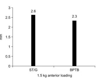
Telos 스트레스 검사상 슬건 군의 15 kg 부하 전방 전 위 정도는 건측과 환측의 비교에서 평균 2.6±1.3 mm의 차이를 보였으며 슬개건 군은 평균 2.3±0.9 mm의 차이 를 보여 두 군간에 통계적인 차이는 없었다(p>0.05) (Fig. 2). Lachman 검사상 두 군 모두 술 전에는 2+
이상의 불안정성을 보였으나 최종 추시시 슬건 군이 20 예(91%), 슬개건 군이 27예(90%)에서 1+ 이하로 호전 되었다(Table 1). Pivot shift 검사상 최종 추시상 1+
Fig. 1. Diagram of the anterior cruciate ligament reconstruction with the hamstring tendon.
Endopearl
Bioscrew
Cancellous screw and Washer
Fig. 2. Telos stress test (side to side difference).
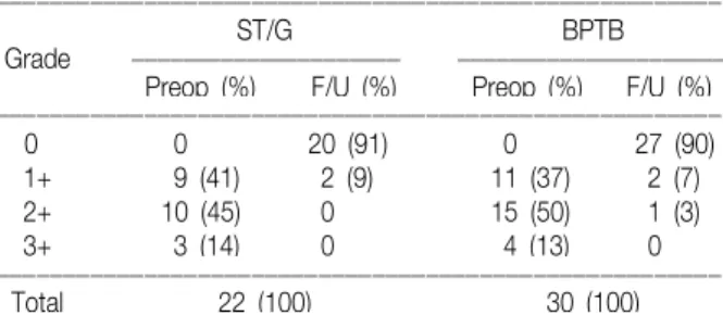
Table 1. Grading of the Lachman test (before surgery vs last follow- up)
ꠏꠏꠏꠏꠏꠏꠏꠏꠏꠏꠏꠏꠏꠏꠏꠏꠏꠏꠏꠏꠏꠏꠏꠏꠏꠏꠏꠏꠏꠏꠏꠏꠏꠏꠏꠏꠏꠏꠏꠏꠏꠏꠏꠏꠏꠏꠏꠏꠏꠏꠏꠏꠏꠏ
ST/G BPTB
Grade ꠏꠏꠏꠏꠏꠏꠏꠏꠏꠏꠏꠏꠏꠏꠏꠏꠏꠏꠏꠏ ꠏꠏꠏꠏꠏꠏꠏꠏꠏꠏꠏꠏꠏꠏꠏꠏꠏꠏꠏ Preop (%) F/U (%) Preop (%) F/U (%) ꠏꠏꠏꠏꠏꠏꠏꠏꠏꠏꠏꠏꠏꠏꠏꠏꠏꠏꠏꠏꠏꠏꠏꠏꠏꠏꠏꠏꠏꠏꠏꠏꠏꠏꠏꠏꠏꠏꠏꠏꠏꠏꠏꠏꠏꠏꠏꠏꠏꠏꠏꠏꠏꠏ
0 0 15 (68) 0 19 (63)
1+ 0 5 (23) 0 8 (27)
2+ 11 (50) 2 (9) 22 (73) 3 (10)
3+ 11 (50) 0 8 (27) 0
ꠏꠏꠏꠏꠏꠏꠏꠏꠏꠏꠏꠏꠏꠏꠏꠏꠏꠏꠏꠏꠏꠏꠏꠏꠏꠏꠏꠏꠏꠏꠏꠏꠏꠏꠏꠏꠏꠏꠏꠏꠏꠏꠏꠏꠏꠏꠏꠏꠏꠏꠏꠏꠏꠏ
Total 22 (100) 30 (100)
ꠏꠏꠏꠏꠏꠏꠏꠏꠏꠏꠏꠏꠏꠏꠏꠏꠏꠏꠏꠏꠏꠏꠏꠏꠏꠏꠏꠏꠏꠏꠏꠏꠏꠏꠏꠏꠏꠏꠏꠏꠏꠏꠏꠏꠏꠏꠏꠏꠏꠏꠏꠏꠏꠏ ST/G, semitendinosus tendon/gracilis; BPTB, bone patellar tendon bone.
이상의 불안정성을 보인 경우는 슬건 군이 2예(9%), 슬개 건 군이 3예(10%)였다(Table 2).
Lysholm 점수는 슬건 군에서 술 전 평균 58점에서 최 종 추시 평균 88점으로, 슬개건 군에서 술 전 평균 56점 에서 최종 추시 평균 91점으로 호전되었으며 87%에서 양호이상의 결과를 보였고 보통은 양 군에서 약 13%를 차지하였다(Table 3). 두 군 간에 통계적인 차이는 없었 다(p>0.05).
Tegner 활동도 점수는 슬건 군에서 술 전 가벼운 산행 을 할 수 있는 평균 2.4점에서 최종 추시시 취미로 배드 민턴을 할 수 있는 평균 5.8점으로, 슬개건 군에서 술 전 수영을 할 수 있는 평균 2.3점에서 최종 추시시 테니스와 야구를 할 수 있는 평균 6.2점으로 호전되었으며, 두 군 간에 통계적인 차이는 없었다(p>0.05).
IKDC 활동도 등급에 의하면 술 전에는 두 군 모두 비 정상(C) 이하였으나 최종 추시시 슬건 군은 정상(A) 2예 (9%), 거의 정상(B) 14예(64%), 비정상(C) 6예(27%)이 었으며 슬개건 군은 정상(A) 3예(10%), 거의 정상(B) 20 예(67%), 비정상(C) 7예(23%)로 두 군 간에 통계적인 차
이는 없었다(p>0.05).
대퇴 사두근 근력 측정 및 슬관절 기능 상태를 보기 위 해 실시한 최종 추시 one-leg hop 검사(거리)상 슬건 군 은 건측 다리에 비해 평균 79%, 슬개건 군은 평균 81%의 한발 멀리 뛰기를 할 수 있었다(Table 4). 대퇴 사두근 위축 정도는 건측에 비해 슬건 군이 1.7 cm, 슬개건 군은 1.6 cm 위축된 양상을 보였다.
무릎을 꿇은 상태에서 나타나는 전방 슬부 동통의 빈도 는 슬건 군이 2예(8%), 슬개건 군이 11예(37%)로 슬개건 군에서 통계적으로 유의성있게 높았다(p<0.05).
최종 추시시 슬관절의 운동 제한은 슬개건 군의 1예에 서만 나타났으며 건측에 비해 10도 내외의 신전 장애가 있었고 그 외 다른 합병증은 없었다.
고 찰
10 mm폭의 골-슬개건-골 이식건의 최고 인장 강도 는 2,376 N으로 전방십자인대에 비해 약 1.1배 정도로 강하며 강성은 812 N/mm로 전방십자인대에 비해 3.2 배, 단면적은 0.8배이고 반건양건과 박건으로 된 4겹의 슬건은 최고 인장 강도가 4,108 N으로 전방십자인대에 비해 약 2배이며 강성은 776 N/mm로 약 3배, 단면적은 약 1.2배로 전방십자인대에 비해 최고 강도 및 강성, 단 면적에서 우수한 이식건이다11).
Aglietti 등은 KT-2000 관절계로 슬관절 안정성을 측 정하였는데 20 lb 전방 부하를 주었을 때 전방 경골 전위 의 건측-환측 차이가 5 mm 이상 나는 빈도가 슬건 군이 13%, 슬개건 군이 10%, 30 lb일 때는 슬건 군이 20%, 슬개건 군이 13%, 수동 최대 검사시는 슬건 군이 23%, 슬개건 군이 20%로 양 군 간에 통계적인 유의성이 없었 다고 하였고1) Marder 등도 KT-1000 관절계(20 lb)로 측정한 결과 건측-환측 차이가 슬건 군이 1.9+1.3 mm, ꠏꠏꠏꠏꠏꠏꠏꠏꠏꠏꠏꠏꠏꠏꠏꠏꠏꠏꠏꠏꠏꠏꠏꠏꠏꠏꠏꠏꠏꠏꠏꠏꠏꠏꠏꠏꠏꠏꠏꠏꠏꠏꠏꠏꠏꠏꠏꠏꠏꠏꠏꠏꠏꠏ
ST/G BPTB
Grade ꠏꠏꠏꠏꠏꠏꠏꠏꠏꠏꠏꠏꠏꠏꠏꠏꠏꠏꠏꠏꠏ ꠏꠏꠏꠏꠏꠏꠏꠏꠏꠏꠏꠏꠏꠏꠏꠏꠏꠏꠏꠏꠏ Preop (%) F/U (%) Preop (%) F/U (%) ꠏꠏꠏꠏꠏꠏꠏꠏꠏꠏꠏꠏꠏꠏꠏꠏꠏꠏꠏꠏꠏꠏꠏꠏꠏꠏꠏꠏꠏꠏꠏꠏꠏꠏꠏꠏꠏꠏꠏꠏꠏꠏꠏꠏꠏꠏꠏꠏꠏꠏꠏꠏꠏꠏ
0 0 20 (91) 0 27 (90)
1+ 9 (41) 2 (9) 11 (37) 2 (7)
2+ 10 (45) 0 15 (50) 1 (3)
3+ 3 (14) 0 4 (13) 0
ꠏꠏꠏꠏꠏꠏꠏꠏꠏꠏꠏꠏꠏꠏꠏꠏꠏꠏꠏꠏꠏꠏꠏꠏꠏꠏꠏꠏꠏꠏꠏꠏꠏꠏꠏꠏꠏꠏꠏꠏꠏꠏꠏꠏꠏꠏꠏꠏꠏꠏꠏꠏꠏꠏ
Total 22 (100) 30 (100)
ꠏꠏꠏꠏꠏꠏꠏꠏꠏꠏꠏꠏꠏꠏꠏꠏꠏꠏꠏꠏꠏꠏꠏꠏꠏꠏꠏꠏꠏꠏꠏꠏꠏꠏꠏꠏꠏꠏꠏꠏꠏꠏꠏꠏꠏꠏꠏꠏꠏꠏꠏꠏꠏꠏ ST/G, semitendinosus tendon/gracilis; BPTB, bone patellar tendon bone.
Table 3. Changes in the Lysholm score between the pre- operative state and the last follow-up
ꠏꠏꠏꠏꠏꠏꠏꠏꠏꠏꠏꠏꠏꠏꠏꠏꠏꠏꠏꠏꠏꠏꠏꠏꠏꠏꠏꠏꠏꠏꠏꠏꠏꠏꠏꠏꠏꠏꠏꠏꠏꠏꠏꠏꠏꠏꠏꠏꠏꠏꠏꠏꠏꠏ
ST/G BPTB
Grade ꠏꠏꠏꠏꠏꠏꠏꠏꠏꠏꠏꠏꠏꠏꠏꠏꠏꠏ ꠏꠏꠏꠏꠏꠏꠏꠏꠏꠏꠏꠏꠏꠏꠏꠏꠏꠏꠏ Preop (%) F/U (%) Preop (%) F/U (%) ꠏꠏꠏꠏꠏꠏꠏꠏꠏꠏꠏꠏꠏꠏꠏꠏꠏꠏꠏꠏꠏꠏꠏꠏꠏꠏꠏꠏꠏꠏꠏꠏꠏꠏꠏꠏꠏꠏꠏꠏꠏꠏꠏꠏꠏꠏꠏꠏꠏꠏꠏꠏꠏꠏ
Excellent 0 5 (23) 0 8 (27)
Good 0 14 (64) 0 18 (60)
Fair 1 (4) 3 (13) 0 4 (13)
Poor 21 (96) 0 30 (100) 0
ꠏꠏꠏꠏꠏꠏꠏꠏꠏꠏꠏꠏꠏꠏꠏꠏꠏꠏꠏꠏꠏꠏꠏꠏꠏꠏꠏꠏꠏꠏꠏꠏꠏꠏꠏꠏꠏꠏꠏꠏꠏꠏꠏꠏꠏꠏꠏꠏꠏꠏꠏꠏꠏꠏ
Mean score 58.2 88.0 56.3 90.6
ꠏꠏꠏꠏꠏꠏꠏꠏꠏꠏꠏꠏꠏꠏꠏꠏꠏꠏꠏꠏꠏꠏꠏꠏꠏꠏꠏꠏꠏꠏꠏꠏꠏꠏꠏꠏꠏꠏꠏꠏꠏꠏꠏꠏꠏꠏꠏꠏꠏꠏꠏꠏꠏꠏ ST/G, semitendinosus tendon/gracilis; BPTB, bone patellar tendon bone.
ꠏꠏꠏꠏꠏꠏꠏꠏꠏꠏꠏꠏꠏꠏꠏꠏꠏꠏꠏꠏꠏꠏꠏꠏꠏꠏꠏꠏꠏꠏꠏꠏꠏꠏꠏꠏꠏꠏꠏꠏꠏꠏꠏꠏꠏꠏꠏꠏꠏꠏꠏꠏꠏꠏ
Grade ST/G (%) BPTB (%)
ꠏꠏꠏꠏꠏꠏꠏꠏꠏꠏꠏꠏꠏꠏꠏꠏꠏꠏꠏꠏꠏꠏꠏꠏꠏꠏꠏꠏꠏꠏꠏꠏꠏꠏꠏꠏꠏꠏꠏꠏꠏꠏꠏꠏꠏꠏꠏꠏꠏꠏꠏꠏꠏꠏ
A (100-90%) 6 (27) 9 (30)
B (90-70%) 14 (64) 18 (60)
C (75-50%) 2 (9) 3 (10)
D (<50%) 0 (0) 0 (0)
ꠏꠏꠏꠏꠏꠏꠏꠏꠏꠏꠏꠏꠏꠏꠏꠏꠏꠏꠏꠏꠏꠏꠏꠏꠏꠏꠏꠏꠏꠏꠏꠏꠏꠏꠏꠏꠏꠏꠏꠏꠏꠏꠏꠏꠏꠏꠏꠏꠏꠏꠏꠏꠏꠏ
Mean 79% 81%
ꠏꠏꠏꠏꠏꠏꠏꠏꠏꠏꠏꠏꠏꠏꠏꠏꠏꠏꠏꠏꠏꠏꠏꠏꠏꠏꠏꠏꠏꠏꠏꠏꠏꠏꠏꠏꠏꠏꠏꠏꠏꠏꠏꠏꠏꠏꠏꠏꠏꠏꠏꠏꠏꠏ ST/G, semitendinosus tendon/gracilis; BPTB, bone patellar tendon bone.
슬개건 군이 1.6 ±1.4 mm로 비슷한 결과를 나타내었다 고 하였다10).
Toritsuka 등은 이식건의 단면적은 슬개건이 41 mm2, 세겹 또는 네겹 슬건이 47.6 mm2 이고 터널의 적합성은 슬개건은 59.2%, 슬건의 적합성이 79.6%여서 슬건이 더 넓은 단면적과 더 나은 이식건-터널 적합성을 가져서 슬 개건이 가지는 골-골 치유의 장점(bone to bone heal- ing)없이도 좋은 결과를 가질 수 있다고 주장하였다16). 저자들의 연구에서도 15 kg 부하 Telos 스트레스 검사 상 슬건 군은 최종 추시상 평균 2.6 mm의 건측・환측 차이를 보였으며 슬개건 군은 평균 2.3 mm의 차이를 보 여 두 군 간에 통계적인 차이는 없었고(Fig. 3) Lachman 검사와 Pivot shift 검사에서도 두 군 모두 우수한 결과를 나타내었다. 하지만 건측-환측 차이가 4 mm 이상 나는 빈도는 슬건 군이 4예(18%), 슬개건 군이 1예(3%)로, 슬 개건 군이 더 고른 안정성을 나타내었다.
그러나 본 연구에서 Lachman 검사, Lysholm 평가, Pivot shift 검사에 비해 IKDC 활동도 평가의 비정상(C) 비율이 슬건 군은 27%, 슬개건 군은 23%로 전반적으로 높은 편인데 이는 IKDC 평가 항목 중 하나라도 낮은 등 급을 받게 된 군이 전 항목의 점수를 결정하며 IKDC 평 가 항목에 환자의 주관적 평가가 반영된 점도 그 원인이 라 생각할 수 있었다.
슬개건을 채취함으로써 생길 수 있는 단점은 공여부에 슬개골 골절과 슬개건 파열, 슬개 대퇴 관절면에 동통(전 방 슬부 동통), 슬개건염이 발생될 수 있으며 복재신경 (saphenous nerve)의 하슬개분지가 손상되어 이상감각 및 감각 둔마가 발생되며 무릎 꿇기가 불편할 수 있다14). 본 연구에서도 무릎을 꿇을 때 나타나는 전방 슬부 동통 은 슬개건 군이 11예(37%)로 슬건 군 2예(8%)보다 많았 으며(p<0.05) 슬개건 군의 5명(16.6%)에서 이학적 검사 시 슬개골 하부의 압통을 관찰할 수 있었다. 전방 십자
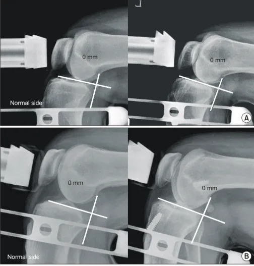
Fig. 3. Telos stress view of the ham- string tendon group and Bone- patellar tendon-bone (BPTB) group:
At the last follow up, the Telos stress view of the injured side showed same stability compared with the normal side in both groups. (A) 29 year old man who sustained an anterior cruciate liga- ment (ACL) and lateral meniscus injury as a result of soccer had an ACL reconstruction with a ham- string tendon. (B) 27 year old man who sustained an ACL and medial meniscus injuries as a result of traffic accident had an ACL recon- struction with a BPTB autograft.
Normal side
0 mm 0 mm
A
B
Normal side
0 mm 0 mm
를 할 경우 나타나는 필연적인 합병증이 아니며 술 후 슬관절의 운동 범위와 깊은 관련이 있다고 주장하였으
나9,13,15) 저자들의 연구에서 슬관절의 10도 이내의 신전
장애가 있었던 환자는 슬개건 군 1예였으며 이 환자 경우 무릎을 꿇을 때 나타나는 전방 슬부 동통이 없었던 점을 고려해보면 저자들의 결과가 위의 주장과 일치하지는 않 았다.
Clatworthy 등3)은 슬개건보다 슬건을 이용할 경우 터 널 확장이 문제가 될 수 있지만 터널 확장은 임상 소견, 슬관절 점수, KT-1000이나 등속성 근력(isokinetic mus- cle strength)과 연관성이 없다고 주장하였다.
Corry 등은 전방 십자 인대 재건술 후 1년째는 슬개건 군이 슬건 군보다 대퇴사두근 위축이 더 심하게 나타나나 술 후 2년째는 대퇴 둘레가 건측과 10 mm 이하 차이가 나는 빈도가 슬건 군이 75%, 슬개건 군이 81%로 통계적 으로 차이가 없다고 하였다4). 본 연구에서도 최종 추시시 대퇴둘레가 10-20 mm 차이가 나는 빈도가 슬건 군이 73%, 슬개건 군이 87%로 두 군 간에 차이가 없었으나 Corry 등의 연구에 비해서 대퇴 사두근 위축이 심한 것을 알 수 있었다. 이는 술 후 적극적인 근육 강화 및 재활 치료의 부족으로 생긴 것으로 생각되었다.
단, 본 연구는 전향적 무작위 연구가 아니라 후향적 연 구여서 시기에 따라 이식건 선택이 다르기 때문에 두 군 간의 평균 추시 기간이 2배 이상 차이를 보여 두 군 간의 비교에 약간의 오차는 예상한 연구였다.
결 론
자가 슬건을 이용한 관절경적 전방십자인대 재건술은 자가 슬개건에 비해 무릎을 꿇을 때 나타나는 전방 슬부 동통의 빈도를 낮출 수 있으며 슬개건과 함께 만족할 만 한 임상적 결과를 얻을 수 있었다. 그러나 슬건 및 슬개건 은 각각의 장점과 단점을 가지므로 나이, 활동도, 직업 등 여러 요인을 고려해서 최적의 이식건을 선택하는 것이 바람직하리라 생각한다.
참고문헌
1. Aglietti P, Buzzi R, Zaccherotti G and De Biase P: Patellar tendon versus doubled semitendinosus and gracilis tendons for anterior cruciate ligament reconstruction. Am J Sports Med, 22:
2. Andersson C, Odensten M and Gillquist J: Knee function after surgical or nonsurgical treatment of acute rupture of the anterior cruciate ligament: a randomized study with a long-term follow-up period. Clin Orthop Relat Res, 264: 255-263, 1991.
3. Clatworthy MG, Annear P, Bulow JU, Bartlett RJ: Tunnel widening in anterior cruciate ligament reconstruction: a pro- spective evaluation of hamstring and patella tendon grafts. Knee Surg Sports Traumatol Arthrosc, 7: 138-145, 1999.
4. Corry IS, Webb JM, Clingeleffer AJ and Pinczewski LA:
Arthroscopic reconstruction of the anterior cruciate ligament. A comparison of patellar tendon autograft and four-strand hamstr- ing tendon autograft. Am J Sports Med, 27: 444-454, 1999.
5. Dye SF, Wojtys EM, Fu FH, Fithian DC and Gillquist I:
Factors contributing to function of the knee joint after injury of reconstruction of the anterior cruciate ligament. Instr Course Lect, 48: 185-198, 1999.
6. Eriksson E: Reconstruction of the anterior cruciate ligament.
Orthop Clin North Am, 7: 167-179, 1976.
7. Feagin JA Jr and Curl WW: Isolated tear of the anterior cruciate ligament: 5-year follow-up study. Am J Sports Med, 4:
95-100, 1976.
8. Johnson RJ, Beynnon BD, Nichols CE and Renstrom PA:
The treatment of injuries of the anterior cruciate ligament. J Bone Joint Surg, 74-A: 140-151, 1992.
9. Kartus J, Magnusson L, Stener S, Brandsson S, Eriksson BI and Karlsson J: Complications following arthroscopic anterior cruciate ligament reconstruction. A 2-5 year follow-up of 604 patients with special emphasis on anterior knee pain. Knee Surg Sports Traumatol Arthrosc, 7: 2-8, 1999.
10. Marder RA, Raskind JR and Carroll M: Prospective evaluation of arthroscopically assisted anterior cruciate ligament reconstruction. Patella tendon versus semitendinosus and gracilis tendons. Am J Sports Med, 19: 478-484, 1991.
11. Miller SL and Gladstone JN: Graft selection in anterior cruciate ligament reconstruction. Orthop Clin North Am, 33:
675-683, 2002.
12. Rosenberg TD, Franklin JL, Baldwin GN and Nelson KA:
Extensor mechanism function after patellar tendon graft harvest for anterior cruciate ligament reconstruction. Am J Sports Med,
20: 519-525, 1992.
13. Sachs RA, Daniel DM, Stone ML and Garfein RF: Patello- femoral problems after anterior cruciate ligament reconstruction.
Am J Sports Med, 17: 760-765, 1989.
14. Safran MR: Graft selection in knee surgery. Current concepts Am J Knee Surg, 8: 168-180, 1995.
15. Shelbourne KD and Trumper RV: Preventing anterior knee
pain after anterior cruciate ligament reconstruction. Am J Sports Med, 25: 41-47, 1997.
16. Toritsuka Y, Horibe S, Mitsuoka T, Nakamura N, Hamada M and Shino K: Comparison between the cross-sectional area of bone-patellar tendon-bone grafts and multistranded hamstring tendon grafts obtained from the same patients. Knee Surg Sports Traumatol Arthrosc, 11: 81-84, 2003.