surgical techniques, and advances in fixation devices, the single bundle ACL reconstruction has provided good clinical outcomes.
However, some studies reported that 10%–30% of the patients with single bundle ACL reconstruction continued to show rota- tional instability and development of osteoarthritis3). It has been assumed that these kinds of problems arise from lack of the pos- terolateral (PL) bundle in the single bundle ACL reconstructed knee. Therefore, double bundle or anatomical single bundle ACL reconstruction, which more closely restores the normal anatomy of the ACL, was proposed for the treatment of ACL injury. How- ever, problems including the development of osteoarthritis after ACL reconstruction still persist despite significant improvement in ACL reconstruction. In this paper, we will review the current trend of ACL reconstruction with regard to surgical techniques, fixation devices, and graft materials.
Operative Techniques for ACL Reconstruction
1. Anatomical Double Bundle ACL Reconstruction
Single bundle ACL reconstruction has been considered the standard technique for restoring anterior instability, especially in flexion, by addressing the anteromeidal (AM) bundle only.
However, 10%–30% of the ACL reconstructed patients complain of a feeling of rotational instability, so-called pivot-shift phenom- enon4). Moreover, several biomechanical studies showed that single bundle reconstruction can restore anterior-posterior stabil-
Current Trends in Anterior Cruciate Ligament Reconstruction
Ha Sung Kim, MD, Jong Keun Seon, MD, and Ah Reum Jo, MD
Department of Orthopaedic Surgery, Center for Joint Disease, Chonnam National University Hwasun Hospital, Hwasun, Korea
The advances in the knowledge of anatomy, surgical techniques, and fixation devices have led to the improvement of anterior cruciate ligament (ACL) reconstruction over the past 10 years. Nowadays, double bundle and anatomical single bundle ACL reconstruction that more closely restores the normal anatomy of the ACL are becoming popular. Although there is still no definite conclusion whether double bundle ACL reconstruction provides better clinical results than single bundle reconstruction, the trend has shifted to anatomic reconstruction regardless of single bundle or double bundle techniques. We could not find any significant differences in the clinical outcomes and stability after ACL reconstruction according to the type of graft or fixation device. Therefore, surgeons should select an ideal ACL reconstruction according to the patient’s condition and surgeon’s experience.
Keywords: Anterior cruciate ligament, Anatomical reconstruction, graft, fixation pISSN 2234-0726 · eISSN 2234-2451
Knee Surgery & Related Research
Received July 15, 2013; Revised October 2, 2013; Accepted October 6, 2013Correspondence to: Jong Keun Seon, MD
Department of Orthopaedic Surgery, Center for Joint Disease, Chonnam National University Hwasun Hospital, 322 Seoyang-ro, Hwasun 519- 763, Korea
Tel: +82-61-379-7676, Fax: +82-61-379-7894 E-mail: [email protected]
Introduction
Rupture of the anterior cruciate ligament (ACL) is one of the most common knee injuries. The annual incidence of the ACL injury ranges between 100,000–200,000 in USA1,2). Due to the unsatisfactory outcomes of conservative treatment for ACL in- juries, reconstruction surgery remains the treatment of choice in most young patients who want to maintain an active lifestyle.
The main aims of ACL reconstruction are to restore intact knee stability and normal knee kinematics after reconstruction. Tra- ditionally, ACL reconstruction has focused on non-anatomical single bundle reconstruction using a transtibial technique, which provides only anterior stability in knee flexion.
Owing to the better understanding of anatomy, improvement in
165
This is an Open Access article distributed under the terms of the Creative Commons Attribution Non-Commercial License (http://creativecommons.org/licenses/by-nc/3.0/) which permits unrestricted non-commercial use, distribution, and reproduction in any medium, provided the original work is properly cited.
Copyright © 2013 KOREAN KNEE SOCIETY www.jksrr.org
ity but not rotational stability, which means it does not restore normal rotational kinematics5).
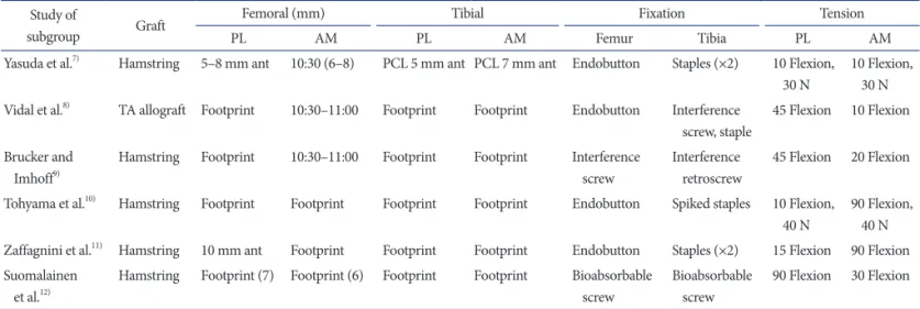
With better knowledge of ACL anatomy, double bundle ACL reconstruction was proposed to closely restore the normal struc- ture of the ACL3,6). Although double bundle reconstruction is more effective than single bundle reconstruction for restoring normal knee kinematics, the operative techniques are various in terms of the fixation angle and device (Table 1)7-12). Moreover, double bundle reconstruction techniques are technically more demanding and necessitate longer operative times and more ex- tensive bone loss, thereby potentially rendering revision surgery more difficult. Although a recent meta-analysis study showed that double bundle reconstruction provides better results in terms of anterior stability and pivot-shift test than single bundle reconstruction13), some studies failed to demonstrate significant comparative advantages of double bundle techniques in terms of clinical outcomes13,14). Moreover, some studies reported 3%–16%
of tears of the PL reconstruction grafts on second-look arthros- copy after double bundle ACL reconstruction15).
According to the literature review, although double bundle re- construction has theoretical advantages over single bundle recon- struction in terms of restoring normal anatomy and kinematics, we could not recommend double bundle reconstruction for all patients with ACL injuries. However, double bundle techniques can be useful for specific cases of substantial rotational instability in hyper-lax knee joints and revision surgery16).
2. Anatomical Single Bundle ACL Reconstruction
With regard to single bundle ACL reconstruction, Woo et al.17) reported that the standard high femoral tunnel in ACL reconstruction resists anterior tibial loading, but it is not suf-
ficient to control combined rotatory loads. Furthermore, several biomechanical studies have shown that the more anatomic low femoral tunnel has some advantages over the high femoral tun- nel in terms of rotational stability3,18). Therefore, anatomical graft placement has been emphasized for restoration of normal knee kinematics in ACL reconstruction. Although anatomical double bundle reconstruction can closely restore the normal ACL anat- omy, it does not provide consistently good results because of the abovementioned disadvantages. In addition, it has been known that 6% of the reconstructions lead to a rupture in the contralat- eral intact knee.
Attention has returned to single bundle reconstruction with grafts placed at the center of anatomical position. Recently, sev- eral biomechanical studies showed that the single bundle ACL grafts placed in the center of their anatomic insertions can pro- vide nearly normal knee kinematics comparable to double bundle reconstruction14,19,20). Sastre et al.19) reported that single bundle ACL reconstruction in anatomical insertion site produced results comparable to those obtained using the double bundle technique, as determined by KT-1000 measurements, International Knee Documentation Committee scores, and pivot shift test results. In a study by Steiner et al.14), a central anatomical single bundle ACL reconstruction was superior to the conventional non-anatomical single bundle ACL reconstruction in restoring normal anterior and rotational knee laxity.
Araki et al.20) reported that anatomical double bundle ACL re- construction showed superior results in stability measured with an electromagnetic system than anatomical single bundle recon- struction but there was no difference in clinical outcome (KT- 1000 measurements, isokinetic peak muscle torque, and Lysholm score). Controversy exists regarding the fact that anatomical fem-
Table 1. Current Outcome Studies of Double Bundle Anterior Cruciate Ligament Reconstruction Study of
subgroup Graft Femoral (mm) Tibial Fixation Tension
PL AM PL AM Femur Tibia PL AM
Yasuda et al.7) Hamstring 5–8 mm ant 10:30 (6–8) PCL 5 mm ant PCL 7 mm ant Endobutton Staples (×2) 10 Flexion, 30 N
10 Flexion, 30 N Vidal et al.8) TA allograft Footprint 10:30–11:00 Footprint Footprint Endobutton Interference
screw, staple
45 Flexion 10 Flexion Brucker and
Imhoff9)
Hamstring Footprint 10:30–11:00 Footprint Footprint Interference screw
Interference retroscrew
45 Flexion 20 Flexion Tohyama et al.10) Hamstring Footprint Footprint Footprint Footprint Endobutton Spiked staples 10 Flexion,
40 N
90 Flexion, 40 N Zaffagnini et al.11) Hamstring 10 mm ant Footprint Footprint Footprint Endobutton Staples (×2) 15 Flexion 90 Flexion Suomalainen
et al.12)
Hamstring Footprint (7) Footprint (6) Footprint Footprint Bioabsorbable screw
Bioabsorbable screw
90 Flexion 30 Flexion PL: posterolateral, AM: anteromeidal, ant: anterior, PCL: posterior cruciate ligament, N: newton (kg·m/s2), TA: tibialis anterior tendon.
oral tunnel placement can be achieved using a transtibial tunnel drilling technique. Giron et al.21) showed that the standard trans- tibial technique in ACL reconstruction could not restore the ana- tomic femoral origin of the ACL despite some technical modifi- cations. To address problems related to a vertical femoral tunnel, some surgeons have advocated performing independent drilling (transportal technique) through an anteromedial portal to place the femoral tunnel in the anatomical position instead of using the standard transtibial drilling technique3,19,22). In addition, Kim et al.22) reported excellent clinical results of anatomical ACL recon- struction using 3 portals by adding a far anteromedial portal to the frequently used 2 portals. While some studies have reported that anteromedial portal drilling could place the femoral tunnel in the anatomical position better than transtibial drilling, other studies reported that modified transtibial drilling technique can place a graft at anatomical position by adjusting flexion or rota- tional angle or using a flexible reamer during femoral drilling3,18). In addition, although the transtibial and transportal techniques have some advantages, outside-in technique has been recently reported as a reliable alternative. Lubowitz and Konicek23) re- ported that the outside-in technique could be performed through a small incision and prevent excessively short femoral tunneling unlike the transportal technique. Seo et al.24) suggested that there was no significant difference between the transtibial and outside- in techniques in the clinical outcome, and the outside-in tech- nique provides superior knee joint rotational stability compare to the transtibial technique.
The standard location of a tibial tunnel was slightly posterior,
which was more close to the PL bundle than the AM bundle to prevent graft impingement at the intercondylar notch. However, in the anatomical single bundle reconstruction, many surgeons make a tibial tunnel at the center of the AM and PL bundles (Fig.
1).
The paradigm of anterior cruciate ligament reconstruction has shifted from isometric reconstruction to anatomic reconstruction using a single bundle or double bundle technique. Due to the questionable advantages of double bundle ACL reconstruction in clinical studies, anatomical single bundle ACL reconstruction in the mid bundle position has received more attention recently.
3. Remnant Preserving ACL Reconstruction
Because of the potential problem including impingement or poor visualization during reconstruction, ACL remnants are totally debrided in traditional ACL reconstruction. However it is well known that tibial remnants contain several types of mecha- noreceptors. These mechanoreceptors may provide positive ef- fects on the proprioceptive function of the knee25,26). It has been suggested that the ACL secondarily functions as a sensory organ providing proprioceptive feedback and initiating protective re- flexes and stabilizing muscular reflexes. In addition, some studies have shown that the ACL remnants provide some biomechanical stability to the knee25,26). Moreover, posterior cruciate ligament (PCL) reconstruction with a remnant preserving technique showed better stability than PCL reconstruction without rem- nant tissue. Hence, some surgeons proposed remnant preserving
Fig. 1. Schematic drawing of the central and standard tunnel positions in anatomical single-bundle anterior cruciate ligament reconstructions.
AM: anteromedial bundle, PL: posterolateral bundle.
Table 2. Differences in the Clinical Outcomes of Remnant Preserving Anterior Cruciate Ligament (ACL) Reconstruction
No. Author Yr No. ofpatients F/U
(mo) Outcomes Study results 1 Ahn
et al.26)2010 68 6.3 MRI Larger ACL grafts No cyclops lesion 2 Ahn
et al.15)2011 63 27.7 MRI Clinical
score
Good clinical results Cyclops lesions↑ (no
clinical significance) 3 Gao
et al.29)2010 235 50 MRI
KT-1000 Very good clinical results 4 Gohil
et al.30)2007 49 12 MRI
KT-1000 No difference 5 Kim
et al.22)2009 27 12 Clinical Remnant preservation could be effective methods 6 Lee
et al.25)2008 42 35.1 Clinical
KT-1000 Good proprioceptive and functional outcomes F/U: follow-up, MRI: magnetic resonance imaging.
Graft Materials for Anterior Cruciate Ligament Re
construction
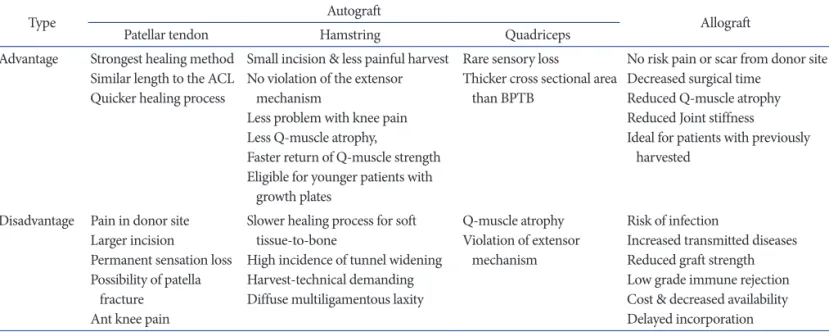
Graft choice for ACL reconstruction is influenced by patient age, activity level, gender, associated injuries, degree of laxity, and planned concomitant operations. As a general guideline, autografts are recommended for young patients because it is pre- sumed that these patients are more active. Allografts are not as strong as autografts and are only indicated in patients undergo- ing revision ACL surgery or in those who only want to return to lower demand activities. Allografts in ACL reconstruction have advantages including decreased operative time, smaller incisions, and less post-operative pain. However, autografts are still pre- ferred because allografts carry the possibility of disease transmis- sion (Table 3).
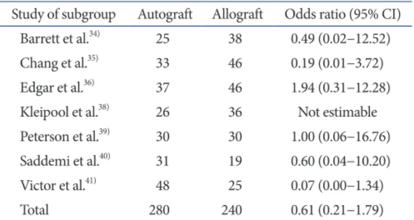
In a meta-analysis study and systematic review on the compari- son of clinical outcomes of ACL reconstruction using autografts and allograft, there was no difference in the outcome scores, lax- ity, clinical failure rates, and return to sports33) (Tables 4, 5)34-41).
The two most commonly used autografts in ACL reconstruc- tion are the patellar tendon (PT) autograft and the four-strand hamstring (HS) tendon autograft consisting of the gracilis and semitendinosus tendons. The other sources of autografts include the quadriceps tendons. Although there has been little research on quadriceps tendon grafts, several recent studies support their use in ACL reconstruction42). To date, there have been a number of prospective and retrospective studies comparing patellar ten- don bone-tendon-bone grafts (BPTB) and four-strand hamstring grafts (DSTG; double semitendinosus and gracilis tendon grafts).
ACL reconstruction. Although the preservation of a remnant ACL stump might lead to incorrect tibial tunnel placement or cyclops formation26), it has many theoretical advantages including accelerated revascularization and ligamentization, preservation of the proprioceptive nerve fibers, enhanced biological environ- ment for healing, and reduced incidence of tibial bone tunnel enlargement5,25-28). Ahn et al.27) reported that remnant preserv- ing ACL reconstruction provided good clinical outcomes and stabilities without compromising accuracy of tunnel position.
However, cyclops lesions were found in 12 out of 48 patients on MRI even though they did not have any limitation in extension (Table 2)15,22,25,26,29,30). Mifune et al.31) reported that remnant pre- serving selective bundle augmentation showed good anterior and rotational stability in patients with only AM or PL tear, and they recommended remnant preserving augmentation for partial ACL tears. In terms of tunnel widening, Zhang et al.32) showed less tibial tunnel enlargement after remnant preserving ACL recon- struction than remnant removing ACL reconstruction, but no difference was seen in clinical outcomes.
Although some studies showed that remnant preserving ACL reconstruction provided promising results in terms of clinical outcomes and tunnel widening, we could not find any literature that this technique improves proprioception or stability and al- lows for rapid rehabilitation compared to traditional remnant re- moving ACL reconstruction. Moreover, there are no prospective randomized studies comparing the remnant preserving and tra- ditional ACL reconstruction. In conclusion, the present literature does not conclusively support the benefits of remnant preserving ACL reconstruction.
Table 3. Summary of Advantages and Disadvantages of Graft Materials
Type Autograft
Allograft
Patellar tendon Hamstring Quadriceps
Advantage Strongest healing method Similar length to the ACL Quicker healing process
Small incision & less painful harvest No violation of the extensor
mechanism
Less problem with knee pain Less Q-muscle atrophy,
Faster return of Q-muscle strength Eligible for younger patients with
growth plates
Rare sensory loss
Thicker cross sectional area than BPTB
No risk pain or scar from donor site Decreased surgical time
Reduced Q-muscle atrophy Reduced Joint stiffness
Ideal for patients with previously harvested
Disadvantage Pain in donor site Larger incision
Permanent sensation loss Possibility of patella
fracture Ant knee pain
Slower healing process for soft tissue-to-bone
High incidence of tunnel widening Harvest-technical demanding Diffuse multiligamentous laxity
Q-muscle atrophy Violation of extensor
mechanism
Risk of infection
Increased transmitted diseases Reduced graft strength Low grade immune rejection Cost & decreased availability Delayed incorporation ACL: anterior cruciate ligament, BPTB: bone-tendon-bone grafts.
Some studies found similar laxity values and functional results between the two types of graft tissues43-45), while others2,46,47) found the patellar tendon graft produced significantly better re- sults in terms of stability, but this did not correlate with the func- tional outcome. The advantage of the hamstring tendon autograft is relatively low overall postoperative pain, especially anterior knee pain. Thus, it would be the preferred choice of graft in ACL reconstruction for patients with a low pain tolerance, a job that requires kneeling, or a history of knee pain. In addition, it would be a better option for patients concerned about aesthetics because it requires a relatively small incision. The disadvantage is that it takes relatively long time for the graft to heal into the tunnel since there is no “bone-to-bone” healing.
Despite the disadvantage, the hamstring autograft has recently been widely used as the primary graft of choice in ACL recon- struction because of the relatively low postoperative knee pain, low comorbidity due to preservation of the extensor mechanism by not violating the patellar tendon and quadriceps tendon, and little clinical and functional difference compared to the BPTB.
Fixation Devices for ACL Reconstruction
The fixation device for graft in ACL reconstruction should be secure and allow graft healing within the tunnel. Because more aggressive rehabilitation program has been adopted in ACL re- construction recently, the strength of fixation device should be enough to allow immediate range of motion exercises, weight bearing, and early return to sports without any loss of fixation strength. Over the past 10 years, significant advances in fixation have led to the development of many different fixation devices for bony and soft tissue graft fixation.
1. Bone Plug Fixation Device
Metal or bio- interference screws are most commonly used fixation device for bone plug in ACL reconstruction. Metal inter- ference screws designed by Kurosaka have been used as the stan- dard fixation device in ACL reconstruction with patellar tendon autograft48). However, with the increasing use of hamstring soft tissue grafts, bioabsorbable interference screws, poly-L-lactic acid (PLLA) screws, and polyglyconate screws49) are becoming more popular. While the bioabsorbable screw has the advantages, such as incorporation into the surrounding tissue, almost no need for implant removal, and less interference with MRI50), it seems to provide clinical results similar to those of metal screws according to a systematic review.
Cross biodegradable or metal pins (Rigid Fix; DePuy Mitek Inc., Raynham, MA, USA) can be used for the fixation of the bone plug. The principal failure mode with cross pins in this uti- lization is bone block fracture, and the cross pin fixation strength improves with larger bone plug diameters. Cross pins showed similar fixation strength as interference fixation screws with bone plugs51). The RetroScrew (Arthrex, Naples, FL, USA), recently designed to be inserted from the articular side of the tibia, may actually increase the graft tension as the screw is advanced19). In a recent biomechanical study, however, the fixation strength and fixation failure load of RetroScrew was not found to be as good as interference screws52).
2. Soft Tissue Fixation Device
Interference screws are also commonly used for the fixation of soft tissue graft. The adequate length of interference screw is required for improved fixation strength (30−35 mm screws).
Cross biodegradable pins (Rigid Fix) and RetroScrews (Arthrex) can also be used for the fixation of soft tissue graft. When used Table 4. Instrumented Laxity Measurement of >5 mm Pooled according
to Graft Source
Study of subgroup Autograft Allograft Odds ratio (95% CI) Barrett et al.34) 25 38 0.20 (0.01−4.02)
Chang et al.35) 22 34 1.03 (0.16−6.74)
Edgar et al.36) 37 46 3.97 (0.40−39.86)
Harner et al.37) 26 64 1.25 (0.21−7.28)
Kleipool et al.38) 26 36 1.42 (0.19−10.77) Peterson et al.39) 30 30 5.35 (0.25−116.31) Saddemi et al.40) 25 18 0.23 (0.01−5.95)
Total 191 266 1.23 (0.52−2.92)
Odd ratio was calculated using the Mantel-Haenszel method and random-effects analysis model.
CI: confidence interval.
Table 5. Clinical Failures Pooled according to Graft Source
Study of subgroup Autograft Allograft Odds ratio (95% CI) Barrett et al.34) 25 38 0.49 (0.02−12.52)
Chang et al.35) 33 46 0.19 (0.01−3.72)
Edgar et al.36) 37 46 1.94 (0.31−12.28)
Kleipool et al.38) 26 36 Not estimable Peterson et al.39) 30 30 1.00 (0.06−16.76) Saddemi et al.40) 31 19 0.60 (0.04−10.20) Victor et al.41) 48 25 0.07 (0.00−1.34)
Total 280 240 0.61 (0.21−1.79)
Odd ratio was calculated using the Mantel-Haenszel method and random-effects analysis model.
CI: confidence interval.
Conflict of Interest
No potential conflict of interest relevant to this article was re- ported.
References
1. Buoncristiani AM, Tjoumakaris FP, Starman JS, Ferretti M, Fu FH. Anatomic double-bundle anterior cruciate ligament reconstruction. Arthroscopy. 2006;22:1000-6.
2. Freedman KB, D’Amato MJ, Nedeff DD, Kaz A, Bach BR Jr.
Arthroscopic anterior cruciate ligament reconstruction: a metaanalysis comparing patellar tendon and hamstring ten- don autografts. Am J Sports Med. 2003;31:2-11.
3. Abebe ES, Moorman CT 3rd, Dziedzic TS, Spritzer CE, Co- thran RL, Taylor DC, Garrett WE Jr, DeFrate LE. Femoral tunnel placement during anterior cruciate ligament recon- struction: an in vivo imaging analysis comparing transtibial and 2-incision tibial tunnel-independent techniques. Am J Sports Med. 2009;37:1904-11.
4. Prodromos CC, Fu FH, Howell SM, Johnson DH, Lawhorn K. Controversies in soft-tissue anterior cruciate ligament re- construction: grafts, bundles, tunnels, fixation, and harvest. J Am Acad Orthop Surg. 2008;16:376-84.
5. Fu FH, Jordan SS. The lateral intercondylar ridge: a key to anatomic anterior cruciate ligament reconstruction. J Bone Joint Surg Am. 2007;89:2103-4.
6. Abebe ES, Kim JP, Utturkar GM, Taylor DC, Spritzer CE, Moorman CT 3rd, Garrett WE, DeFrate LE. The effect of femoral tunnel placement on ACL graft orientation and length during in vivo knee flexion. J Biomech. 2011;44:1914- 20.
7. Yasuda K, Kondo E, Ichiyama H, Kitamura N, Tanabe Y, Tohyama H, Minami A. Anatomic reconstruction of the anteromedial and posterolateral bundles of the anterior cru- ciate ligament using hamstring tendon grafts. Arthroscopy.
2004;20:1015-25.
8. Vaquero J, Vidal C, Cubillo A. Intra-articular traumatic dis- orders of the knee in children and adolescents. Clin Orthop Relat Res. 2005;(432):97-106.
9. Brucker PU, Imhoff AB. Functional assessment after acute and chronic complete ruptures of the proximal hamstring tendons. Knee Surg Sports Traumatol Arthrosc. 2005;13:411- 8.
10. Tohyama H, Kondo E, Hayashi R, Kitamura N, Yasuda K.
Gender-based differences in outcome after anatomic double- in ACL reconstruction using a hamstring tendon, they produce
clinical results that can be comparable to those of reconstruction using interference screws and the EndoButton22,51,53).
The EndoButton CL (Smith & Nephew Endoscopy, Andover, MA, USA), an extra cortical suspensory fixation device, has been widely used as a fixation device for the hamstring graft on the femoral side. Although EndoButton has a higher failure load and less stiffness than interference screws, it induces some micro motion of the graft within the bone tunnel during loading, and can be a cause of tunnel widening49). Baumfeld et al.54) reported that 2 cross pin fixation resulted in less femoral tunnel widening than the EndoButton fixation. On the other hand, Kong et al.55) suggested that the clinical results were comparable between the cross pin fixation and EndoButton fixation and there was no sig- nificant difference in femoral tunnel widening between the two fixation devices. An advantage, however, of the same is that extra cortical fixation creates a long bone-tendon interface, making it suitable for many types of ACL reconstruction techniques as well as single- and double-bundle reconstruction. Suture tying around the screw post is also another established technique. This fixation system has adequate strength for graft fixation and also has ad- vantages of tendon healing into the bony tunnel.
In summary, most of modern fixation devices have enough strength to fix the graft in ACL reconstruction regardless of graft materials. All systems have their specific advantages and disad- vantages. Therefore, the choice of a fixation device should be based on the type of graft or quality of bone.
Conclusions
Although there are still no definite conclusions whether double bundle ACL reconstruction can provide better clinical results than single bundle technique, the main trend for ACL recon- struction has shifted to anatomic reconstruction regardless of single bundle or double bundle techniques during the past 10 years. In the literature, the type of graft or fixation device did not make significant differences in clinical outcomes or stabil- ity of ACL reconstruction. Because there are a variety of options available today, selection of optimum combination should be individualized to the patient’s condition and the experience of the surgeon. Further advances in surgical techniques should continue to be developed so as to restore near normal knee kinematics and anatomy.
bundle anterior cruciate ligament reconstruction with ham- string tendon autografts. Am J Sports Med. 2011;39:1849-57.
11. Zaffagnini S, Bruni D, Marcheggiani Muccioli GM, Bonanz- inga T, Lopomo N, Bignozzi S, Marcacci M. Single-bundle patellar tendon versus non-anatomical double-bundle ham- strings ACL reconstruction: a prospective randomized study at 8-year minimum follow-up. Knee Surg Sports Traumatol Arthrosc. 2011;19:390-7.
12. Suomalainen P, Jarvela T, Paakkala A, Kannus P, Jarvinen M.
Double-bundle versus single-bundle anterior cruciate liga- ment econstruction: a prospective randomized study with 5-year results. Am J Sports Med. 2012;40:1511-8.
13. Xu M, Gao S, Zeng C, Han R, Sun J, Li H, Xiong Y, Lei G.
Outcomes of anterior cruciate ligament reconstruction us- ing single-bundle versus double-bundle technique: meta- analysis of 19 randomized controlled trials. Arthroscopy.
2013;29:357-65.
14. Steiner ME, Battaglia TC, Heming JF, Rand JD, Festa A, Baria M. Independent drilling outperforms conventional transtibial drilling in anterior cruciate ligament reconstruc- tion. Am J Sports Med. 2009;37:1912-9.
15. Ahn JH, Choi SH, Wang JH, Yoo JC, Yim HS, Chang MJ.
Outcomes and second-look arthroscopic evaluation after double-bundle anterior cruciate ligament reconstruction with use of a single tibial tunnel. J Bone Joint Surg Am.
2011;93:1865-72.
16. Seon JK, Park SJ, Lee KB, Yoon TR, Seo HY, Song EK. Sta- bility comparison of anterior cruciate ligament between double- and single-bundle reconstructions. Int Orthop.
2009;33:425-9.
17. Woo SL, Kanamori A, Zeminski J, Yagi M, Papageorgiou C, Fu FH. The effctiveness of reconstruction of the anterior cruciate ligament with hamstrings and patellar tendon: a ca- daveric study comparing anterior tibial and rotational loads.
J Bone Joint Surg Am. 2002;84:907-14.
18. Rue JP, Lewis PB, Parameswaran AD, Bach BR Jr. Single- bundle anterior cruciate ligament reconstruction: technique overview and comprehensive review of results. J Bone Joint Surg Am. 2008;90 Suppl 4:67-74.
19. Sastre S, Popescu D, Nunez M, Pomes J, Tomas X, Peidro L. Double-bundle versus single-bundle ACL reconstruc- tion using the horizontal femoral position: a prospective, randomized study. Knee Surg Sports Traumatol Arthrosc.
2010;18:32-6.
20. Araki D, Kuroda R, Kubo S, Fujita N, Tei K, Nishimoto K, Hoshino Y, Matsushita T, Matsumoto T, Nagamune K, Ku-
rosaka M. A prospective randomised study of anatomical single-bundle versus double-bundle anterior cruciate liga- ment reconstruction: quantitative evaluation using an elec- tromagnetic measurement system. Int Orthop. 2011;35:439- 46.
21. Giron F, Cuomo P, Edwards A, Bull AM, Amis AA, Aglietti P. Double-bundle “anatomic” anterior cruciate ligament re- construction: a cadaveric study of tunnel positioning with a transtibial technique. Arthroscopy. 2007;23:7-13.
22. Kim MK, Lee BC, Park JH. Anatomic single bundle anterior cruciate ligament reconstruction by the two anteromedial portal method: the comparison of transportal and transtibial techniques. Knee Surg Relat Res. 2011;23:213-9.
23. Lubowitz JH, Konicek J. Anterior cruciate ligament femo- ral tunnel length: cadaveric analysis comparing antero- medial portal versus outside-in technique. Arthroscopy.
2010;26:1357-62.
24. Seo SS, Kim CW, Kim JG, Jin SY. Clinical results compar- ing transtibial technique and outside in technique in single bundle anterior cruciate ligament reconstruction. Knee Surg Relat Res. 2013;25:133-40.
25. Lee BI, Kwon SW, Kim JB, Choi HS, Min KD. Comparison of clinical results according to amount of preserved remnant in arthroscopic anterior cruciate ligament reconstruction us- ing quadrupled hamstring graft. Arthroscopy. 2008;24:560- 8.
26. Ahn JH, Lee YS, Yoo JC, Chang MJ, Koh KH, Kim MH.
Clinical and second-look arthroscopic evaluation of repaired medial meniscus in anterior cruciate ligament-reconstructed knees. Am J Sports Med. 2010;38:472-7.
27. Ahn JH, Lee YS, Ha HC. Anterior cruciate ligament recon- struction with preservation of remnant bundle using ham- string autograft: technical note. Arch Orthop Trauma Surg.
2009;129:1011-5.
28. Kim SJ, Jo SB, Kim TW, Chang JH, Choi HS, Oh KS. A modified arthroscopic anterior cruciate ligament double- bundle reconstruction technique with autogenous quad- riceps tendon graft: remnant-preserving technique. Arch Orthop Trauma Surg. 2009;129:403-7.
29. Gao K, Chen S, Wang L, Zhang W, Kang Y, Dong Q, Zhou H, Li L. Anterior cruciate ligament reconstruction with LARS artificial ligament: a multicenter study with 3- to 5-year follow-up. Arthroscopy. 2010;26:515-53.
30. Gohil S, Annear PO, Breidahl W. Anterior cruciate ligament reconstruction using autologous double hamstrings: a com- parison of standard versus minimal debridement techniques
using MRI to assess revascularisation: a randomised pro- spective study with a one-year follow-up. J Bone Joint Surg Br. 2007;89:1165-71.
31. Mifune Y, Ota S, Takayama K, Hoshino Y, Matsumoto T, Kuroda R, Kurosaka M, Fu FH, Huard J. Therapeutic advan- tage in selective ligament augmentation for partial tears of the anterior cruciate ligament: results in an animal model.
Am J Sports Med. 2013;41:365-73.
32. Zhang Q, Zhang S, Cao X, Liu L, Liu Y, Li R. The effect of remnant preservation on tibial tunnel enlargement in ACL reconstruction with hamstring autograft: a prospective randomized controlled trial. Knee Surg Sports Traumatol Arthrosc. 2012 Dec 15. [Epub]. http://dx.doi.org/10.1007/
s00167-012-2341-7.
33. Carey JL, Dunn WR, Dahm DL, Zeger SL, Spindler KP. A systematic review of anterior cruciate ligament reconstruc- tion with autograft compared with allograft. J Bone Joint Surg Am. 2009;91:2242-50.
34. Barrett G, Stokes D, White M. Anterior cruciate ligament re- construction in patients older than 40 years: allograft versus autograft patellar tendon. Am J Sports Med. 2005;33:1505- 12.
35. Chang SK, Egami DK, Shaieb MD, Kan DM, Richardson AB. Anterior cruciate ligament reconstruction: allograft ver- sus autograft. Arthroscopy. 2003;19:453-62.
36. Edgar CM, Zimmer S, Kakar S, Jones H, Schepsis AA. Pro- spective comparison of auto and allograft hamstring tendon constructs for ACL reconstruction. Clin Orthop Relat Res.
2008;466:2238-46.
37. Harner CD, Olson E, Irrgang JJ, Silverstein S, Fu FH, Silbey M. Allograft versus autograft anterior cruciate ligament re- construction: 3- to 5-year outcome. Clin Orthop Relat Res.
1996;(324):134-44.
38. Kleipool AE, Zijl JA, Willems WJ. Arthroscopic anterior cruciate ligament reconstruction with bone-patellar tendon- bone allograft or autograft: a prospective study with an average follow up of 4 years. Knee Surg Sports Traumatol Arthrosc. 1998;6:224-30.
39. Peterson RK, Shelton WR, Bomboy AL. Allograft versus autograft patellar tendon anterior cruciate ligament recon- struction: a 5-year follow-up. Arthroscopy. 2001;17:9-13.
40. Saddemi SR, Frogameni AD, Fenton PJ, Hartman J, Hart- man W. Comparison of perioperative morbidity of anterior cruciate ligament autografts versus allografts. Arthroscopy.
1993;9:519-24.
41. Victor J, Bellemans J, Witvrouw E, Govaers K, Fabry G.
Graft selection in anterior cruciate ligament reconstruction:
prospective analysis of patellar tendon autografts compared with allografts. Int Orthop. 1997;21:93-7.
42. DeAngelis JP, Fulkerson JP. Quadriceps tendon: a reliable al- ternative for reconstruction of the anterior cruciate ligament.
Clin Sports Med. 2007;26:587-96.
43. Markolf KL, Hame SL, Hunter DM, Oakes D, Gause P. Bio- mechanical effects of femoral notchplasty in anterior cruci- ate ligament reconstruction. Am J Sports Med. 2002;30:83-9.
44. Shaieb MD, Kan DM, Chang SK, Marumoto JM, Richard- son AB. A prospective randomized comparison of patellar tendon versus semitendinosus and gracilis tendon autografts for anterior cruciate ligament reconstruction. Am J Sports Med. 2002;30:214-20.
45. Struewer J, Ziring E, Oberkircher L, Schuttler KF, Efe T. Iso- lated anterior cruciate ligament reconstruction in patients aged fifty years: comparison of hamstring graft versus bone- patellar tendon-bone graft. Int Orthop. 2013;37:809-17.
46. Biau DJ, Tournoux C, Katsahian S, Schranz PJ, Nizard RS.
Bone-patellar tendon-bone autografts versus hamstring autografts for reconstruction of anterior cruciate ligament:
meta-analysis. BMJ. 2006;332:995-1001.
47. Goldblatt JP, Fitzsimmons SE, Balk E, Richmond JC. Recon- struction of the anterior cruciate ligament: meta-analysis of patellar tendon versus hamstring tendon autograft. Arthros- copy. 2005;21:791-803.
48. Matsumoto A, Yoshiya S, Muratsu H, Matsui N, Yagi M, Kuroda R, Kurosaka M. Mechanical evaluation of a soft tis- sue interference screw with a small diameter: significance of graft/bone tunnel cross-sectional area ratio. Knee Surg Sports Traumatol Arthrosc. 2006;14:330-4.
49. Stener S, Ejerhed L, Sernert N, Laxdal G, Rostgard-Chris- tensen L, Kartus J. A long-term, prospective, randomized study comparing biodegradable and metal interference screws in anterior cruciate ligament reconstruction surgery:
radiographic results and clinical outcome. Am J Sports Med.
2010;38:1598-605.
50. Cheung P, Chan WL, Yen CH, Cheng SC, Woo SB, Wong TK, Wong WC. Femoral tunnel widening after quadrupled hamstring anterior cruciate ligament reconstruction. J Or- thop Surg (Hong Kong). 2010;18:198-202.
51. Harilainen A, Sandelin J, Jansson KA. Cross-pin femoral fixation versus metal interference screw fixation in anterior cruciate ligament reconstruction with hamstring tendons:
results of a controlled prospective randomized study with 2-year follow-up. Arthroscopy. 2005;21:25-33.
52. Chang HC, Nyland J, Nawab A, Burden R, Caborn DN.
Biomechanical comparison of the bioabsorbable RetroScrew system, BioScrew XtraLok with stress equalization ten- sioner, and 35-mm Delta Screws for tibialis anterior graft- tibial tunnel fixation in porcine tibiae. Am J Sports Med.
2005;33:1057-64.
53. Harilainen A, Sandelin J. A prospective comparison of 3 hamstring ACL fixation devices: Rigidfix, BioScrew, and Intrafix: randomized into 4 groups with 2 years of follow-up.
Am J Sports Med. 2009;37:699-706.
54. Baumfeld JA, Diduch DR, Rubino LJ, Hart JA, Miller MD, Barr MS, Hart JM. Tunnel widening following anterior cruciate ligament reconstruction using hamstring auto- graft: a comparison between double cross-pin and suspen- sory graft fixation. Knee Surg Sports Traumatol Arthrosc.
2008;16:1108-13.
55. Kong CG, In Y, Kim GH, Ahn CY. Cross pins versus en- dobutton femoral fixation in hamstring anterior cruciate ligament reconstruction: minimum 4-year follow-Up. Knee Surg Relat Res. 2012;24:34-9.