graft ruptures, residual instability, and limited sports performance after surgery remain unresolved issues. To resolve these, preser- vation of the remnant tissue of ACL has been attracting attention of late. ACL remnants may potentially enhance revascularization and cell proliferation and promote the recovery of high-quality proprioception and stability1-5), and to achieve these, a remnant- preserving ACL reconstruction technique was recently developed with good outcomes6-8). However, poor arthroscopic visualization makes accurate socket placement during remnant-preserving ACL reconstruction difficult.
Since 2007, we have used a three-dimensional (3D) fluoroscopy- based navigation system to position the femoral socket accurately and reproducibly during anatomical ACL reconstruction using hamstring tendon grafts or a bone-patellar tendon-bone (BTB) graft9,10). This method is particularly appropriate for remnant- preserving ACL reconstruction because it enables visualization of
Fluoroscopic Navigation System
Shuji Taketomi, MD
1, Hiroshi Inui, MD
1, Takaki Sanada, MD
1, Kensuke Nakamura, MD
1,
Ryota Yamagami, MD
1, Hironari Masuda, MD
2, Sakae Tanaka, MD
1, and Takumi Nakagawa, MD
21Department of Orthopaedic Surgery, Faculty of Medicine, The University of Tokyo, Tokyo; 2Department of Orthopaedic Surgery, Teikyo University School of Medicine, Tokyo, Japan
Introduction: Recently, remnant-preserving anterior cruciate ligament (ACL) reconstruction has been increasingly performed to achieve revascularization, cell proliferation, and recovery of high-quality proprioception. However, poor arthroscopic visualization makes accurate socket placement during remnant-preserving ACL reconstruction difficult. This study describes a surgical technique used to create an anatomical femoral socket with a three-dimensional (3D) fluoroscopy based navigation system during technically demanding remnant-preserving ACL reconstruction.
Surgical Technique: After a reference frame was attached to the femur, an intraoperative image of the distal femur was obtained, transferred to the navigation system and reconstructed into a 3D image. A navigation computer helped the surgeon visualize the entire lateral wall of the femoral notch and lateral intercondylar ridge, even when the remnant of the ruptured ACL impeded arthroscopic visualization of the bone surface. When a guide was placed, the virtual femoral tunnel overlapped the reconstructed 3D image in real time; therefore, only minimal soft tissue debridement was required.
Materials and Methods: We treated 47 patients with remnant-preserving ACL reconstruction using this system. The center of the femoral socket aperture was calculated according to the quadrant technique using 3D computed tomography imaging.
Results: The femoral socket locations were considered to be an anatomical footprint in accordance with previous cadaveric studies.
Conclusions: The 3D fluoroscopy-based navigation can assist surgeons in creating anatomical femoral sockets during remnant-preserving ACL reconstruction.
Keywords: Anterior cruciate ligament, Remnant, Computer-assisted surgery, Three-dimensional computed tomography
Received January 20, 2014; Revised (1st) March 30, 2014;
(2nd) July 7, 2014; Accepted August 6, 2014 Correspondence to: Shuji Taketomi, MD
Department of Orthopaedic Surgery, Faculty of Medicine,
The University of Tokyo, 7-3-1 Hongo, Bunkyo-ku, Tokyo 113-0033, Japan Tel: +81-3-3815-5411, Fax: +81-3-3818-4082
E-mail: [email protected]
168
This is an Open Access article distributed under the terms of the Creative Commons Attribution Non-Commercial License (http://creativecommons.org/licenses/by-nc/3.0/) which permits unrestricted non-commercial use, distribution, and reproduction in any medium, provided the original work is properly cited.
Copyright © 2014 KOREAN KNEE SOCIETY www.jksrr.org
Introduction
The anterior cruciate ligament (ACL) is a site injured frequent- ly; consequently, ACL reconstructions are widely performed.
Despite recent improvements in ACL reconstruction, subsequent
the lateral wall and roof of the femoral intercondylar notch, both of which are less easily visualized by arthroscopy. Using this sys- tem, the surgeon can identify the lateral intercondylar ridge (LIR), which is an important topographical landmark used to identify the femoral attachment of the ACL with minimal debridement of the remnant11).
The present report aimed to describe remnant-preserving ACL reconstruction using a 3D fluoroscopy-based navigation system and determine the usefulness of this system in the successful cre- ation of an accurately placed femoral socket.
Surgical Technique
1. Image Data Acquisition and Reconstruction
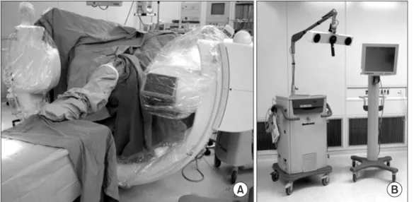
The reference frame (Orthopaedic Frame HC; Medtronic Inc., Louisville, CO, USA) was attached rigidly to the femur with two half-pins at the beginning of surgery. Intraoperative 3D images were acquired with the C-arm of Arcadis Orbic 3D (Siemens AG, Erlangen, Germany) (Fig. 1A). The C-arm of the image intensifier was equipped with a wireless tracker (Stealth Active wireless tracker S/N 130, Medtronic Inc.) to allow image capture and automatic registration by the navigation computer. The ac- quired image data were downloaded to the navigation computer (StealthStation TRIA plus, Medtronic Inc.) and a 3D image of the distal femur was reconstructed on the computer screen (Fig. 1B).
The medial half of the 3D-reconstructed distal femur was deleted using a specific function of the computer software to provide a better view of the lateral wall and the roof of the femoral inter- condylar notch (Fig. 2).
2. Graft Selection, Harvesting, and Preparation
In most cases, both BTB and hamstring grafts were available.
Grafts were selected by a surgeon who took into consideration the activity of patients, the types of sports they may be involved in, and patient preference. BTB graft was selected primarily for young male or collision/contact athletes, while hamstring grafts were selected for the others during the study period. BTB grafts were 10 mm in width and harvested with bone plugs at both ends from the central portion of the patellar tendon. The femoral bone plug for a rectangular socket was usually 5×10×15 mm as described by Shino et al.12). It was connected to an EndoButton (Smith & Nephew Endoscopy, Andover, MA, USA) using No. 5 and No. 2 FiberWires (Arthrex Inc., Naples, FL, USA). For the hamstring tendon grafts, the semitendinosus tendon was primar- ily harvested. When the length or diameter of semitendinosus
Fig. 1. Image data acquisition and recon- struction. (A) Intraoperative three-di- mensional images are acquired with the C-arm. (B) The acquired image data are downloaded to the navigation computer.
Fig. 2. Navigation views of the right knee. The surface of the lateral wall of the intercondylar notch in an orthogonal projection is visualized on the navigation computer screen. Concurrent axial, coronal, and sagittal two-dimensional images of any point can also be referenced.
5-mm in diameter. The intraosseous graft length should be at least 14 mm. Each graft was connected to an EndoButton CL (Smith & Nephew Endoscopy) in the usual manner.
3. Computer Navigation-Assisted Femoral Socket Preparation When the remnant tissue of ACL was scarred to the posterior cruciate ligament (PCL) or reattached to either the roof of the notch or the lateral wall, the minimal amount of the residual remnant was cleaned from the lower side to place the guide at the anatomical femoral attachment site using a thermal device.
designated location.
When the remnant tissue impeded guide placement during positioning of the femoral guide through the far AM portal, the remnant tissue of ACL was carefully retracted medially through the medial portal. Image-interactive navigation enabled the sur- geon to confirm the position of the tip of the femoral guide on the 3D reconstructed image in real time (Fig. 3). After the tip of the guide was placed at the target point (center of a socket), and after the position of the guide tip was verified on the computer screen, a guide was inserted slightly, the femoral guide tip was
Fig. 3. Arthroscopic and navigation views of the femoral guide. (A) Arthroscopic view of the lateral wall of the femoral intercon- dylar notch can be obtained through the anteromedial (AM) portal with the knee in 90o flexion. The tip of the femoral guide is placed within the anatomical femoral foot- print through a far AM portal. (B) Naviga- tion view of the lateral wall and roof of the femoral intercondylar notch on the three- dimensional reconstructed image. The white arrow shows the tip of the femoral guide.
Fig. 4. Navigation view of the right knee. (A) The three-dimensional image is rotated 90o on the navigation screen in order to evalu- ate the risk of a back wall blowout. (B) The image is rotated 180o to show the lateral aspect of the distal femur.
maintained within the femoral footprint, and the knee was fully flexed. Accordingly, displacement of the guide tip caused by knee flexion could be mostly prevented. On the navigation computer screen, the surgeon could then identify the entire image of the lateral wall of the femoral notch and LIR, even when arthroscopic visualization of the lateral wall of the intercondylar notch was impeded because of the presence of the remnant on deep knee flexion (Fig. 3). If the guide tip was displaced and was not ar- throscopically recognized, the surgeon could recognize it on the navigation monitor. Next, the 3D image was rotated 90o on the
navigation screen where the risk of a back wall blowout could be evaluated (Fig. 4A). Finally, the 3D image was rotated 180o to reveal the lateral aspect of the distal femur on the navigation screen (Fig. 4B). The total length of the femoral tunnel could thus be evaluated at once. When the BTB graft was used, two paral- lel guide wires were inserted with a 5-mm distance in the center of the femoral insertion along its long axis (Fig. 5). Conversely, when double-bundle reconstruction with hamstring grafts was performed, two guide wires for the AM and PL sockets were in- serted separately at the center of each footprint. After insertion
Fig. 5. Intraoperative arthroscopic views of the right knee. (A) The triangles show residual remnant of the torn anterior cruci- ate ligament (ACL). (B) The ACL remnant is retracted medially by a probe. (C) The femoral guide (white arrow) is placed with- in the anatomical femoral footprint through a far anteromedial portal. (D) Two parallel guide wires are inserted within the femoral insertion of the ACL. The triangles show the residual remnant of the torn ACL and the black arrow shows the end of the guide pin.
Fig. 6. Intraoperative arthroscopic views of the right knee. (A) Rectangular socket for bone-patellar tendon-bone graft (BTB) grafting. (B) The BTB graft is covered by the preserved remnant of the torn anterior cruciate ligament.
of the guide wires during BTB grafting, they were overdrilled for an appropriate length using a 5-mm cannulated drill, the two tunnels were interconnected using a dilator (Smith & Nephew Endoscopy), and the lateral femoral cortex was drilled through the center of the two tunnels using an EndoButton drill (Smith &
Nephew Endoscopy) (Fig. 6). When using hamstring tendon(s), each guide wire was overdrilled for an appropriate length by a cannulated drill with a diameter 0.5 mm smaller than that of the graft, each socket was dilated using a dilator with a diameter similar to that of the graft, and the distant cortex was breached with the EndoButton drill.
The tibial insertion site was arthroscopically determined in reference to the ACL remnant, the medial tibial eminence, the anterior horn of the lateral meniscus, the intermeniscal ligament, and the PCL14). Tibial tunnel creation and graft passage were performed roughly according to the method described by Ochi et al.15). Briefly, a No. 11 scalpel blade was used to make a longi- tudinal slit in an ACL remnant tissue. This method allowed not only visualization of the tips of the guide pins or the drill during creation of tibial tunnels but also smooth graft passage. The tibial tunnels were placed in the center of the AM and PL footprints for double-bundle reconstruction. Meanwhile, two parallel guide wires were positioned within the tibial footprint and overdrilled for the entire length using a cannulated drill, and a dilator (Smith
& Nephew Endoscopy) was used to interconnect the two tunnels for rectangular reconstruction using a BTB graft. After creation of the tibial tunnel, a No. 3 or No. 5 polyester braided suture was passed through the eyelet of a passing pin, which was inserted into the femoral tunnel through the longitudinal slit in the rem- nant tissue of ACL. Subsequently the suture was retrieved by an arthroscopic grasper into the tibial tunnel. The graft was passed through the remnant tissue and the EndoButton loop was flipped outside the femoral cortex in the usual manner. Creation of a
longitudinal slit, gradual hooking and pulling up the graft by a probe, and rounding of sharp corners of the bone plug were es- sential to allow technically challenging graft passage of the BTB through the remnant tissue.
Tibial fixation of the hamstring autografts was accomplished over a suture after fixation using a fully threaded 6.5-mm cancel- lous screw and washer (Meira Inc., Nagoya, Japan). The AM and PL bundle grafts were fixed at full knee extension and the BTB graft was also fixed at full knee extension. After graft passage, when residual remnant impingement on the roof or the lateral wall of the femoral notch or the PCL was arthroscopically dem- onstrated, the impinging part of the remnant was shaved off (Fig.
7).
Materials and Methods
Between January 2011 and December 2013, we treated 47 pa- tients with remnant-preserving ACL reconstruction using the navigation system described above, with the aim of creating a femoral socket with sufficient remnant tissue from the tibia to the femoral notch or the PCL. Eighteen patients underwent remnant- preserving ACL reconstruction using a rectangular BTB graft, while 29 underwent double-bundle reconstruction using ham- string tendon grafts. Remnant-preserving ACL reconstruction was performed in patients with type 1, 2, or 3 remnant tissue, as described by Crain et al.16). They classified the ACL remnants into 4 morphological patterns: type 1, bridging between the PCL and tibia; type 2, bridging between the roof of the intercondylar notch and tibia; type 3, bridging between the lateral wall of the intercondylar notch and tibia; and type 4, no substantial ACL remnants. Of the 47 patients (22 female and 25 male), 18 had type 1 remnant tissue, 10 had type 2 remnant tissue, and 19 had type 3 remnant tissue.
covered by the remnant of the torn anterior cruciate ligament. (A) The triangles show the graft for the anteromedial bundle. (B) The black arrow indicates the graft for the postero lateral bundle.
There were no patients with partial ACL rupture, including iso- lated AM or PL bundle rupture. The mean age of the patients was 31 years (range, 16 to 55 years) at the time of surgery. The height and weight [means±standard deviations (SDs)] of patients were 166±9 cm and 62±12 kg, respectively. The median duration be- tween injury and surgery was 4 months (range, 1 to 348 months).
3D computed tomography (CT) imaging of the operated knee was performed 1 week after surgery and the center of the femoral sock- et aperture was calculated according to the quadrant technique, as described by Bernard et al.17). The incidence of a back wall blowout
and short femoral tunnel (<25 mm) was evaluated. The Institu- tional Review Board approved this retrospective study. Patients (and their families) were informed that their data would be submitted for publication, and all gave their consent for the same.
Results
Data in text are given as means±standard deviations (range, minimum to maximum). In ACL reconstruction using a rect- angular BTB graft, the center of the femoral socket aperture was
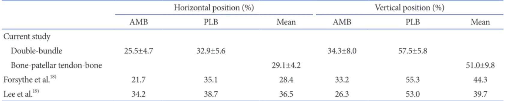
Fig. 8. Femoral socket(s) on three-dimensional computed tomography (3D CT) after surgery. Medial 3D CT view of the reconstructed distal femur at 1 week after surgery shows accurately placed femoral sockets for hamstring tendon graft (A) and an accurately placed rectangular femoral socket for a bone-patellar tendon-bone graft (BTB) (B). Morphometric assessment of femoral tunnel positioning was performed using the quadrant technique, as described by Bernard et al.17). The horizontal position of the femoral tunnel center is defined as the percentage distance from the most posterior contour in reference to the total length of the lateral condyle, whereas its vertical position is defined as the percentage distance from Blumensaat’s line in reference to the total lateral intercondylar notch height. For the rectangular tunnel for the BTB grafts, the center of the ellipse by which the rectan- gular tunnel aperture was approximated was defined as the center of the femoral tunnel for the BTB graft. The black square shows the location of the center of the anteromedial socket, the black triangle shows the location of the posterolateral socket, and the black dot shows the center of the socket location in anterior cruciate ligament reconstruction using a BTB graft.
Table 1. Comparison Between the Cadaver Studies and the Current Study Using the Quadrant Method
Horizontal position (%) Vertical position (%)
AMB PLB Mean AMB PLB Mean
Current study
Double-bundle 25.5±4.7 32.9±5.6 34.3±8.0 57.5±5.8
Bone-patellar tendon-bone 29.1±4.2 51.0±9.8
Forsythe et al.18) 21.7 35.1 28.4 33.2 55.3 44.3
Lee et al.19) 34.2 38.7 36.5 26.3 53.0 39.7
Values are presented as mean±standard deviation or number.
AMB: anteromedial bundle, PLB: posterolateral bundle.
(range, 48.5% to 72.0%) in the horizontal and vertical direction, respectively. The femoral socket locations were considered to be anatomical in accordance with previous cadaveric studies exam- ining the positions of the ACL femoral insertion site18,19) (Fig. 8 and Table 1). Neither back wall blowout nor short femoral tunnel was observed.
Discussion
Computer-assisted surgery has recently been introduced to improve the accuracy and reproducibility of socket placement during ACL reconstruction20-23). We have been using a 3D fluo- roscopy-based navigation system for accurate and reproducible placement of the femoral socket through a far AM portal9,10). Us- ing this system, surgeons can identify the LIR, which is an impor- tant landmark for ACL femoral insertion not only on arthroscopy but also on 3D images of the navigation system during primary ACL reconstruction. In remnant-preserving ACL reconstruc- tion, arthroscopic imaging alone is insufficient to evaluate tunnel placement because the residual remnant impedes visualization of the femoral bone surface. Despite poor arthroscopic visualiza- tion, this system enables surgeons to visualize the orientation of the lateral wall and the roof of the femoral intercondylar notch.
The principle finding of our research was that morphometric analysis of femoral socket placement on postoperative 3D CT im- ages using the quadrant method in this report revealed anatomi- cal femoral socket placements that were similar to cadaveric data previously reported18,19). According to a cadaveric study described by Forsythe et al., the average positions of the center of the AM bundle were 21.7% in the horizontal direction and 33.2% in the vertical direction, while those of the PL bundle were 35.1% and 55.3% in the respective directions. Calculated from this data, the centers between the AM and PL bundles were located at 28.4%
in the horizontal direction and 44.3% in the vertical direction.
Our data were compared with the cadaveric data using 3D CT images in previous studies (Table 1). Considering these cadaveric data, it was clear that the femoral sockets were anatomically and reproducibly created using 3D imaging-based navigation in the
rather, it is based on the fact that the remnant tissue only has a supplementary effect and that the aim of remnant-preserving re- construction is coverage by the conserved ACL tissue. Therefore, the position of the remnant fibers does not affect femoral socket placement using the current technique. Furthermore, decreasing the remnant volume by using the double-bundle reconstruction procedure is acceptable, and the current procedure can be ap- plied to patients in whom the remnant is reattached to the PCL or the roof of the notch.
The current report did not analyze the clinical results of rem- nant-preserving ACL reconstruction using the navigation sys- tem. Further follow-up is required to confirm whether or not this technology will result in good clinical outcomes. It will also be necessary to observe whether or not this procedure is associated with complications such as Cyclops syndrome or pain and/or limited range of knee motion due to impingement on the femoral intercondylar roof, lateral wall, or PCL.
A major problem with the navigation system is the existence of inaccuracy. One of the elements that cause the inaccuracy is a registration error. In the navigation system described in this re- port, registration based on digital fluoroscopic images which are acquired during surgery is automatically performed; therefore, the registration error can be avoided. According to a laboratory study, 3D fluoroscopic navigation can be performed with an overall application accuracy of 0.47±0.21 mm24).
There are other disadvantages associated with the navigation system described in this report. In this system, a reference frame must be fixed to the lateral femur with two half-pins, which ne- cessitates additional skin incisions and drill holes in the femur.
Other disadvantages include exposure of the patient and medical staff to radiation at the beginning of the procedure in addition to increased medical costs.
Conclusions
3D fluoroscopy-based navigation can assist surgeons in creat- ing anatomical femoral sockets during technically challenging remnant-preserving ACL reconstruction.
Conflict of Interest
No potential conflict of interest relevant to this article was re- ported.
References
1. Schultz RA, Miller DC, Kerr CS, Micheli L. Mechanorecep- tors in human cruciate ligaments. A histological study. J Bone Joint Surg Am. 1984;66:1072-6.
2. Georgoulis AD, Pappa L, Moebius U, Malamou-Mitsi V, Pappa S, Papageorgiou CO, Agnantis NJ, Soucacos PN.
The presence of proprioceptive mechanoreceptors in the remnants of the ruptured ACL as a possible source of re-in- nervation of the ACL autograft. Knee Surg Sports Traumatol Arthrosc. 2001;9:364-8.
3. Adachi N, Ochi M, Uchio Y, Iwasa J, Ryoke K, Kuriwaka M.
Mechanoreceptors in the anterior cruciate ligament contrib- ute to the joint position sense. Acta Orthop Scand. 2002;73:
330-4.
4. Ochi M, Iwasa J, Uchio Y, Adachi N, Sumen Y. The regen- eration of sensory neurones in the reconstruction of the an- terior cruciate ligament. J Bone Joint Surg Br. 1999;81:902-6.
5. Ahn JH, Lee SH, Choi SH, Lim TK. Magnetic resonance im- aging evaluation of anterior cruciate ligament reconstruction using quadrupled hamstring tendon autografts: comparison of remnant bundle preservation and standard technique.
Am J Sports Med. 2010;38:1768-77.
6. Ahn JH, Wang JH, Lee YS, Kim JG, Kang JH, Koh KH. An- terior cruciate ligament reconstruction using remnant pres- ervation and a femoral tensioning technique: clinical and magnetic resonance imaging results. Arthroscopy. 2011;27:
1079-89.
7. Gohil S, Annear PO, Breidahl W. Anterior cruciate ligament reconstruction using autologous double hamstrings: a com- parison of standard versus minimal debridement techniques using MRI to assess revascularisation: a randomised pro- spective study with a one-year follow-up. J Bone Joint Surg Br. 2007;89:1165-71.
8. Yasuda K, Kondo E, Kitamura N, Kawaguchi Y, Kai S, Ta- nabe Y. A pilot study of anatomic double-bundle anterior cruciate ligament reconstruction with ligament remnant tis- sue preservation. Arthroscopy. 2012;28:343-53.
9. Nakagawa T, Takeda H, Nakajima K, Nakayama S, Fukai A, Kachi Y, Kawano H, Miura T, Nakamura K. Intraoperative 3-dimensional imaging-based navigation-assisted anatomic
double-bundle anterior cruciate ligament reconstruction.
Arthroscopy. 2008;24:1161-7.
10. Taketomi S, Nakagawa T, Takeda H, Nakajima K, Nakayama S, Fukai A, Hirota J, Kachi Y, Kawano H, Miura T, Fukui N, Nakamura K. Anatomical placement of double femoral tunnels in anterior cruciate ligament reconstruction: antero- medial tunnel first or posterolateral tunnel first? Knee Surg Sports Traumatol Arthrosc. 2011;19:424-31.
11. Shino K, Suzuki T, Iwahashi T, Mae T, Nakamura N, Nakata K, Nakagawa S. The resident’s ridge as an arthroscopic land- mark for anatomical femoral tunnel drilling in ACL recon- struction. Knee Surg Sports Traumatol Arthrosc. 2010;18:
1164-8.
12. Shino K, Nakata K, Nakamura N, Toritsuka Y, Horibe S, Nakagawa S, Suzuki T. Rectangular tunnel double-bundle anterior cruciate ligament reconstruction with bone-patellar tendon-bone graft to mimic natural fiber arrangement. Ar- throscopy. 2008;24:1178-83.
13. Shino K, Horibe S, Hamada M, Nakamura N, Nakata K, Mae T, Toritsuka Y. Allograft anterior cruciate ligament re- construction. Tech Knee Surg. 2002;1:78-85.
14. Ferretti M, Doca D, Ingham SM, Cohen M, Fu FH. Bony and soft tissue landmarks of the ACL tibial insertion site:
an anatomical study. Knee Surg Sports Traumatol Arthrosc.
2012;20:62-8.
15. Ochi M, Abouheif MM, Kongcharoensombat W, Nakamae A, Adachi N, Deie M. Double bundle arthroscopic Anterior Cruciate Ligament reconstruction with remnant preserving technique using a hamstring autograft. Sports Med Arthrosc Rehabil Ther Technol. 2011;3:30.
16. Crain EH, Fithian DC, Paxton EW, Luetzow WF. Varia- tion in anterior cruciate ligament scar pattern: does the scar pattern affect anterior laxity in anterior cruciate ligament- deficient knees? Arthroscopy. 2005;21:19-24.
17. Bernard M, Hertel P, Hornung H, Cierpinski T. Femoral insertion of the ACL. Radiographic quadrant method. Am J Knee Surg. 1997;10:14-21.
18. Forsythe B, Kopf S, Wong AK, Martins CA, Anderst W, Tashman S, Fu FH. The location of femoral and tibial tun- nels in anatomic double-bundle anterior cruciate ligament reconstruction analyzed by three-dimensional computed to- mography models. J Bone Joint Surg Am. 2010;92:1418-26.
19. Lee JK, Lee S, Seong SC, Lee MC. Anatomy of the anterior cruciate ligament insertion sites: comparison of plain ra- diography and three-dimensional computed tomographic imaging to anatomic dissection. Knee Surg Sports Traumatol
navigation system in anterior cruciate ligament reconstruc- tion. Knee Surg Sports Traumatol Arthrosc. 2012;20:1503- 10.
22. Tensho K, Kodaira H, Yasuda G, Yoshimura Y, Narita N,
2012;1:e95-9.
24. Nolte LP, Beutler T. Basic principles of CAOS. Injury. 2004;
35 Suppl 1:S-A6-16.