약학희 지 제 52 권 제 3 호 165~171 (2008)
Yakhak Hoeji
Vol. 52, No. 3황 후 총 학
B16 Melanoma
세포에서 돌나물 추출몰의 엘라닌 생성 저해 효과심 관 섬* • 김 진 화 • 이 범 천 * • 이 동 환
■
이 근 수 • 표 형 배 한불화 장품(주) 기술연구소, *Dalhousie University (Received December 3, 2007; Revised June 8, 2008)Inhibitory Effects on Melanin Production in B16 Melanoma Cells of Sedum sarmentosum
Gwan Sub S in /, Jin Hwa Kim, Bum Chun Lee*, Dong Hwan Lee, Geun Soo Lee and Hyeong Bae Pyo
R&D Center, Hanbul Cosmetics Co, Ltd,, 72-7, Yongsung-ri, Samsung-myun, Umsung-kun, Chungbuk 369-834, Korea
^Department of Biochemistry and Molecular Biology, Faculty of Medicine, Tkpper Medical Building, Dalhousie University, Halifax, Nova Scotia B3H 1X5, Canada
Abstract — To develop effective skin whitening agents, we tested natural herbal extracts for their melanogenic inhibitory activities.
Sedum samentosum
was selected for its inhibitory effect on melanogenesis in B16 melanoma cells. Ethanolic extract of 5.samentosum
(SSE) was evaluated for antioxidative effect and tyrosinase inhibitory activity of melanogenesis.We investigated the changes in protein level and mRNA level of tyrosinase, tyrosinase related protein (TRP)-l and TRP- 2 by using western blotting and RT-PCR, respectively. SSE showed scavenging activities of free radicals and reactive oxygen species (ROS) with the IC5Q of 342.7 |ig/m/ against 1,1 -diphenyl-2-picrylhydrazyl (DPPH) radical and 64.69 |ig/m/ against superoxide radicals in the xanthine/xanthine oxidase system, respectively. SSE treatment suppressed the biosynthesis of melanin up to 46% and reduced tyrosinase activity up to 51% at 100 ^g/m/ in B16 melanoma cells. The tyrosinase activity and tyrosinase expression in B16 melanoma cells were reduced in a dose-dependent manner by SSE. Also, SSE was able to significantly inhibit tyrosinase and TRP-1 expression in mRNA level. These results suggest that SSE inhibited melanin production which may be dependent on tyrosinase activity and expression in B16 melanoma cells, and an effective whitening agent for the skin.
Keywords □
Sedum sarmentosum,
B16 melanoma, melanin, tyrosinase, tyrosinase related protein괴부의 색은 주로 피부 속에 존재하는 델라닌(melanin)이라는 색소의 함량에 의해 결정된다. 이러한 델라닌은 피부의 기저층 에 존재하는 델라닌 생성세포(melanocytes)에 의해서 생합성되 고 세포질 돌기률 통 하 표 괴 의 기저층에서 각질층으로 각질형 성세포(kerationcytes)의 각화과정에 의해서 이동하게 된다.^' 표 피에 존재하는 멜라닌은 태양광선으로부터 들어오는 자외선을
차단하는 색소로서 델라닌이 국소적^ 과도하게 합성되거나,
노화 등에 의해 피부의 생리기능이 떨어지게 되면 멜라닌이 피부 표면에 침착되어 기미, 주근깨 및 다양한 색소 침착을 유발하게 되는 것으로 알려져 있다.2> 피부에서 델라닌 생합성은 델라닌 생 성세포에서 cascade 효소 반응에 의해 생성된다. 델라닌은 델라 닌 생성세포의 델라노좀(melanosome)에서 합성되며, 델라노좀에
후본 논문에 관한 문의는 저자서1게로 (견화) 043-879-2283 (팩스) 043-881-2128 (E-mail) [email protected]
는 정상적인 델라닌을 합성하는데 필요한 특이적인 효소들을 함 유하고 있다. 이 효소돌 중 가장 잘 알려진 것으로 tyrosinase, TRP-1 파 dopachrome tautomerase(DCT) 등어 있 다 .폐 이들 중 tyrosinase는 melanogenesis의 속도결정단계인 초기 반응에 작 용하는 효소로서, tyrosine을 3,4-dihydroxyphenylalanin(DOPA) 로 견 환 하 는 tyrosine hydroxylase 활 성 과 DOPA를 DOPA quinone으로 산화히는 DOPA oxidase 활성을 모두 가지고 었다.
TRP-1 은 5,6~dihydroxyindole-2*<:ait)oxyIic acid(DHICA) 를 indole- 5,6-quinone-2-carboxylic acid로 산화하는 효소이 다 .rDCT는 초 기에 TRP-2로 불려졌던 효소로서 dopachrome을 DHICA로 이 성화하는 효소이다. 델라닌은 흑 , 같색의 eumlanin파 적 , 노랑색 의 phaeomelanin이 있다. 특히 tyrosinase 는 이들 두 가지 타입 의 melanin합성에 필요하며, TRP-1 과 DCT는 eumelanin의 합 성에 더 많이 관여하는 것으로 알려져 있다.^^ 지금까지 알려져 있는 미백물질둘은 작용 기견에 따라 자외선 흡수제나 산란제, 알부틴 (arbutin), 코직산(kojic acid) 등과 같은 tyrosinase 활성 저
165
심관섭 • 김진화 ■이범천 ■이동환 ■이5 수 • 표형배
해제 , 활성산소종을소거하는아스코르빈산(ascorbic acid) 및유 도체, coenzyme Q10, 토코페롤 등으로 분류할수있다. 또한상 백피 , 당귀 , 고삼, 감초, 백작약, 유기노, 삼륭등의다양한식물 추출물이 미백효과가었는것으로나타나고있다.6
돌나물(SedMW
sarmentosum
Bunge)은돌나물과에속하는다 년생초로써 전국산 이 게 분포하고식용, 약용, 관상용으로쓰 이며, 불 갑 초 휘 , 수분초(종^^¥:)라고도불리운다. 돌나물의 함유성분으로는 sedoheptulose, sucrose, fructose 등의 당질과 특히 비타민 C, 철분및칼슘등을풍부하게함유하고있다. 이 러한돌나물의 생리활성연구로는 terpenoids에의한간보호효 과,®' 돌나물에서분리한 flavonoids인 quercetin, isorhamnetin과 kaempferol 등의 배당체들에 의한 angiotensin convertingenzyme 저해효과,® 돌나물 추출물이 각질형성세포에서의
hyaluronan 생성촉진작용이 있다는것이보고되었다.^®
본 연구에서는천연물로부터의 미백제를개발하기 위하식수 중의천연물을검색하였으며 돌나물의델라닌생성저해효과룰
발견하고돌나물추출물의활성산소소거휠성및 B16 melanoma
세포를이용하식 델라닌생성 저해효파를연구하였다.
실험재료 및 방법
시료의 추출
본실험에서시용한돌나물(SafeoM
sammtosum
Bunge)은국내 익초(경동)시장에서구입하였다. 그늘진곳에서건조한돌나물1 0 0 g 을분쇄하여 70% EtOH IE . 환류하면서 3시간씩 2희반복추출 하였다. 아를강합농측, 동결건조S]여그분말을' 실험에사§된m .세포 및 시약
B16F10& 쥐의 melanoma 세포주로서울대학교한국세포주 은행에서구입하였다. 구입한세포는 5% fetal bovine serum (Bio Whittaker, MD, USA), 1% penicillin-streptomycin(Gibco BRL, MD, USA)을 함유한 Dulbecco's modified Eagle's medium (DMEM) 배지에 100nM a-melanocyte stimulating hormone (MSH, Sigma, USA)를첨가하식 37°C, 5% CO^ 배양기에서 배 앙하였다. Tyrosinase, TRP-1, TRP-2, P-actin 항체는 Santa Cruz Biotechnology(CA, USA)에서구입하여사용하였다.
DPPH radical 소거 효과
항산화활성은 l,l-diphenyl-2-picrylhydrazyl(DPPH, Aldrich, USA)를이용하쉬시료의라디칼소거효과를측정하는 Blois법피 을활용하였다. 0.1 mM DPPH methanol 용액에동일량의시료 를가하여 vortex mixer로잘혼합한후, 실온에 1 0분동안반 응하였다. 이후 spectrophotometer률이용하여 565배에서 흡 광도를측정하였다.
Superoxide radical 거 효과
Xanthine/xanthine oxidase 반응에서 형성된 superoxide radical 소거효과는 nitroblue tetrazolium (NBT) 방법에의해측 정하였다.^' 0.05 M N3 2C0 3 buffer(pH 10.2) 에 3mM xanthine, 3 mM EDTA, 0.72 mM NBT와시료를가한후 25°C에서 10분 간번움하였다. 이반응액에 0.25 U/m/ xanthine oxidase를가하 고 25°C에서 25분동안반응후 superoxide radical 소거효과률
565nm에서흡광도률측정하였다.
세포 생존율 측정
3-(4,5-dimethythiazol-2-yl)-2-5-diphenytetxazolium bromide (MTT) 정량은 Mosmaim데의방법을변형하여실시하였다. B16 melanoma 세포를 1x10크 cellsAvell 농도로 24 well plate에분주 한세포에시료를처리하고 48시간동안배양하였다. MTT 용 액(5
\xgfnm
:첨가하고 3시간후원심분리하여상등액을제거하 고 100 |o/ acid-isopropanol(0.04 N HCI in isopropanol)을 첨가 한후 565 nm에서흡광도률측정하였다.엘라닌 정량
델라닌정량은 Hosoi^^> 등의방법을변형하식시용하였다. 6 well plate에 3xl0®cells/well로세포률분주하였고, 시료를처리 하고 48시간동안 37°C CO2 배양기에서배양하였다. 세포률수 집하여 세포수를 측정하고, 1200rpm에서 5분간원심 분리 하여 첨전한 후
, 1ml
homogenization buffer(50 mM Sodium phosphate pH 6.5, 1% Triton X-100, 2 mM PMSF)로용해시켰 다. 여기서 얻은 pellet에 IN NaOH(10% DMSO) 200^를첨 가하고 vortex 후 405nm에서 O.D^os 값을측정하였다. 델라닌 표준^(Sigma, USA)으로얻은표준검량선을이용하여각 well 에서생성된델라닌잉을신출하였다. 멜라닌은단위세포(l(y* cells) 에서의 델라닌생성량을비교하였다.세포내 tyrosinase 활성 측정
세포내 tyrosinase 활성측정법은 Pawelk파 Pomerantz고크'도® 방 법을사용하였다. 6 well plate에 5x10® cells/well로세포를분 주하고하루동안배양한후시료를처리하였다. 24시간후, 세 포를 수집하여 용해시킨후 0.2% L-DOPA가 첨가된 0.1 M sodium phosphate buffer(pH 6.8>를넣고 37°C에서 2시간동안 배양하고 490nm에서흡광도를측정하였다.
Western blot analysis
시료를 48시간 처리한 B16 melanoma 세포를 RIPA buffer (10 mM sodium fluoride, 0.1% SDS, 1% NP-40, 1 mM DTT, 500 |oM sodium orthovanadate, 10
jig/ml
aprotinin, 10ng/ml
leupeptin, Im M PMSF)로용해하고원심분러하였다. 여기서
i:의 델라닌생성 저해효과 167
얻은상층액을 12% SDS-PAGE룰이용해 전기영동하고 이를
nitrocellulose membrane으로이견시켰다. 이를 3% skim milk 가함유된 tris-buffer에서 tyrosinase(sc-7833), TRP-l(sc-10443), TRP-2(sc-10452), p-actin(sc-1616) 항체와 각각 반응시킨 후, alkaline phosphatase가 결합된 항체를 가한 후, 5-bromo-4- chloro-3-indoly-l-phophate/nitro blue tetrazolium(BCIP/NBT) 을가하여발색시켰다. Western blot 결괴는 Calibrated densito
meter GS-800(Biorad, CA, USA)률이용하여분석하였다.
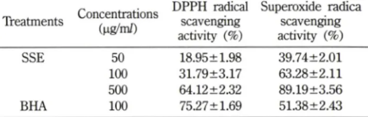
Table I - Anti-oxidant activity of SSE in DPPH and NBT assay
Treatments Concentrations (나 g/m/)
DPPH radical scavenging activity (%)
Superoxide radica scavenging activity (%)
SSE 50 18.95 ±1.98 39.74±2.01
1 0 0 31.79±3.17 63.28±2.11
500 64.12±2.32 89.19±3.56
BHA 1 0 0 75.27 ±1.69 51.38±2.43
The results were expressed as the average of triplicate samples with S.D.
RNA 분러및RT-PCR
Total RNA 추출은 RNeasy mini kit(Qiagen, MD, USA)을 이용하였다. cDNA 합성은 l|ig의 total RNA를 oligo(dT)15 primer, dNTP(0.5 |iM), 1 unit RNase inhibitor 그러고 4 unit Omniscript reverse transcriptase(Qiagen, Hilden, Germany)로 37°C에서 60분, 93X에서 5분 heating 시킴으로서반응을중지시 켰다. Polymerase chain reaction(PCR)은■ cDNA로부터 tyrosinase, TRP-1, TRP-2, p-actin을증폭하기 위하여 cDNA, 0.5 ^실의 5' 과 3'primei; 10xbuffer(10 mM Tris-HCl, pH 8.3, 50 mM KCl, 0.1% Triton X-100), 200 nM dNTR 25 mM MgClg, 2.5 unit
Taq
polymerase(Qiagen, Hilden, Germany)률이용하여 PCR을 실시하였다. PCR 증폭은 94°C 0.5분, 50~55°C 0.5분, 72°C 1분, 20-29 cycles로반응시켰다. pcR에의하여 생성된산물을 1.5% agarose gel에서전기영동하여 tyrosinase, TRP-1, TRP-2 와 P-actin 유전자의 발현을 image analyzer(BIS303PC, DNR Imaging Systems Ltd, UK)로확인하였으며,
각 band의 density는 densitometric prograin(NIH Image software, MD, USA)i:이용하 였다. Tyrosinase, TRP-1, TRP-2의 oligonucleotide 서열은 ty- rosinase: sense; 5'-GAGAAGCGAGTCTTGATTAG-3', antisense;5'-TGGTGCTTCATGGGCAAAATC-3', TRP-1: sense: 5'-GCTG- CAGGAGCCTTCTTTCTC-3', antisense; 5'-AAGACGCTGCAC- TGCTGGTCT-3', TRP-2: sence: 5'-CCTGTCTCTCCAGAAGTT- TG-3', antisence: 5'-CGTCTGTAAAAGAGTGGAGG-3'이며, p- actin의 oligonucleotide 서열은 sense; 5-ATGAGAAGGAGATC- ACTGC-3', antisense; 5-CTGCGCAAOTTAGGTTTTGT-3미다.
자료분석 및통계처리
모든실험결과는평균±표준편차로표기하였고, 통계적 유의 성은 Student's Mest로하였으며
p
값이 0.05 미만일때통계적 으로유의하다고관단하였다.실험결과 및 고찰
둘나울 추출물의 함산화 효과
DPPH는 free radical의안정된모델로번응중 DPPH의감소
는 free radical의소거반응의진행됨을알수었고지질과산화의 초기반응의 억제정도를예측할수있다. 유해산소라불려지는 활성산소는세포생체막의구성성분인불포화지방•상을공격하여 지질과산화반응을일으켜체내 과산화지질을축적함으로인해 생체기능이 저하되고동시에 색소침착, 노화및성인병 질환을
유발하는것으로알려져 있다.^^^ 돌나물추출물의 항산화작용
이있는지률확인하기위하여 DPPH를이용한유리라디칼소
거반응을관찰하였다. 돌나물 추출물을 50, 100, 500 ^ig/m/의 농도로처리한경우각 DPPH radical 소거능은 18.95%, 31.79%,
64.12% 소거효과를나타내었다. 양성대조군으로는항산화효과
가알려진 3-f-butyl-4~hydroxyanisole(BHA)을■이용하여 돔나물 추출물의항산화효과룰비교하였다. 그결과 BHA는 100 |ig/m/
에서 75.27%의 DPPH radical을소거하였으며 , 돌나물추출물은 투여 농도 의존적으로 DPPH radical 소거작용을 나타내었다 (Table I).
Xanthine/xanthine oxidase의효소에 의한 superoxide 옴이온 저해작용은 superoxide 옴이온소거작용과 xanthine oxidase 효 소저해에 의해 나타난다. Xanthine oxidase에의해 형성되는 superoxide anion의생성저해의결과는 Table I에나타내었다. 양 성대조군으로 BHA를이용하여 돌나물 추출물의 superoxide
radical 소거효과를비교하였다. 그결과돌나물추출물은 투여
농도의존적으로 superoxide radical 소거작용을나타내 50, 100, SOOng^m/의농도로처리한경우각 superoxide radical 소거능 은 39.74%, 63.28%, 89.19%로우수한 superoxide radical 소거 효과를 나타내었다. 양성 대조군인 BHA는 100 ng/m/에서 51.38%의 superoxide radical을소거하였다.
틀나물 추출물의 세포 독성
돌나물추출물이 세포득성에 미처는농도를조사하여 미백 실험에사용될농도범위결정을 위해서 MTT assay률시행하 였다. B16 melanoma 세포에대한돌나물주줄물의 세포득성을
측정한결과, 돌나물추출물은 1 0 0
^g/ml
이하의 농도로 처리시세포생존율이 90% 이상으로나타났으며, 그이상의 농도에 서는생존율이 저하되었다(Fig. 1). IC50 값 413 ng/m/로나타 났다.
Vol. 52, No. 3, 2008
25 50 100 200 500 Concentration (|jg/ml)
Fig. 1 - Relative cell viability of SSE on B16 melanoma cells by MTT assay. The cells were treated with various con
centration of SSE for 48 h. The cell viability was measured by the MTT method. Data are normalized by taking 100%
as viability of non-txeated cells. Results are means ± S.D.
from 3 separated experiments.
Melanin 생성 저해 효과
돌나물추출물식델라닌합성에미치는영향을확인하기위해
B16 melanoma세포룰이용하여 델라닌생성저해효과를측정
하였다. 준비된세포에돌나물추출물을농도별로처리하고 48
시간동안 배양하였다. 세포수를수집하식 델라닌양을측정한 결과, 돌나물추출물처리군모두가농도에비례하여 델라닌합 성이저해되었으며 lOOng/m/ 농도에서 약 46% 델라닌 생성저
Control Arbutin 100 50 Concentration (ug/ml)
25
Fig. 2 Effect of SSE on tyrosinase activity in B16 melanoma cells.
The cells were incubated with SSE for 24 h. Results are means ± S.D. from 3 separate experiments. */?<0,05 com
pared with control. Arbutin (100 ug/m/).
SSE (fjg/m l)
Control Arbutin 100 50
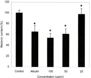
Fig. 3 - Effect of SSE on melanin production in B16 melanoma cells.
The cells were incubated with SSE for 48 h. Melanin content is quantified by absorption at 405 nm calibrated with synthetic melanin as standard SSE decreased the intracellular melanin contents at treated concentration.
Results are means ± S.D. from 3 separate experiments.
Arbutin (100 나g/m/).
해효과가 있었다(Fig. 2). 양성대조군으로사용한 알부틴은
100 ng/m/에서약 35% 델라닌감소효과를나타냈다. 또한델라 닌합성억제효과를육안으로관찰하기위하여각시료당 1x10®
개의세포를수집하쉬관찰하였다. 대조군으로서 델라닌생성을 촉진시키는호르몬인 a-MSH률처리한세포군에서는델라닌이 상당히중가되었옴을확인하였다. 돌나물추출물처리군은대조 군보다델라닌색소가감소하였고, 특히 100 ng/m/ 농도에서현 저히감소하였다(Fig. 3).
120
Control Arbutin 100 50 Concentration (ug/ml)
Fig. 4 - Inhibitory effect of SSE on melanin accumulation in cult
ured B16 melanoma cells. Cells were cultured for 48 h in medium containing 100, 50, 25 나g/m/ of SSE. Picture of cellular melanin solubilized with 2 M NaOH. Arbutin (100
심관섬 • 김진화 • 이범천 • 이동환 • 이근수 • 표형배
0^0002어864
OOJlcooioo/J준!qe!> 10
Concentration (pg/ml)
Fig. 6 - Effect of SSE on tyrosinase, TRP-1, TRP-2 mRNA expres
sion in B16 melanoma cells. B16 melanoma cells were treated for 24 h with SSE. Total RNA extracted from B16 melanoma cells was analyzed by RT-PCR.
3-actin
g.
140
120 100
80
60
40
20
■ Tyrosinase ■ TRP-1 ■ TRP-2
Control 100 50 25
Concentration (Mg/ml)
Fig. 5 - Effect of SSE on tyrosinase, TRP-1 and TRP-2 expression in B16 melanoma cells. B16 melanoma cells were treated for 48 h with SSE. Solubilized total protein (50 |ig) was electrophoresed in 12% SDS-PAGE gels and transferred to nitrocellulose membrane. Specific detection of proteins was perfomed with the polyclonal antibody against tyrosinase, TRP-1 and TRP-2. Similar results were observed in three independent experiments.
Vol. 52, No. 3, 2008
추출물의 델라닌생성 저해효과
세포내 tyrosinase 저해효과
델라닌합성은 tyrosinase에의해조절되는일련의효소적 반 응에의해조절되며, tyrosine을기질로하여 dopa룰 생성시키 고다시 dopaquinone^.?- 산화시키는연속적인효소적 산화가 진행된후각 생성물의 중합빈움에 의해 이루어진다고알려져 었다. 델라닌 생성에 관련하는세포내 tyrosinase의활성 저해 효과를죽정하였다. 돌나물주출물을처러한세포를수집하여 용해시킨후, 0.2% L-dopa가첨가된 0.1 M sodium phosphate buffer(pH 6.8)룰넣:il 37X에서배양한후흡광도를측정하였
다. Fig. 4의결과와같이돌나물추출물을처리한설험군에서
는농도의존적으로 tyrosinase의활성이저해되었으며 , 100 ^ig/
m/에서 tyrosinase 활성을약 51% 저해효파가있는것으로나 타났다.
SSE (Mg/ml)
50 25
Tyrosinase
TRP-1
TRP-2
P-actin
Tyrosinase, TRP-1, TRP-2 단택질발현저해효과
세포내 tyrosinase 저해실험의결과로돌나물추출물은세포 내 tyrosinase활성을저해하여 melanin 생성을감소시켰다. 이러 한결과가델라닌생성에관련된단백질발현과도연관성이 있 는지를확인하기위해 tyrosinase, TRP-1, TRP-2의항체를이용 한 western blot으로관련단백질의 발현량번화에 대하여조사 하였다(Fig. 5). 돌나물추출물은 tyrosinase, T'RP-1 의발현을저 해하는것으로나타났으며 TRP-2의발현량에는 영향을머치지 않았다. 또한이에 대한 density률죽정한결과, 100|ig/m/에서 tyrosinase발현을 29%, TRP-1 의발현을
13%
감소시키는것으 로나타났고, TRP-2 발현량은번화가없었다. Yoon 등 에 의한 tyrosinase와 TRP-1 의발현저해는제주조릿대잎열수추출물이a-MSH에의한세포신호전달경로중지속적인 ERK 활성화를
저해함으로써 tyrosinase와 TRP-1 의발현을저해하는것으로보 고되고있다. 따라서, 돌나물추출물은 a-MSH에의해유도된 extracellular signal-regulated kinase(ERK)의활성화률 저해함 으로써델라닌생합성에직접적으로관여하는효소인 tyrosinase 와 TRP-1 의발현을저해하는것 로 사료된다.
S S E (|ig/ml)
TRI TRf Tyrosini
u
o
o
o
o
o
o J-
2
o
8
6
4
2
111
{%}AllssJU
! 9>UBIeJJ
25 00 50
Control
170 심관섭 • 김진화 • 이범천 • 이동환 • 이근수 • 표형배
Tyrosinase, TRP-1, TRP-2 mRNA 발현저해효과 돌나물추출물이 델라닌생합성에 관여하는유전자의 발현에
머처는영향을확인하기 위하식 RT-PCR을수행하였다. 실험결
과돌나물추출물은 tyrosinase, TRP-1 의발현을저해하는것으 로나타났지만 TRP-2의발현량에는영향을미처지않았다. 또한 이에대한 density률죽정한결과, lOOjig^m/에서 tyrosinase 발 현을 21%, TPR-1 의발현을 12% 감소시키는것^ 나타났고, TRP-2 발현량은변화가없었다(Fig. 6).
델라닌 생합성은 세포내 여러 신호 견달 경로룰 통하여 tyrosinase 발현이중가되어 일어나며, 이러한 tyrosinase 생성에 중요한 견사인자인 microphthalmia-associated transcription factorOVHTF)의발현에의해조절된다고알려져있다. MITF
는 델라닌생합성에관여하는신호전달경로중 a-MSH에의한
cAMP 경로를 통하여 발현이 중가되고, extracellular signal- regulated kinase(ERK)에의헤인산화되어발현이저해된다.오크■혜 따라서, tyrosinase와 TRP-1 유전자발현을저해한돌나물추출
물은 MITF를조절하는신호전달과정에관여하여 델라닌생성
을저해하는것으로추측되며 향후에추가적인연구가필요하다 고 생각된다.
결 론
둘나물추출물이델라닌생성에미치는영향을조사하기위하
여 항산화 효과 및 B16 melanoma 세포룰 이용하여 세포내
tyrosinase 저해효과, 델라닌생성저해효과, tyrosinase와관련단 백질및유전자에 머처는영향을연구하였다. 돌나물추출물의 DPPH 소거효파와 superoxide radical 소거효과가우수하게 나 타났으며, B16 melanoma세포률이용한세포내 tyrosinase 활성 은 51% 저해하였고, 델라닌생성은 100 ng/m/ 농도률처리한실 험군에서 46% 감소하였다. 또한 tyrosinase 단백질발현량은 29%
감소, TRP-1도 13% 감소시켰으나 TRP-2에대한저해효과는나
타나지않았다. RT-PCR을이용한유견자발현을평가한결과
tyrosinase, TRP-1 의 mRNA 발현도감소하였으나, TRP-2의발 현은번화가없었다. 돌나물추출물은항산화효과및델라닌생 합성에관여하는단백질과유전자의발현조절경로를통하식 델 라닌생합성 저해효과률나타내는것으로보이며 향후미백제 로응용할수 있을것으로생각된다.
참고문헌
1) Seiberg, M. : Kerationcyte-melanocyte interaction during melanosome transfer.
Piment Cell Res.
14, 236 (2001).2) Hill, H. Z., Li, W, Xin, E and Mitchell, D, L. : Melanin: a two edged sword?
Pigment Cell Res.
10, 158 (1997).3) Cabanes, J., Chazarra, S. and Garda-Carmona, E : Kojic acid, a cosmetic skin whitening agent, is a slow-binding inhibitor of catecholase activity of tyrosinase. /.
Pharm. Pharmacol.
46,982 (1994).
4) Marmol, V and Beermann, E : Tyrosinase and related protein in mammalian pigmentation.
FEBS Letters
381, 165 (1996).5) Hearing, V J. and Tsukamoto, K. : Biochemical control of melanogenesis and melanosomal organization.
J. Invest Dermatol Symp, Proc. 4,
24 (1999).6) Lee, K. E., Sim, G,
S.,
Kim, J. H., Park, S. M., Lee. B. C., Yun, Y. R, Zhang, Y. H. and Pyo, H. B .: Effects of the Scirpi rhizoma on antixodiation and melanogenesis.Yakhak Hoeji
48, 323 (2004).7) Cho, Y. R , Lee, B. C., Kim, J. H., Kim, J. H., Pyo, H. B., Zhang, Y. H. and Park, H. D. : Effect of
Aetemisia anomala
S.Moore on antioxidant activity and melanogenesis.
Korean J.
Pharmacognosy
36, 273 (2005).8) Aimin, H., Mingshi, W, Hongyan, H., Decheng, Z. and Lee, K. H, : Hepatoprotectice triterpenes from
Sedum sarmentosum. Phytochemistry
49, 2607 (1998).9) Oh, H. C., Kang, D. G., Kwon, J. W, Kwon, T. 0., Lee, S. Y., Lee, D. B. and Lee, H. S .: Isolation of angiotensin converting enzyme (ACE) inhibitory flavonoids from
Sedum sarmentosum.
Biol Pharm, Bull.
27, 2035 (2004).10) Sim, G. S., Kim, J. H., Lee, D. H.,
Na, Y.,
Lee, G. S. and Pyo, H. B .:Sedum sarmentosum
enhances hyaluronan synthesis in transformed human keratinocytes and increases water content in human skin.J. Soc. Cosmet Scientists Korea
33, 17 (2007).11) Blois, M. S. : Antioxidation determination by the use of a stable free radical.
Nature
181, 1199 (1958).12) Furuno, K., Akasako, T. and Sugihara, N .: The contribution of the pyrogallol moiety to the superoxide radical scavenging activity of flavonoids.
Biol Pharm. Bull
25, 19 (2002) 13) Mosmaim, T.: Rapid colorimetric assay for the cellular growthand survival: application to proliferation and cytotoxic assay.
J, Immun. Methos
65, 55 (1983).14) Hosoi, J., Abe, E., Suda, T. and Kuroki, T : Regulation of melanin synthesis of B16 mouse melanoma cells by 1 alpha, 25-dihydroxyvitamin D3 and retinoic acid.
Cancer Res.
45,1474 (1985).
15) Pawelk, J. : Melanoma cells in culture.
Methods Enzymol
58,564 (1978).
16) Pomerantz, S. H. : Separation, purification, and properties of two tyrosinase from hamster melanoma.
J. BioL Chem.
238,2351 (1963).
17) Hatano, T : Constituents of natural medicines with scavenging effects on active oxygen species tannins and related polyphenols.
Natural Medicines
49, 357 (1995).18) Yoo, H. S., Kim, J, K. and Kim, S. J. : The inhibitory effect on
돌나물 추출물의 델라닌생성 저해효과 171
the melanin synthesis in B16/F10 mouse melanoma cells by Sosa quelpaertensis leaf extract.
J. Life Sciences
17, 873 (2007).19) Bentley, N. J., Eisen, T. and Coding, C. R .: Melanocyte-specific expression of the human tyrosinase promoter: activation by the microphthalmia gene product and role of the initiator.
Mol Cell. Biol.
14, 7996 (1994).20) Fisher; D. E. : Microphthalmia: A signal responsive
transcriptional regulator in development.
Pigment Cell Res.
24) 13(SuppL 8), 145 (2000).21) Steingrimsson, E., Copeland, N. G. and Jenkins, N. A. :
Melanocytes and the microphtalmia transcription factor network.
Annu. Rev. Genet
38, 365 (2004).22) Tachibana, M. : MITF: A stream flowing for pigment cells.
Pigment Cell Res.
13, 230 (2000).23) Bertolotto, C., Abbe, R, Hemesath, T. J., Bille, K., Fisher, D. E., Ortonne, J. P and Ballotti, R. : Microphthalmia gene product as a signal transducer in cAMP-induced differentiation of melanocytes.
J. Cell Biol
142, 827 (1998).Busca, R. and Ballotti, R .: Cyclic AMP a key messenger in the regulation of skin pigmentation.