Copyright © 2018 Korean Stroke Society
This is an Open Access article distributed under the terms of the Creative Commons Attribution Non-Commercial License (http://creativecommons.org/licenses/by-nc/4.0/) which permits unrestricted non-commercial use, distribution, and reproduction in any medium, provided the original work is properly cited.
Original Article
Background and Purpose The pathophysiology of post-stroke depression (PSD) is complex and may differ according to an individual’s mood immediately after stroke. Here, we compared the therapeutic response and clinical characteristics of PSD at a later stage between patients with and without depression immediately after stroke.
Methods This study involved a post hoc analysis of data from EMOTION (ClinicalTrials.gov NCT01278498), a placebo-controlled, double-blind trial that examined the efficacy of escitalopram (10 mg/day) on PSD and other emotional disturbances among 478 patients with acute stroke.
Participants were classified into the Baseline-Blue (patients with baseline depression at the time of randomization, defined per the Montgomery-Asberg Depression Rating Scale [MADRS] ≥8) or the Baseline-Pink groups (patients without baseline depression). We compared the efficacy of escitalopram and predictors of 3-month PSD (MADRS ≥8) between these groups.
Differences in Therapeutic Responses and Factors Affecting Post-Stroke Depression at a Later Stage According to Baseline Depression
Eun-Jae Lee,
aJong S. Kim,
aDae-Il Chang,
bJong-Ho Park,
cSeong Hwan Ahn,
dJae-Kwan Cha,
eJi Hoe Heo,
fSung-Il Sohn,
gByung-Chul Lee,
hDong-Eog Kim,
iHahn Young Kim,
jSeongheon Kim,
kDo-Young Kwon,
lJei Kim,
mWoo-Keun Seo,
nJun Lee,
oSang-Won Park,
pSeong-Ho Koh,
qJin Young Kim,
rSmi Choi-Kwon,
sMin-Sun Kim,
tJi Sung Lee,
ufor the EMOTION Investigators
aDepartment of Neurology, Asan Medical Center, University of Ulsan College of Medicine, Seoul, Korea
bDepartment of Neurology, Kyung Hee University Medical Center, Kyung Hee University School of Medicine, Seoul, Korea
cDepartment of Neurology, Myongji Hospital, Goyang, Korea
dDepartment of Neurology, Chosun University Hospital, Chosun University College of Medicine, Gwangju, Korea
eDepartment of Neurology, Dong-A University Hospital, Dong-A University College of Medicine, Busan, Korea
fDepartment of Neurology, Severance Hospital, Yonsei University College of Medicine, Seoul, Korea
gDepartment of Neurology, Keimyung University Dongsan Medical Center, Keimyung University School of Medicine, Daegu, Korea
hDepartment of Neurology, Hallym University Sacred Heart Hospital, Hallym University College of Medicine, Anyang, Korea
iDepartment of Neurology, Dongguk University Ilsan Hospital, Dongguk University College of Medicine, Goyang, Korea
jDepartment of Neurology, Konkuk University School of Medicine, Seoul, Korea
kDepartment of Neurology, Kangwon National University School of Medicine, Chuncheon, Korea
lDepartment of Neurology, Korea University Asan Hospital, Korea University College of Medicine, Asan, Korea
mDepartment of Neurology, Chungnam National University Hospital, Chungnam National University School of Medicine, Daejeon, Korea
nDepartment of Neurology, Samsung Medical Center, Sungkyunkwan University School of Medicine, Seoul, Korea
oDepartment of Neurology, Yeungnam University Medical Center, Yeungnam University College of Medicine, Daegu, Korea
pDepartment of Neurology, Daegu Fatima Hospital, Daegu, Korea
qDepartment of Neurology, Hanyang University Guri Hospital, Hanyang University College of Medicine, Guri, Korea
rDepartment of Psychiatry, Hyundai Hospital, Eumseong, Korea
sThe Research Institute of Nursing Science, Seoul National University College of Nursing, Seoul, Korea
tCollege of Medicine, Michigan State University, East Lansing, MI, USA
uClinical Research Center, Asan Medical Center, University of Ulsan College of Medicine, Seoul, Korea
Correspondence: Jong S. Kim Department of Neurology, Asan Medical Center, University of Ulsan College of Medicine, 88 Olympic-ro 43-gil, Songpa-gu, Seoul 05505, Korea Tel: +82-2-3010-3440 Fax: +82-2-474-4691 E-mail: [email protected] Received: November 17, 2017 Revised: May 10, 2018 Accepted: May 11, 2018
Introduction
Post-stroke depression (PSD) is common1,2 and negatively ef- fects the course of stroke recovery in many patients.3 PSD has a dynamic natural course;4-8 while some patients develop de- pression immediately following a stroke, others develop it later.
This is partly because neurological deficits, which are strongly associated with PSD,9 change dynamically after stroke, espe- cially in the acute/subacute stage.10
In the acute stage of stroke, depressive symptoms are fairly common. According to our recent EMOTION (the efficacy of escitalopram on post-stroke emotional disturbances and neu- rologic dysfunction) trial,11 more than half of all acute stroke patients experience depression (per a Montgomery-Asberg De- pression Rating Scale [MADRS] score ≥8)12 immediately follow- ing the stroke. Although escitalopram was not effective in re- ducing the prevalence of moderate to severe PSD (MADRS ≥16) at 3 months, it successfully decreased the rate of moderate PSD (MADRS 8–15) at 3 months. Notably, escitalopram was less effective at preventing PSD in the subacute stage in pa- tients with baseline depressive symptoms (the Baseline-Blue group) than in those without (the Baseline-Pink group). These results suggest that the pathogenic mechanism(s) underlying depressive symptoms may differ between these groups. Con- sidering the complex pathophysiology of PSD and the dynamic changes in associated factors such as neurological deficits, we hypothesized that patient response to antidepressants, risk fac- tors, and clinical characteristics of PSD may differ by baseline depression status. In the present study, rather than severe PSD (MADRS ≥16), we employed a PSD defined by a MADRS ≥8 as the primary outcome of interest. This score had previously been found to be effectively reduced by escitalopram in a post hoc analysis of our data.11
Methods
Study design and participants
This is a post hoc analysis of the EMOTION trial.11 EMOTION was a 24-week, double-blind, placebo-controlled, multicenter trial, which assessed the efficacy of escitalopram on PSD and other emotional disturbances in patients who had experienced acute stroke. In this study, either escitalopram (10 mg/day) or placebo was randomly administered to the patients for 3 months. The placebo was identical to escitalopram in color, shape, and size. Randomization was done in a 1:1 ratio using a web-based system with randomly permuted blocks of four to six and was stratified by center. Patients were followed until 6 months post-stroke (3 months after discontinuation of the study medication). Detailed inclusion and exclusion criteria were described previously.11 Briefly, patients older than 20 with an acute stroke history (within 21 days of study onset) and a modified Rankin Scale score ≥2 were enrolled by investigators at each participating center. Patients who had a history of de- pression prior to the index stroke were excluded. Intention-to- treat analysis, which included all randomized participants, was used in this post hoc study.
The EMOTION study (ClinicalTrials.gov NCT01278498) was conducted according to Good Clinical Practice guidelines and the Declaration of Helsinki and was approved by the Institu- tional Review Boards of all participating centers. Informed consent was obtained from all participants.
In this study, we categorized patients into two groups: the Baseline-Blue group, which included patients who experienced depression that developed immediately after the index stroke (MADRS ≥8) at the time of randomization, and the Baseline- Pink group, which included those who did not.
Results There were 203 Baseline-Pink and 275 Baseline-Blue patients. The efficacy of escitalopram in reducing PSD risk was more pronounced in the Baseline-Pink than in the Baseline-Blue group (P for interaction=0.058). Several risk factors differentially affected PSD development based on the presence of baseline depression (P for interaction <0.10). Cognitive dysfunction was an independent predictor of PSD in the Baseline-Blue, but not in the Baseline-Pink group, whereas the non-use of escitalopram and being female were more strongly associated with PSD in the Baseline-Pink group.
Conclusions Responses to escitalopram and predictors of PSD 3 months following stroke differed based on the presence of baseline depression. Our data suggest that PSD pathophysiology is heterogeneous; therefore, different therapeutic strategies may be needed to prevent PSD emergence following stroke.
Keywords Depression; Stroke; Escitalopram; Anger; Emotional incontinence
Assessments
In the EMOTION study, patients’ depressive symptoms were eval- uated with MADRS, which consists of a 10-item questionnaire (apparent sadness, reported sadness, inner tension, reduced sleep, reduced appetite, concentration difficulties, lassitude, in- ability to feel, pessimistic thoughts, and suicidal thoughts) where each item yields a score from 0 to 6. A higher MADRS score indi- cates more severe depression. PSD was defined as a MADRS score ≥8.12 Emotional incontinence was evaluated by Kim’s crite- ria,2 anger proneness by the modified Spielberger trait anger scale (range from 0 to 40, a higher score indicates higher anger proneness),13 neurologic deficits by the National Institute of Health Stroke Scale (NIHSS),14 and cognitive function by the Montreal Cognitive Assessment (MoCA) (score range from 0 to 30; a higher score denotes higher cognitive function).15 Changes in NIHSS scores during the initial month were calculated (“NI- HSS score at baseline” – “NIHSS score at 1 month”) and used in the analysis of PSD risk factors. In the case of missing data (e.g., patients who dropped out before study termination), we used the most recently available records.
Statistical analyses
For the univariate analysis, chi-square tests, Fisher’s exact tests, Student’s t-tests, or Mann-Whitney U tests were used. Baseline characteristics and clinical variables were compared between the Baseline-Pink group and the Baseline-Blue group. In addition, the efficacy of escitalopram 3 months post-stroke was analyzed
within each group as were interactions between the effect of escitalopram and the presence of baseline depression. Next, pre- dictors of PSD at 3 months were identified using a multivariable logistic regression model. First, we devised a model including all patients and then compared the odds ratio for each risk factor between the Baseline-Pink and Baseline-Blue groups, also con- sidering interactions with the presence of baseline depression. To explore independent risk factors for PSD at 3 months, we con- ducted univariate analyses and selected, for a final multivariable model, variables which: (1) were associated with baseline de- pression, (2) were associated with PSD at 3 months. or (3) dem- onstrated an interaction with baseline depression for the devel- opment of PSD at 3 months. Variables with P-values <0.10 in any of these analyses were included in multiple logistic regres- sion models. In a multivariable model for the full study cohort, the presence of baseline depression (MADRS ≥8) was also in- cluded in the analysis to adjust for its effect.
In addition, to examine whether characteristics of PSD are different according to baseline depression, emotional and neu- rologic disturbances were compared between patients with PSD in the Baseline-Pink and the Baseline-Blue groups. A two- tailed t-test value of P<0.05 was considered statistically sig- nificant. For interactions, 0.05≤P<0.10 was regarded as a sig- nificant trend.16 All statistical analyses were performed using SPSS version 22.0 (IBM Corp., Armonk, NY, USA) and SAS ver- sion 9.4 (SAS Institute Inc., Cary, NC, USA).
488 Patients in the EMOTION cohort
203 Included in analysis 275 Included in analysis
185 Completed evaluation at 3 months 243 Completed evaluation at 3 months 18 Lost to follow-up
11 Withdrew consent 4 Violated protocol 3 Could not be reached
10 Had no baseline evaluation 8 Withdrew consent 2 Met exclusion criteria
32 Lost to follow-up 2 Died
15 Withdrew consent 11 Violated protocol
3 Were considered to be treated for depression 1 Could not be reached
203 Not depressive at baseline 275 Depressive at baseline
478 Had baseline evaluation
Baseline
3 months
Figure 1. Trial profile. EMOTION, the efficacy of escitalopram on post-stroke emotional disturbances and neurologic dysfunction.
Results
Between January 27, 2011 and June 30, 2014, a total of 488 patients were considered for their eligibility, 10 of whom were not included (Figure 1). Given this, 478 patients were included in the present intention-to-treat population and these analy- ses. Their mean age was 64.0±12.1 years, and 187 (39.1%) were women. The median (interquartile range [IQR]) duration from stroke onset to study randomization was 7 days (IQR, 4 to 10). At baseline, 275 patients (57.5%) had depression as de- fined per the MADRS ≥8 (Baseline-Blue), while 203 did not (Baseline-Pink). Compared with the Baseline-Pink, the Base- line-Blue patients were older, more often females, less often smokers, and more likely to have experienced emotional incon-
tinence, severe stroke, and cognitive dysfunction (Table 1).
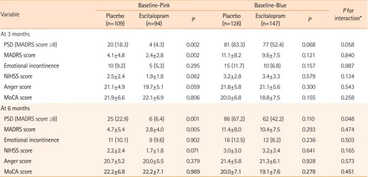
In the Baseline-Pink and Baseline-Blue patients, clinical vari- ables at baseline were well-balanced between the escitalopram and placebo users except for a higher prevalence of emotional incontinence in the placebo arm of the Baseline-Pink group (Supplementary Table 1). Patient responses to escitalopram at 3 months after initiation of therapy were stratified by group (Table 2). In the Baseline-Pink group, patients who were ran- domized to escitalopram showed significantly lower MADRS scores and less frequent PSD (MADRS ≥8) at 3 months. In addi- tion, NIHSS and anger scores tended to be lower among esci- talopram users. In the Baseline-Blue group, although the esci- talopram group was less likely to have PSD (MADRS ≥8) with a marginal statistical significance, escitalopram generally did not Table 1. Baseline characteristics of patients with initial depressive symptoms (Baseline-Blue) and those without (Baseline-Pink)
Characteristic All patients (n=478) Baseline-Pink (n=203) Baseline-Blue (n=275) P *
Demographic and risk factors
Age (yr) 64.0±12.1 62.4±12.1 65.2±12.0 0.013
Female sex 187 (39.1) 63 (31.0) 124 (45.1) 0.002
Hypertension 360 (75.3) 156 (76.8) 204 (74.2) 0.504
Diabetes 204 (42.7) 78 (38.4) 126 (45.8) 0.106
Hyperlipidemia 235 (49.2) 98 (48.3) 137 (49.8) 0.739
Coronary artery disease 63 (13.2) 24 (11.8) 39 (14.2) 0.451
Smoking 230 (48.1) 112 (55.2) 118 (42.9) 0.008
Time-to-randomization 7 (4—10) 7 (4—10) 7 (5—11) 0.206
Lesion side 0.357
Right 239 (50.0) 95 (46.8) 144 (52.4)
Left 208 (43.5) 96 (47.3) 112 (40.7)
Both 31 (6.5) 12 (5.9) 19 (6.9)
Lesion location
Anterior cortex 126 (26.4) 46 (22.7) 80 (29.1) 0.115
Thalamus 43 (9.0) 21 (10.3) 22 (8.0) 0.376
Medulla 39 (8.2) 16 (7.9) 23 (8.4) 0.849
Cerebellum 37 (7.7) 13 (6.4) 24 (8.7) 0.347
Posterior cortex 24 (5.0) 7 (3.4) 17 (6.2) 0.176
Subcortex 226 (47.3) 96 (47.3) 130 (47.3) 0.997
Pons+midbrain 106 (22.2) 44 (21.7) 62 (22.5) 0.821
Clinical variables
MADRS 10.7±8.2 3.4±2.4 16.0±6.7 <0.001
NIHSS 4.8±2.9 4.2±2.3 5.3±3.1 <0.001
Emotional incontinence 32 (6.7) 6 (3.0) 26 (9.5) 0.005
Anger proneness 22.8±5.7 22.6±5.4 22.9±5.9 0.528
MoCA 18.4±6.9 19.8±6.5 17.4±7.1 <0.001
Values are presented as mean±standard deviation, number (%), or median (interquartile range).
MADRS, Montgomery-Asberg Depression Rating Scale; NIHSS, National Institute of Health Stroke Scale; MoCA, Montreal Cognitive Assessment.
*P via two-tailed t-test for Baseline-Pink vs. Baseline-Blue groups.
improve emotional and neurological disturbances to a signifi- cant degree. The favorable effect of escitalopram on patients’
depressive symptoms in the Baseline-Pink group was persistent up to 6 months after the index stroke (i.e., 3 months after medication cessation). Of note, there was a trend interaction between baseline depression and the effect of escitalopram on PSD prevention at 3 months (P for interaction=0.058), which was significant at 6 months (P for interaction=0.048).
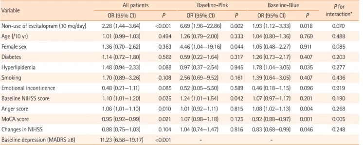
Next, we analyzed independent predictors of 3-month PSD and explored whether they would be different based on the presence of baseline depression. Candidate variables were se- lected as those with a P<0.10. Differences between the Base- line-Pink and the Baseline-Blue groups were significant in terms of age, female percentage, smoking rates, NIHSS, emo- tional incontinence rates, and MoCA scores (Table 1). PSD at 3 months was associated with old age, being female, a diagnosis of diabetes or hyperlipidemia, non-use of escitalopram, baseline NIHSS, anger levels, and MoCA scores (Table 3). The variables showing an interaction with baseline depression for 3-month PSD were non-use of escitalopram, anger scores, MoCA scores, and changes in NIHSS score (0 to 1 month) (Supplementary Ta- ble 2). The presence of baseline depression (MADRS ≥8) was also adjusted via a multivariable model for all patients.
In the final model (Table 4) including all patients, the pres- ence of baseline depression (MADRS ≥8) was most strongly as-
sociated with PSD at 3 months. In addition, higher baseline NI- HSS scores and anger scores, cognitive dysfunction, and non- use of escitalopram were identified as independent predictors of the development of PSD at 3 months. Changes in NIHSS scores were marginally significant. We then assessed whether the effect of each risk factor on PSD at 3 months would differ according to the presence of baseline depression. In the Base- line-Pink group, female sex, baseline NIHSS score, and non-use of escitalopram significantly increased the odds of PSD at 3 months, while in the Baseline-Blue group, hyperlipidemia, higher anger scores, cognitive dysfunction (MoCA scores), changes in NIHSS scores, and non-use of escitalopram were significantly associated with PSD at 3 months. Among these variables, cognitive dysfunction, being female, and non-use of escitalopram demonstrated statistically significant interactions or trends towards interactions with baseline depression (P for interaction <0.10). Although non-use of escitalopram was an independent predictor of 3-month PSD in both the Baseline- Pink and Baseline-Blue groups, the PSD odds ratio was sub- stantially greater in the Baseline-Pink group than in the Base- line-Blue group.
Figure 2 illustrates changes in MADRS scores throughout the study period. In the Baseline-Pink group, placebo and escitalo- pram users displayed different patterns; patients randomized to the placebo treatment showed a gradual increase in MADRS Table 2. Efficacy of escitalopram according to baseline depressive symptoms
Variable
Baseline-Pink Baseline-Blue
P for interaction*
Placebo
(n=109) Escitalopram
(n=94) P Placebo
(n=128) Escitalopram
(n=147) P
At 3 months
PSD (MADRS score ≥8) 20 (18.3) 4 (4.3) 0.002 81 (63.3) 77 (52.4) 0.068 0.058
MADRS score 4.1±4.8 2.4±2.8 0.002 11.1±8.2 9.6±7.5 0.121 0.840
Emotional incontinence 10 (9.2) 5 (5.3) 0.295 15 (11.7) 10 (6.8) 0.157 0.987
NIHSS score 2.5±2.4 1.9±1.8 0.062 3.2±2.8 3.4±3.3 0.579 0.134
Anger score 21.1±4.9 19.7±5.1 0.059 21.8±5.8 21.1±5.6 0.300 0.543
MoCA score 21.9±6.6 22.1±6.9 0.806 20.0±6.8 18.8±7.5 0.155 0.258
At 6 months
PSD (MADRS score ≥8) 25 (22.9) 6 (6.4) 0.001 86 (67.2) 62 (42.2) 0.110 0.048
MADRS score 4.7±5.4 2.8±4.0 0.005 11.4±8.0 10.4±7.5 0.293 0.474
Emotional incontinence 11 (10.1) 9 (9.6) 0.902 16 (12.5) 12 (8.2) 0.236 0.503
NIHSS score 2.2±2.4 1.7±1.8 0.071 3.0±3.0 3.2±3.4 0.641 0.165
Anger score 20.7±5.2 20.0±5.5 0.379 21.4±5.8 21.3±6.1 0.928 0.573
MoCA score 22.2±6.8 22.2±7.1 0.969 20.0±7.1 19.1±7.6 0.278 0.451
Values are presented as number (%) or mean±standard deviation.
PSD, post-stroke depression; MADRS, Montgomery-Asberg Depression Rating Scale; NIHSS, National Institute of Health Stroke Scale; MoCA, Montreal Cogni- tive Assessment.
*P for interaction according to the presence of baseline depressive symptoms (MADRS ≥8).
scores over time while those randomized to escitalopram dem- onstrated an improvement; a significant difference in MADRS scores between treatment groups was demonstrated at 3 months and maintained at 6 months. In the Baseline-Blue group, however, both placebo and escitalopram users showed a uniform decrease in MADRS scores for the first 3 months with- out significant difference between treatment groups. Improve- ments were most prominent during the first month and gradu- ally diminished thereafter.
Lastly, clinical characteristics were compared between PSD (at 3 months) patients in the Baseline-Pink and Baseline-Blue groups (Table 5). At baseline, all clinical variables, except for depressive symptoms and cognitive dysfunction, were compa-
rable. At 3 months, depressive symptoms (MADRS score) and cognitive dysfunction (MoCA score) remained diminished while emotional incontinence was more prevalent in PSD patients in the Baseline-Pink group.
Discussion
We examined various clinical characteristics and factors asso- ciated with PSD according to the depression levels immediately following a stroke. As the data were obtained as part of a ran- domized controlled study, risk factors and clinical variables were prospectively collected and well balanced between the two treatment arms. We found that responses to escitalopram Table 3. Risk factors for post-stroke depression (MADRS ≥8) at 3 months in all the patients
Variable PSD at 3 months
Without (n=296) With (n=182) P
Demographic and risk factors
Age (yr) 62.5±11.8 66.4±12.4 0.001
Female sex 107 (36.1) 80 (44.0) 0.089
Hypertension 75 (25.3) 43 (23.6) 0.673
Diabetes 116 (39.2) 88 (48.4) 0.049
Hyperlipidemia 136 (45.9) 99 (54.4) 0.073
Coronary artery disease 38 (12.8) 25 (13.7) 0.778
Smoking 145 (49.0) 85 (46.7) 0.628
Lesion side 0.131
Right 144 (48.6) 95 (52.2)
Left 137 (46.3) 71 (39.0)
Both 15 (5.1) 16 (8.8)
Lesion location
Anterior cortex 72 (24.3) 54 (29.7) 0.198
Thalamus 29 (9.8) 14 (7.7) 0.435
Medulla 25 (8.4) 14 (7.7) 0.770
Cerebellum 20 (6.8) 17 (9.3) 0.305
Posterior cortex 13 (4.4) 11 (6.0) 0.422
Subcortex 135 (45.6) 91 (50.0) 0.350
Pons+Midbrain 61 (20.6) 45 (24.7) 0.293
Clinical variables at baseline
Non-use of escitalopram (10 mg/day) 160 (54.1) 81 (44.5) 0.043
MADRS 7.1±5.9 16.4±8.2 < 0.001
NIHSS score 4.4±2.6 5.5±3.1 < 0.001
Changes in NIHSS (0–1 mo) 1.5±1.6 1.3±1.5 0.147
Emotional incontinence 21 (7.1) 11 (6.0) 0.655
Anger score 22.1±5.8 23.9±5.2 < 0.001
MoCA score 19.7±6.3 16.3±7.3 < 0.001
Values are presented as mean±standard deviation or number (%).
MADRS, Montgomery-Asberg Depression Rating Scale; PSD, post-stroke depression; NIHSS, National Institute of Health Stroke Scale; MoCA, Montreal Cogni- tive Assessment.
and predictors for PSD at 3 months differed between the Base- line-Blue and Baseline-Pink groups.
The efficacy of escitalopram was more marked in the Baseline- Pink than in the Baseline-Blue group (Table 2). PSD risk and MADRS scores at 3 months were significantly lower in the esci- talopram users than in the placebo users only in the Baseline- Pink group. Of note, in reducing the PSD at 3 months, there was a trend interaction between baseline depression and escitalo- pram use. Moreover, non-use of escitalopram was a more impor- tant predictor of PSD at 3 months in the Baseline-Pink than in the Baseline-Blue groups (Table 4). Perhaps, in the Baseline-Blue group, factors such as neurologic recovery from severe deficits and a relatively low medication dose might have contributed to the relatively diminished effectiveness of escitalopram. Given
higher baseline MADRS scores (mean of 16) in the Baseline-Blue group, a higher dose of escitalopram (>10 mg/day) might have been necessary to reduce PSD risk at 3 months.
Consistent with a previous report,17 baseline neurological dis- ability, as assessed by the NIHSS, was a significant predictor of PSD in all participants in our study. However, the degree of neu- rologic improvement (changes in NIHSS scores) during the first month was not an important determinant of PSD levels at 3 months, although there was a trend relationship between neuro- logic improvement and the decreased risk of PSD (odds ratio of 0.88 according to one-point improvement of NIHSS score). In a previous report from our group, we reported that changes in NI- HSS scores during an initial 3 months of treatment were closely associated with changes in MADRS scores in patients with base- line depression (baseline MADRS ≥16 [r=0.206, P=0.040] or a baseline MADRS score of 8 to 15 [r=0.171, P=0.049]), but not in those without baseline depression (baseline MADRS <8 [r=
–0.023, P=0.76]).11 Based on these findings, we speculated that patients with severe baseline depression and severe neurological deficits largely reflect a form of “reactive depression” that im- proves over time along with neurological recovery, masking the efficacy of escitalopram. In this post hoc analysis, however, changes in NIHSS were not clearly identified as an independent risk factor for PSD in patients with baseline depression. Different time periods employed in our current and earlier studies may have affected the diverging results. In this study, we used a shorter time period of 1 month after the index stroke (rather than 3 months) to evaluate the predictive effect of NIHSS score change on the development of PSD at 3 months.
Follow-up (wk)
Mean of MADRS
0 0 5 10 15 20
4 12 24
Baseline-Pink: placebo Baseline-Pink: escitalopram Baseline-Blue: placebo Baseline-Blue: escitalopram
Figure 2. Changes in Montgomery-Asberg Depression Rating Scale (MADRS) scores during the study period. *P<0.001.
Table 4. Independent predictors of post-stroke depression (MADRS ≥8) at 3 months
Variable All patients Baseline-Pink Baseline-Blue P for
interaction*
OR (95% CI) P OR (95% CI) P OR (95% CI) P
Non-use of escitalopram (10 mg/day) 2.28 (1.44—3.64) <0.001 6.69 (1.96—22.86) 0.002 1.93 (1.12—3.33) 0.018 0.070
Age (/10 yr) 1.01 (0.99—1.03) 0.494 1.26 (0.79—2.00) 0.333 1.04 (0.80—1.36) 0.769 0.488
Female sex 1.36 (0.70—2.62) 0.363 4.46 (1.04—19.16) 0.044 1.05 (0.48—2.27) 0.911 0.085
Diabetes 1.14 (0.72—1.80) 0.569 0.59 (0.22—1.64) 0.317 1.26 (0.73—2.17) 0.407 0.203
Hyperlipidemia 1.48 (0.94—2.33) 0.088 0.97 (0.37—2.54) 0.945 1.78 (1.04—3.05) 0.035 0.277
Smoking 1.70 (0.89—3.26) 0.108 2.56 (0.69—9.52) 0.161 1.39 (0.64—3.05) 0.407 0.436
Emotional incontinence 0.48 (0.21—1.11) 0.085 0.52 (0.05—5.50) 0.589 0.46 (0.18—1.15) 0.096 0.919 Baseline NIHSS score 1.10 (1.01—1.20) 0.025 1.24 (1.01—1.54) 0.042 1.07 (0.97—1.17) 0.201 0.190
Anger score 1.06 (1.01—1.10) 0.010 1.01 (0.92—1.11) 0.815 1.08 (1.02—1.13) 0.004 0.268
MoCA score 0.95 (0.92—0.99) 0.021 1.07 (0.98—1.18) 0.125 0.92 (0.88—0.97) 0.001 0.005
Changes in NIHSS 0.88 (0.75—1.03) 0.104 1.04 (0.74—1.47) 0.816 0.83 (0.68—0.99) 0.046 0.248
Baseline depression (MADRS ≥8) 11.23 (6.58—19.17) <0.001 - -
MADRS, Montgomery-Asberg Depression Rating Scale; OR, odds ratio; CI, confidence interval; NIHSS, National Institute of Health Stroke Scale; MoCA, Mon- treal Cognitive Assessment.
*P for interaction according to the presence of baseline depressive symptoms (MADRS ≥8).
We found that baseline cognitive dysfunction, measured by MoCA, was associated with PSD at 3 months across all patients (Table 4). Cognitive dysfunction, as measured by tools such as Mini-Mental State examination18 and Abbreviated Memory Test,19 has been shown to be associated with PSD.8 However, we also found that baseline cognitive dysfunction, which was more common in the Baseline-Blue group, was a significant predictor for PSD only among Baseline-Blue patients and not among Baseline-Pink patients (Table 4). In addition, among pa- tients with PSD at 3 months, cognitive function was worse in the Baseline-Blue group than in the Baseline-Pink group (Table 5). The association between cognitive decline and persistent depression may, in part, be related to shared diagnostic items such as “difficulty in concentration.” Alternatively, both depres- sion and cognitive dysfunction may be common manifestations of damage in certain structures such as the frontal lobes or multiple small vessels which cause white matter changes. Un- fortunately, this study was not able to explore the impact of
small vessel disease on depression and cognitive dysfunction.
Generally, females are known to have a higher risk of PSD than males.20 Less social support after the index stroke may re- sult in this outcome, considering that female patients are re- ported to receive relatively little support from both health care providers and the broader community.21,22 In our study, being female was not an independent predictor of PSD in all of the patients. However, being female increased the risk of PSD in the Baseline-Pink group but not in the Baseline-Blue group.
The reason for this remains elusive. We speculate that, in the Baseline-Blue group there were other strong predictors (e.g., more severe neurologic deficits and cognitive dysfunction) of PSD and thus the contribution of sex to PSD might have been masked by our use of a multiple variable model.
Our results suggest that different approaches are necessary to treat/prevent PSD based on the presence of baseline depres- sion. In patients without baseline depression, depressive symp- toms gradually worsened over time if a placebo was given but Table 5. Group-based differences in the clinical characteristics of patients with post-stroke depressive symptoms 3 months following stroke
Characteristic PSD at 3 months
Baseline-Pink (n=24) Baseline-Blue (n=158) P Demographics
Age (yr) 62 (51—78) 69 (59—76) 0.344
Female sex 10 (41.7) 70 (44.3) 0.808
Lesion location
Anterior cortex 6 (25.0) 48 (30.4) 0.591
Thalamus 1 (4.2) 13 (8.2) 0.698
Medulla 1 (4.2) 13 (8.2) 0.698
Cerebellum 1 (4.2) 16 (10.1) 0.704
Posterior cortex 1 (4.2) 10 (6.3) >0.999
Subcortex 14 (58.3) 77 (48.7) 0.381
Pons+Midbrain 6 (25.0) 39 (24.7) >0.999
Clinical variables at baseline
MADRS 4 (2—6) 18 (12—24) <0.001
NIHSS score 4 (3—7) 5 (3—7) 0.831
Emotional incontinence 1 (4.2) 10 (6.3) >0.999
Anger score 24 (19—26) 24 (21—28) 0.280
MoCA score 23 (15—27) 16 (11—21) 0.002
Clinical variables at 3 months
MADRS 11 (9—14) 14 (11—20) 0.004
NIHSS score 3 (1—5) 3 (1—6) 0.265
Emotional incontinence 6 (25.0) 14 (8.9) 0.030
Anger score 23 (18—26) 23 (20—26) 0.517
MoCA score 27 (18—28) 19 (11—23) <0.001
Values are presented as median (interquartile range) or number (%).
PSD, post-stroke depression; MADRS, Montgomery-Asberg Depression Rating Scale; NIHSS, National Institute of Health Stroke Scale; MoCA, Montreal Cogni- tive Assessment.
improved with escitalopram (Figure 2). Escitalopram appears to be effective in this group of patients (Table 4) and therefore active pharmacological prevention might be a reasonable ap- proach, especially in female patients or those with a mild stroke. Our findings align with a recent meta-analysis that re- ported on the efficacy of selective serotonin reuptake inhibitors (SSRIs) in preventing depression among initially non-depressed stroke patients.23 However, given a low rate of PSD develop- ment at 3 months (approximately 18%) in the placebo users of the Baseline-Pink group, the cost-effectiveness and feasibility of this approach needs to be further evaluated. In contrast, in the Baseline-Blue group, escitalopram was found to be less ef- fective and PSD was more closely related to neurological im- provements. Given this, early and active rehabilitation may be necessary to promote the improvement of neurologic deficits and consequently of depressive symptoms in this group of pa- tients. However, it should be further noted that, we used fixed doses (10 mg) of escitalopram for a set duration (3 months).
Therefore, it remains to be studied whether a higher dose and/
or longer duration of escitalopram administration,24 or the use of another class of antidepressant, would be more effective in treating Baseline-Blue patients.
Finally, among patients with PSD at 3 months, patients with- out baseline depression were more likely to have newly-devel- oped emotional incontinence as compared to those with base- line depression (Table 5). It has been reported that emotional incontinence was closely associated with damages to subcorti- cal structures (e.g., the basal ganglia or internal capsule).2,25 Because there are abundant serotonergic fibers in these ar- eas,26 altered neurotransmission after ischemic brain damage may play a role in the development of emotional incontinence.9 The more frequent development of emotional incontinence and relatively good response rates to escitalopram suggest that de- pressive symptoms in the Baseline-Pink group may be more closely associated with neurochemical changes27 due to brain damage than those in the Baseline-Blue group.
There are a number of limitations in the present study. First, we utilized post hoc analyses data from a randomized con- trolled trial. Therefore, baseline variables could not be balanced with the efficacy of escitalopram in each study group. Second, all patients were ethnically Korean, and the generalizability of our results to other ethnic populations may be limited. Third, in this clinical trial, we did not compile information on items such as patient socioeconomics, job status, or familial/social sup- port, which may play roles in the development of depression.27 Finally, we examined the efficacy of escitalopram with a fixed dosage (10 mg/day) for a limited period.
Despite these limitations, our results demonstrate that pa-
tient responses to escitalopram and predictors of PSD at 3 months may vary according to the presence of depression im- mediately following stroke. These findings highlight the het- erogeneity of PSD pathophysiology and suggest the require- ment for tailored therapeutic approaches in stroke patients who are at risk of PSD.
Supplementary materials
Supplementary materials related to this article can be found online at https://doi.org/10.5853/jos.2017.02712.
Disclosure
Jong S. Kim has received grants from Dong-A Pharmaceutical Company and from the Ministry for Health, Welfare, and Family Affairs, South Korea. All other authors declare no competing in- terests.
Acknowledgments
This study was supported by the Ministry for Health, Welfare, and Family Affairs, Republic of Korea (HI14C1985).
References
1. Robinson RG, Jorge RE. Post-stroke depression: a review. Am J Psychiatry 2016;173:221-231.
2. Kim JS, Choi-Kwon S. Poststroke depression and emotional incontinence: correlation with lesion location. Neurology 2000;54:1805-1810.
3. Parikh RM, Robinson RG, Lipsey JR, Starkstein SE, Fedoroff JP, Price TR. The impact of poststroke depression on recovery in activities of daily living over a 2-year follow-up. Arch Neurol 1990;47:785-789.
4. House A, Dennis M, Mogridge L, Warlow C, Hawton K, Jones L. Mood disorders in the year after first stroke. Br J Psychiatry 1991;158:83-92.
5. Wade DT, Legh-Smith J, Hewer RA. Depressed mood after stroke. A community study of its frequency. Br J Psychiatry 1987;151:200-205.
6. Aström M, Adolfsson R, Asplund K. Major depression in stroke patients. A 3-year longitudinal study. Stroke 1993;24:976- 982.
7. Farner L, Wagle J, Engedal K, Flekkøy KM, Wyller TB, Fure B.
Depressive symptoms in stroke patients: a 13 month follow- up study of patients referred to a rehabilitation unit. J Affect Disord 2010;127:211-218.
8. Ayerbe L, Ayis S, Rudd AG, Heuschmann PU, Wolfe CD. Natural history, predictors, and associations of depression 5 years after stroke: the South London Stroke Register. Stroke 2011;42:
1907-1911.
9. Kim JS. Post-stroke mood and emotional disturbances: phar- macological therapy based on mechanisms. J Stroke 2016;18:
244-255.
10. Naess H, Kurtz M, Thomassen L, Waje-Andreassen U. Serial NIHSS scores in patients with acute cerebral infarction. Acta Neurol Scand 2016;133:415-420.
11. Kim JS, Lee EJ, Chang DI, Park JH, Ahn SH, Cha JK, et al. Effi- cacy of early administration of escitalopram on depressive and emotional symptoms and neurological dysfunction after stroke: a multicentre, double-blind, randomised, placebo- controlled study. Lancet Psychiatry 2017;4:33-41.
12. Kearns NP, Cruickshank CA, McGuigan KJ, Riley SA, Shaw SP, Snaith RP. A comparison of depression rating scales. Br J Psy- chiatry 1982;141:45-49.
13. Kim JS, Choi S, Kwon SU, Seo YS. Inability to control anger or aggression after stroke. Neurology 2002;58:1106-1108.
14. Brott T, Adams HP Jr, Olinger CP, Marler JR, Barsan WG, Biller J, et al. Measurements of acute cerebral infarction: a clinical examination scale. Stroke 1989;20:864-870.
15. Nasreddine ZS, Phillips NA, Bédirian V, Charbonneau S, Whitehead V, Collin I, et al. The Montreal Cognitive Assess- ment, MoCA: a brief screening tool for mild cognitive im- pairment. J Am Geriatr Soc 2005;53:695-699.
16. Higgins SL, Hummel JD, Niazi IK, Giudici MC, Worley SJ, Sax- on LA, et al. Cardiac resynchronization therapy for the treat- ment of heart failure in patients with intraventricular con- duction delay and malignant ventricular tachyarrhythmias. J Am Coll Cardiol 2003;42:1454-1459.
17. Kutlubaev MA, Hackett ML. Part II: predictors of depression after stroke and impact of depression on stroke outcome: an updated systematic review of observational studies. Int J
Stroke 2014;9:1026-1036.
18. Tombaugh TN, McIntyre NJ. The mini-mental state examina- tion: a comprehensive review. J Am Geriatr Soc 1992;40:922- 935.
19. Jitapunkul S, Pillay I, Ebrahim S. The abbreviated mental test:
its use and validity. Age Ageing 1991;20:332-336.
20. Poynter B, Shuman M, Diaz-Granados N, Kapral M, Grace SL, Stewart DE. Sex differences in the prevalence of post-stroke depression: a systematic review. Psychosomatics 2009;50:563- 569.
21. Reeves MJ, Bushnell CD, Howard G, Gargano JW, Duncan PW, Lynch G, et al. Sex differences in stroke: epidemiology, clinical presentation, medical care, and outcomes. Lancet Neurol 2008;7:915-926.
22. Glader EL, Stegmayr B, Norrving B, Terént A, Hulter-Asberg K, Wester PO, et al. Sex differences in management and out- come after stroke: a Swedish national perspective. Stroke 2003;34:1970-1975.
23. Salter KL, Foley NC, Zhu L, Jutai JW, Teasell RW. Prevention of poststroke depression: does prophylactic pharmacotherapy work? J Stroke Cerebrovasc Dis 2013;22:1243-1251.
24. Bech P, Andersen HF, Wade A. Effective dose of escitalopram in moderate versus severe DSM-IV major depression. Phar- macopsychiatry 2006;39:128-134.
25. Poeck K. Pathophysiology of emotional disorders associated with brain damage. In: Vinken PJ, Bruyn GW. Handbook of Clinical Neurology. New York, NY: Elsevier, 1969:343-367.
26. Kim JS. Post-stroke emotional incontinence after small len- ticulocapsular stroke: correlation with lesion location. J Neu- rol 2002;249:805-810.
27. Choi-Kwon S, Han K, Choi S, Suh M, Kim YJ, Song H, et al.
Poststroke depression and emotional incontinence: factors re- lated to acute and subacute stages. Neurology 2012;78:1130- 1137.
Supplementary Table 1. Baseline characteristics of patients (placebo vs. escitalopram)
Characteristic Baseline-Pink Baseline-Blue
Placebo (n=109) Escitalopram (n=94) P Placebo (n=128) Escitalopram (n=147) P At baseline
Age (yr) 63.0±12.5 61.7±11.8 0.452 64.9±11.3 65.4±12.7 0.746
Female sex 32 (29.4) 31 (33.0) 0.578 54 (42.2) 70 (47.6) 0.367
Hypertension 80 (73.4) 76 (80.9) 0.209 97 (75.8) 107 (72.8) 0.572
Diabetes 47 (43.1) 31 (33.0) 0.139 55 (43.0) 71 (48.3) 0.376
Hyperlipidemia 57 (52.3) 41 (43.6) 0.217 62 (48.4) 75 (51.0) 0.669
Coronary artery disease 12 (11.0) 12 (12.8) 0.699 18 (14.1) 21 (14.3) 0.958
Smoking 59 (54.1) 53 (56.4) 0.747 51 (39.8) 67 (45.6) 0.338
Lesion side 0.097 0.739
Right 55 (50.5) 40 (42.6) 70 (54.7) 74 (50.3)
Left 45 (41.3) 51 (54.3) 49 (38.3) 63 (42.9)
Both 9 (8.3) 3 (3.2) 9 (7.0) 10 (6.8)
Clinical variables
MADRS 3.6±2.3 3.2±2.5 0.320 16.6±6.7 15.5±6.7 0.222
NIHSS 4.2±2.6 4.2±2.1 0.922 5.2±3.0 5.3±3.2 0.686
Emotional incontinence 6 (5.5) 0 0.021 12 (9.4) 14 (9.5) 0.966
Anger proneness 22.9±5.1 22.1±5.7 0.278 23.2±5.6 22.6±6.1 0.405
MoCA 20.0±6.4 19.6±6.6 0.656 18.1±6.9 16.8±7.2 0.145
Values are presented as mean±standard deviation or number (%).
MADRS, Montgomery-Asberg Depression Rating Scale; NIHSS, National Institute of Health Stroke Scale; MoCA, Montreal Cognitive Assessment.
Supplementary Table 2. Risk factors of post-stroke depressive symptoms at 3 months by the presence of baseline depression
Variable Crude odds ratio (95% CI) P for interaction with
baseline depression Baseline-Pink (n=296) Baseline-Blue (n=182)
Demographic and risk factors
Age (/10 yr) 1.01 (0.97—1.05) 1.03 (1.01—1.05)* 0.372
Female sex 1.70 (0.71—4.06) 0.93 (0.57—1.50) 0.235
Hypertension 0.89 (0.33—2.39) 1.34 (0.78—2.31) 0.479
Diabetes 0.78 (0.32—1.92) 1.58 (0.98—2.57) 0.174
Hyperlipidemia 1.08 (0.46—2.54) 1.64 (1.01—2.66)† 0.403
Coronary artery disease 0.30 (0.04—2.29) 1.22 (0.61—2.44) 0.199
Smoking 1.16 (0.49—2.74) 1.22 (0.75—1.97) 0.923
Lesion side 0.879
Left vs. Right 0.99 (0.41—2.40) 0.82 (0.50—1.36)
Both vs. Right 1.53 (0.30—7.90) 2.00 (0.68—5.85)
Lesion location
Anterior cortex 1.16 (0.43—3.11) 1.16 (0.68—1.97) 0.999
Thalamus 0.35 (0.04—2.70) 1.08 (0.44—2.61) 0.320
Medulla 0.48 (0.06—3.77) 0.96 (0.41—2.27) 0.540
Cerebellum 0.61 (0.08—4.87) 1.54 (0.63—3.72) 0.421
Posterior cortex 1.25 (0.14—10.88) 1.06 (0.39—2.88) 0.891
Subcortex 1.66 (0.70—3.93) 1.15 (0.71—1.85) 0.467
Pons+Midbrain 1.24 (0.46—3.33) 1.34 (0.75—2.40) 0.892
Clinical variables at baseline
Non-use of escitalopram (10 mg/day) 5.00 (1.67—14.29)* 1.56 (0.96—2.56) 0.058
MADRS 1.19 (0.98—1.44) 1.16 (1.11—1.22)* 0.791
NIHSS score 1.25 (1.05—1.48)† 1.07 (0.99—1.16) 0.117
Changes in NIHSS (0–4 wk) 1.12 (0.86—1.46) 0.86 (0.73—1.00)† 0.082
Emotional incontinence 1.51 (0.17—13.53) 0.43 (0.19—0.98)† 0.289
Anger score 1.00 (0.92—1.08) 1.09 (1.04—1.14)* 0.053
MoCA score 1.02 (0.95—1.09) 0.92 (0.88—0.95)* 0.008
CI, confidence interval; MADRS, Montgomery-Asberg Depression Rating Scale; NIHSS, National Institute of Health Stroke Scale; MoCA, Montreal Cognitive Assessment.
*P<0.01; †P<0.05.