I n t rod u c t i o n
H e i l m a n
1first described an unusual form of tremor involving the legs and trunk, which was present only when standing. He called this move-
ment disorder “orthostatic tremor”. Over 100 cases have since been reported. However there have been some confusions about orthostatic tremor in several aspects. Therefore the defini- tion and the classification criteria of the ortho- static tremor were not clear yet. For the past ten years, we have observed 4 patients with ortho- static tremor. In each case tests were performed to investigate the following three important areas of inquiry. Firstly, whether this disorder is an independent diagnostic entity or a variant of essential tremor. Secondly, whether the progress
길입성 진전의 병태생리: 다증 증례 연구
전북대학교 의과대학 신경과학교실, 서울대학교 의과대학 신경과학교실*
서 만 욱・이 광 우*
Pathophysiology of orthostatic tremor: a multiple case study
Man-Wook Seo, M.D., Kwang-Woo Lee, M.D.*
Department of Neurology, College of medicine, Jeonbuk National University Department of Neurology, College of medicine, Seoul National University*
Introduction : Orthostatic tremor develops in the legs while standing up with no weakness, pain or imbalance in the leg and the tremor is characteristically not observed when walking. However there have been some confusions about orthostatic tremor in several aspects. For the past ten years, we have observed 4 patients with orthostatic tremor. In each case tests were performed to investigate the following three important areas of inquiry about orthostatic tremor. Firstly, whether this disorder is an independent diagnostic entity or a variant of essential tremor. Secondly, whether the progress of this disorder is specifically related with standing posture. Lastly, the nature of the pathophysiologic mechanism behind the appearance of the tremor when standing after the lapse of a certain latent period and its disappearance upon the commencement of walking.
Methods : Our 4 cases of orthostatic tremor were studied clinically, electrophysiologically, and pharmacologically.
Electrophysiological tests included tremor spectrum test and electromyography.
Results : We observed the presence of this tremor in several other tonic postures, as well as its absence, in a vertically lifted position from all our cases. Our cases registered a variable tremor frequency between 5 and 12 Hz according to the tremor spectrum test and EMG. Furthermore all our 4 cases demonstrated patterns of both synchronous EMG activi- ty and alternating EMG activity at various times in homologous muscles of both legs. Orthostatic tremor was improved significantly with propranolol as well as clonazepam.
Conclusions : From the results of our study we drew the following conclusions. It is probable that orthostatic tremor is simply a variant of essential tremor rather than being an independent diagnostic entity and that in most cases its development is specifically related with muscle contraction rather than merely with the act of standing. Furthermore we discovered a clue in the previously described neural control mechanism that the nuclear bag fibers in the muscle spindle have lag time of several seconds in their response to muscle strength and that their baseline does not reset fully in rapid- ly moving muscle. This neural control mechanism could offer sufficient explanation for the phenomena of tremor appearance when standing and disappearance when walking in orthostatic tremor.
Key Words : Orthostatic tremor, Pathophysiology, EMG
대한임상신경생리학회지 4(1):000~000, 2002 ISSN 1229-6414
Address for correspondence Man-Wook Seo, M.D.
Department of Neurology,
Medical School, Chonbuk National University, Chonju, Chonbuk, 561-712, Korea
Tel : + 82-652-250-1895 Fax : +82-652-251-9363 E-mail : [email protected]
of this disorder is specifically related with stand- ing posture. Lastly, the nature of the pathophysi- ologic mechanism behind the appearance of the tremor when standing after the lapse of a certain latent period and its disappearance upon the commencement of walking. The presentation of our case results is followed by literature review involving the comparison and analysis of pub- lished results with those of our own study.
M e t h od s
O r t hostatic tremor develops in the legs while standing up with no weakness, pain or imbalance in the leg and the tremor is characteristically not observed when walking. All our cases satisfied with this Kelly and Sharbrough’ s diagnostic criteria of orthostatic tremor.
2Our 4 cases of orthostatic tremor were studied clinically, electrophysiologi- cally, and pharmacologically. Electrophysiological tests included tremor spectrum test and elec- tromyography. In the use of a Motus I gyroscope (Bioengineering) for the former, the motus angular rate sensors were securely fastened using plaster to the chin, dorsum of the hand, and lower extremity to minimize the development of artifacts during recording. The bandwidth of the sensor was DC 30 Hz. The measured data were analyzed into root mean squared(RMS) values using soft- ware developed by the gyroscope manufacturer, MOTUS I, in order to obtain an accurate angular rate. The peak frequency was determined through spectrum analysis with fast Fourier transforms.
Electromyography was performed by placing sur- face electrodes at bilateral tibialis anterior as homologous muscles of both legs.
Re s u l t s
Case 1
A 20-year old man visited our hospital in May 1992 with a history of several years of slowly pro- gressive unsteadiness and trembling in his legs when standing still. His family demonstrated an autosomal trait(Fig. 1) and had a history of essen- tial tremor, with 7 of 65 members presenting with leg predominant tremors. The case satisfied B r i t t o n’s criteria of primary orthostatic tremor ( P O T )
3, except with regard to frequency. He had no vertiginous or presyncopal symptoms, no history
of trauma, infection, drug usage, or toxin expo- sures, and his tremor was unaffected by coffee and alcohol. Neurological examination was normal except for the tremor. Thyroid function test, EEG, and brain CT were normal. In addition to stand- ing, the leg tremor was also observed in any posi- tion involving periods of sustained muscle con- traction including sitting, lying down, and leaning.
The tremor appeared a few seconds after muscle contraction and its severity gradually increased.
Furthermore, the tremor gradually reappeared with slow rocking movements of the feet but dis- appeared with fast rocking movements. When the patient was seated and relaxed, no EMG activity was noted. But upon standing, synchronous EMG activity and alternating EMG activity of about 10 Hz phase appeared at various times between left and right tibialis anterior upon EMG examination of the lower extremity. There was a delay between standing and onset of EMG activity. EMG activity disappeared when the patient was lifted vertically off the floor but reappeared when he stood up. A 9 Hz tremor independent of the leg tremor was recorded from the outstretched arm. To evaluate the pharmacological effect, the patient was placed on a course of medication comprising propra- nolol(up to 80 mg/day), primidone(up to 750 mg/
day), and clonazepam(up to 4 mg/day). For the latter, a dose of 2 mg/day was found to be effec- tive, in showing clinical improvement. However other drugs were not effective.
Case 2
A 56-year old man from a family with a history of essential tremor visited our hospital in October 1994 primarily due to tremors of the head, upper
Figure 1. The pedigree of case 1. His family demonstrated an autosomal trait and had a history of essential tremor, with 7 of 65 members presenting with leg predominant tremors.
extremities, and lower extremities. He had a his- tory of tremor of the hands starting when he was 40, tremor of the lower extremities commencing several years later, and head tremor from his early 50s as observed by his family. Lower extre- mity tremor was observed when the patient was standing but not when sitting or lifted up(Fig. 2).
The tremor disappeared when the patient walked fast but reappeared when he walked slowly.
Tremor spectrum test revealed tremors of peak frequency 5 Hz in the head, 11 Hz in the upper extremity, and 8 Hz in the lower extremity. Syn- chronous EMG activity and alternating EMG acti- vity appeared at various times between left and righ tibialis anterior upon EMG examination of the lower extremity. Lower extremity tremor decreased to the point of not interfering with daily activities with medication consisting of pro- pranolol at a dose of 80 mg/day.
Case 3
A 68-year old man visited our hospital in April 1999 primarily due to tremors of the upper extre- mities and lower extremities. He had no family history of tremor, and his tremors had developed gradually in the lower and upper extremities after 60 years of age. Neurological examination revealed normal findings except for the tremors. Findings of thyroid function test were normal. Lower extremity tremor was observed when the patient was stand- ing but not when sitting or lifted up. Tremor spec- trum test revealed a 7 Hz peak frequency postural tremor and a 5 Hz peak frequency kinetic tremor in the right upper extremity. A 5.5 Hz peak frequency
tremor was observed in standing posture but no tremor in sitting posture in the lower extremities (Fig. 3). The patient in a standing position exhibit- ed a tremor in the weight bearing leg with EMG activity only demonstrated in the weight bearing leg (Fig. 4). Electromyography performed in the lower extremity showed synchronous EMG activi- ty(90%) and alternating EMG activity(10%) between
Figure 2. EMG findings of case 2. Lower extremity tremor was observed when the patient was standing but not when lift- ed up. Furthermore the tremor disappeared when the patient walked fast but reappeared when he walked slowly. First line:
left tibialis anterior, second line: right tibialis anterior.
Figure 3. The angular rate strip chart of case 3. Tremor spec- trum test revealed a 5.5 Hz peak frequency tremor in standing posture but no tremor in sitting posture in the lower extremi- ties. A 7 Hz peak frequency postural tremor and a 5 Hz peak frequency kinetic tremor were observed in the right upper extremity.
Figure 4. EMG findings of case 3. The patient in a standing position exhibited a tremor in the weight bearing leg with EMG activity only demonstrated in the weight bearing leg. First line:
left tibialis anterior, second line: right tibialis anterior.
left and right tibialis anterior at various times.
Although upper extremity tremor was effectively decreased with the combination of clonazepam at a dose of 4 mg/day and propranolol at 80 mg/day, lower extremity tremor was not improved signifi- cantly with the same dosage of drugs.
Case 4
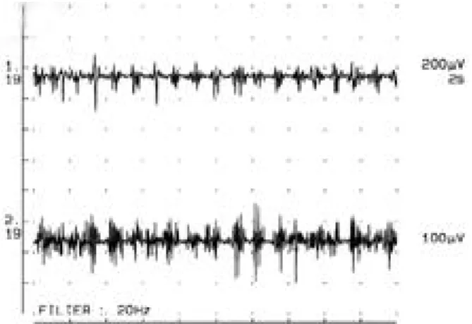
A 69-year old woman visited our hospital in April 2000 primarily due to a leg tremor when standing. The tremor was observed while stand- ing but not when sitting or lifted up. Tremor spectrum showed a 5.5 Hz peak frequency tremor when standing but no tremor when sitting. As in Case 3, the tremor only appeared in the weight bearing leg and was not seen when the patient was lifted up. Electromyography performed in the lower extremity showed synchronous EMG activi- ty(80%) and alternating EMG activity(20%) between left and right tibialis anterior at various times(Fig. 5). The tremor showed definite improvement with clonazepam at 2 mg/day.
Discussions
Our first question was whether orthostatic tremor is a variant of essential tremor or an independent diagnostic entity. Essential tremor is a common monosymptomatic disorder of unknown etiology. It is characterized by postural and kinetic tremors of the upper extremities with a tendency to be inherited as an autosomal domi- nant trait.
4Essential tremor may include a num- ber of variants such as focal task specific tremor,
kinetic predominant essential tremor, and isolat- ed head, face, tongue, chin or voice tremor.
5T h e lower limbs are seldom affected in essential tremor, but in rare cases, the legs are predomi- nantly involved.
6After Heilman’ s first descrip- tion, several authors regarded orthostatic tremor as a variant of essential tremor.
7 - 1 0H o w e v e r , Thompson et al.
1 1insisted that it should be differ- entiated from essential tremor because of its high frequency and orthostatic specificity. Following T h o m p s o n’s line, Britton et al.
3introduced a new term to classify orthostatic tremor as an inde- pendent disease entity. They selected 25 cases from previously reported cases of orthostatic tremor based on their own criteria, defined these as POT and insisted that POT and essential tremor are different in several aspects. We ana- lyzed the 25 POT cases classified by Britton et al.
3in detail and investigated the validity of their diagnostic criteria. We found the following seri- ous problems with their diagnostic criteria.
I. On clinical manifestations
In Britton’ s opinion, the predominant manifes- tations of POT were unsteadiness when standing with a fine rippling movement of the leg muscles.
Firstly however, leg tremor was observed with many tonic postures as well as when standing in several of the POT cases.
1 2 - 1 4And secondly, a fine rippling movement of the leg reflects merely high frequency leg tremor that might be due to partial fusion of the individual muscle contractions at a high frequency of tremor.
1 1II. On electrophysiological findings
Britton et al.
3considered synchronous high fre- quency tremor(around 16 Hz) in homologous mus- cles of both legs as the characteristic electro- physiological finding of POT. Firstly however, the range of tremor frequencies varied and was wide (7~32 Hz) in the reported orthostatic tremor c a s e s ,
1 - 1 4among which 3 POT cases showed a fre- quency below 12 Hz. The case reported by Kim and Lee
1 5also exhibited a low frequency tremor (4~5 Hz), while our own cases registered a vari- able tremor frequency between 5 and 12 Hz according to the tremor spectrum test and EMG.
Secondly, synchronous EMG patterns did not match the findings of orthostatic tremor. Several cases of POT showed an alternating pattern
3 , 1 1 , 1 2 , 1 6Figure 5. EMG findings of case 4. Electromyography per- formed in the lower extremity showed synchronous EMG activity(80%) and alternating EMG activity(20%) between left and right tibialis anterior at various times. First line: left tib- ialis anterior, second line: right tibialis anterior.
and synchronous bursts were also frequently observed in essential tremor.
1 7Both synchronous and alternating patterns of interaction were also observed in the same patient.
1 8All our 4 cases demonstrated patterns of both synchronous EMG activity and alternating EMG activity at various times in homologous muscles of both legs. This finding suggests that EMG patterns have no sig- nificant diagnostic value in orthostatic tremor.
III. On pharmacological features
Britton et al.
3nominated clonazepam as a specific pharmacotherapeutic agent of POT. Firstly howev- er, clonazepam was not only effective against POT but also some variants of essential tremor.
7 , 1 9 , 2 0 , 2 1Secondly, other anticonvulsants such as primidone and phenobarbital also improved the POT condition as well as that of essential tremor.
8 , 1 6 , 2 2 , 2 3A l t h o u g h significance cannot be placed on our study due to the small number of patients, it is apparent that clonazepam may not be the only drug specifically effective for treatment of orthostatic tremor given that propranolol effectively decreased lower ex- tremity tremor in 1 patient.
Considering these results, we concluded that the attempt by Britton et al.
3to classify orthosta- tic tremor as an independent diagnostic entity was not reasonable. In addition to these results, other clinical features of POT also contradicted B r i t t o n’s hypothesis. Some of the POT patients had a family history of essential tremor of the arms, and a majority showed a postural tremor of the arms, similar to that of essential tremor.
7O n e of Heilman’ s cases had concurrent postural hand tremor and a family history of essential tremor.
We also observed either head or upper extremity tremor in conjunction with lower extremity tremor in 3 of our 4 cases. Furthermore, Cases 1 and 2 displayed orthostatic tremor in the families of essential tremor. Case 3 also presented tremors
in both the upper extremities and lower extremi- ties; suggesting to us that this tremor could have been sporadic essential tremor. Thus, these find- ings support the opinion that orthostatic tremor may be a variant of essential tremor rather than an independent diagnostic entity. We believe that the finding that the activity of both cerebella increases upon PET test is further suggestive of POT as a variant of essential tremor.
2 4The second question we had in mind was whether orthostatic tremor is a tremor specifical- ly related with standing posture. Heilman intro- duced the term “orthostatic tremor”by simply observing the abnormal movements of the trunk when standing. Thereafter, many authors found that this tremor was not standing posture-spe- c i f i c .
2 , 1 4 , 2 5They noticed that tremor was induced when patients pushed a stationary object with their feet. Deuschl et al.
1 4described a patient with a 16 Hz leg tremor during all types muscle activi- ty involved in sitting, lying or standing.
Furthermore Kelly and Sharough
2found that although abnormal EMG activities disappeared when patients were in a supine position while not bearing weight, the abnormal firing pattern supervened when patients leaned forward while bearing weight on the leg even in a sitting posi- tion. The cases reported by Kelly and Sharough
2could represent typical POT cases showing high frequency synchronous EMG activity.
Nevertheless, in these cases tremor developed with muscle contraction regardless of the posture.
Based on our clinical experiences, we have also formed the impression that orthostatic tremor is not standing posture-specific. We observed the presence of this tremor in several other tonic postures, as well as its absence, in a vertically lifted position from all our cases. Considering the results of the present study and our literature review, we conclude that orthostatic tremor is not
Table 1. Clinical, electrophysiological, and parmacological characteristics of the present cases.Case Sites of tremor Familial tendency Clin. Charact. MTF(Hz) EMG pattern Drug Resp.
1 head, arms, legs ET family no SPS arm 9, leg 10 Syn. & Alt. CZP(+)
2 head, arms, legs ET family no SPS head 5, arm 11, leg8 Syn. & Alt. IND(+)
3 arms, legs sporadic no SPS arm 7, leg 5.5 Syn. & Alt. CZP & IND(-)
4 legs sporadic no SPS leg 5.5 Syn. & Alt. CZP(+)
Abbreviations. Clin. Charact.: Clinical characteristics, SPS: standing posture-specificity, MTF: mean tremor frequency, Syn.: syn- chronous EMG activity, Alt.: alternating EMG activity, Drug Resp.: Drug response, CZP: clonazepam, IND: propranolol,(+): effec- tive,(-): not effective.
standing posture-specific, but rather is a non- specific tremor related simply with muscle con- traction. When we encounter tremor cases in the future with tremor developing only in a standing posture, and not at all during periods of muscle contraction or when the patient is in non-weight bearing condition, we could consider these cases to be classic orthostatic tremor.
Our third question was on the nature of the pathophysiologic mechanism behind tremor development in a standing position after the lapse of a certain latent period and its disappearance when walking. We noticed a pattern of tremor development in all our cases in which the tremor appeared with the patient standing and disap- pearing when walking. Especially, the tremor disappeared when the patient walked fast but reappeared when walking slowly in Case 2. We also observed gradual reappearance of the tremor with slow rocking movements of the feet and tremor disappearance with fast rocking move- ments in Case 1. We believe that this tremor pat- tern is pathophysiologically similar to the mecha- nism of tremor disappearance when walking. We conducted a careful literature review on the physiologic control mechanisms between muscles and nerves to determine the underlying mecha- nisms behind these phenomena. We discovered a clue in the neural control mechanism described in the study by Brooks
2 6who ascertained that the nuclear bag fibers in the muscle spindle have lag time of several seconds in their response to mus- cle strength and that their baseline does not reset fully in rapidly moving muscle. We suspect that these physiologic phenomena are involved in the explanation for tremor appearance after a certain latent period when the patient is standing and disappearance when walking in orthostatic tremor.
From the results of our study we drew the fol- lowing conclusions. It is probable that orthostatic tremor is simply a variant of essential tremor rather than being an independent diagnostic entity and that in most cases its development is specifically related with muscle contraction rather than merely with the act of standing.
Furthermore, the neural control mechanism pro- posed by Brooks
2 6could offer sufficient explana- tion for the phenomena of tremor appearance when standing and disappearance when walking.
REFERENCES
01. Heilman KM. Orthostatic tremor. Arch Neurol 1984;41:880-881.
02. Kelly JJ, Sharbrough FW. EMG in orthostatic tremor.
Neurology 1987;1434.
03. Britton TC, Thompson PD, Kamp W. Primary orthostatic tremor: Further observations in six cases. J Neurol 1992;239:209-217.
04. Rajput AH, Offord, KP, Beard C. Essential tremor in Rochester, Minnesota : A 45-year study. J Neuro Neurosurg Psychiatr 1984;47:466-470.
05. Lou JS, Jankovic J. Essential tremor: Clinical correlates in 350 patients. Neurology 1991;41:234-238.
06. Veilleux M, Sharbrough FW, Kelly JJ. Shaky-legs syn- drome. J Clin Neurophysiol 1987;4:304-305.
07. Wee AS, Subramony AH, Currier RD. Orthostatic tremor in familial essential tremor. Neurology 1986;36:1241- 1245.
08. Gabelllini AS, Martinelli P, Gulli MR. Orthostatic tremor : Essential and symptomatic cases. Acta Neurol Sand 1990;81:113-117.
09. Gates P, Thyagarajan D. Orthostatic tremor: A cause of postural instability in the elderly. Med J Austral 1990;152:373-384.
10. Cristea RL, Goren H. Orthostatic tremor. Arch Neurol 1991;81:1119-1124.
11. Thompson PD, Rothwell JC, Day BL. The physiology of orthostatic tremor. Arch Neurol 1986;43:584-587.
12. FitzGerald PM and Jankovic J. Orthostatic tremor: An asso- ciation with essential tremor. Mov Disord 1991;6:60-64.
13. Uncini A, Onofri M, Basciani M. Orthostatic tremor;
Report of two cases and an electrophysiological study.
Acta Neurol Scan. 1989;79:119-122.
14. Deuschl G, Lucking CH, Quintern J. Orthostatic tremor:
Clinical signs, pathophysiology and therapy. Z EEg-EMG 1987;18:13-19.
15. Kim CS, Lee JH, Orthostatic Tremor. J Kor Neurol Assoc 1990;8(1):151-153.
16. Papa SM, Gershanik OS. Orthostatic tremor: An essential tremor variant. Mov Disord 1988;3:97-108.
17. Shahani BT, Young RR. Physiological and pharmacologi- cal aids in the differential diagnosis of tremor. J Neurol Neurosurg Psychiatr 1976;47:446-470.
18. Elble RJ. Physiologic and essential tremor. Neurology 1986;36:225-231.
19. Cleeves L, Cowan J, Findley LJ. Orthostatic tremor : Diagnostic entity or variant of essential tremor. J Neurol Neurosurg Psychiatr 1987;52:130-131.
20. Biary N, Koller WC. Kinetic predominant essential tremor: Successful treatment with clonazepam. Neurology 1987;37:471-473.
21. Koller WC, Biary N, Cone S. Disability in essential
tremor: Effect of treatment. Neurology 1986;36:1001- 1004.
22. Walker FO, Mccormick GM, Hunt VP. Isometric features of orthostatic tremor: An electromyographic analysis.
Muscle & Nerve 1990;13:918-922.
23. Zwan A, Verway JC, Gijn V. Relief of orthostatic tremor by primidone. Neurology 1988;38:1332.
24. Wills AJ, Thompson PD, Findley LJ, Brooks DJ. A
positron emisson tomography study of primary orthostatic tremor. Neurology 1996;46:747-752.
25. Boroojerdi B, Ferbert A, Foltys H. Evidence for a non- orthostatic origin of orthostatic tremor. J Neurol Neurosurg Psychiatry 1999;66:284-288.
26. Brooks VB. The neural basis of motor control. Oxford University Press, New York, 1986.