대한핵의학회지 : 제 33 권 제 6 호, 1999 8)
Received Apr. 16, 1999; revision accepted Oct. 8, 1999 Corresponding Author: Seok Kil Zeon, M.D., Department of Nuclear Medicine, Keimyung University School of Medicine, 194 Dong San Dong, Taegu, 700-712, Korea Tel: 82-53-250-7764, Fax: 82-53-250-7132
E-mail: [email protected]
Introduction
Radionuclide renal imaging agents such as technetium-99m dimercaptosuccinic acid (Tc-99m DMSA)1) and Tc-99m glucoheptonate (GHA)2,3) have been used in evaluating abnormalities of the renal cortex or parenchyme. In contrast, Tc-99m mercaptoacetyltriglycine (MAG3), an imaging agent
이식 신장에서 시행한 Tc-99m MAG3 SPECT
계명대학교병원 핵의학과
류종걸・김 순・전석길
Tc-99m MAG3 SPECT on Transplanted Kidney
Jong Gul Ryu, M.D., Soon Kim, M.D. and Seok Kil Zeon, M.D.
Department of Nuclear Medicine, Keimyung University School of Medicine, Taegu, Korea Abs t ra c t
Purpose: This study was designed to evaluate the usefulness of a technetium-99m mercaptoacetyltriglycin (Tc-99m MAG3) single photon emission computed tomography (SPECT) performed on transplanted kidney Materials and Methods: Thirty renal transplant patients were included in this study. Planar scan wa performed for 30 minutes using 555 MBq Tc-99m MAG3. A post-voiding SPECT scan was acquired on th third, seventh, fourteenth and twenty eighth day after transplantation. Results: SPECT scan showe interpretable image quality in 26 of 30 patients (86.7%) and 84 in 120 scans (70%). Fourteen of 26 patien with interpretable SPECT image showed decreased or increased radioactivity, but only 5 had abnorma findings on the planar scan. Focal SPECT defects were seen in allografts with normal function (n=3), acu tubular necrosis (n=3), and acute rejection (n=2). The defects are thought to reflect focally underperfuse renal parenchyme or, in normal allografts, an artifact from uneven radioactivity distribution. Four of 1 patients with renal arterial variation showed focally decreased radioactivity and SPECT helped guide furthe studies that confirmed the exact cause. Five of 10 patients with acute tubular necrosis or acute rejectio showed focally decreased radioactivity, but its relation to the patients' clinical course was not clear. Focal increased radioactivity was observed in 5 allografts with normal function and 1 with double ureter in whic local clearance delay was observed. Conclusion: Tc-99m MAG3 SPECT renal scan can detect additiona focal abnormalities compared to planar scan. Further study is necessary to elucidate the exact clinica significance of the SPECT findings. (Korean J Nucl Med 1999;33:519-26)
Key Words: Tc-99m MAG3, SPECT, Renal transplant, Parenchymal abnormality
520 대한핵의학회지 : 제 33 권 제 6 호 1999
excreted through renal tubule, has been used for evaluating global renal function.4,5) Occasionally, a renal parenchymal abnormality can be seen on the Tc-99m MAG3 planar scan.6) Since resolution of the single photon emission computed tomography (SPECT) scan is far superior to that of the planar scan, SPECT scan is more beneficial in the evaluation of renal parenchymal abnormality that can be detected on Tc-99m MAG3 planar scan.
Thus, this study was designed to analyze whe ther the Tc-99m MAG3 SPECT image of a trans planted kidney can consistently offer interpretable image quality and/or additional information to planar scan.
Materials and Methods
One hundred and twenty planar and SPECT scans on 30 patients who had undergone rena transplantation were performed prospectively on the third, seventh, fourteenth and twenty eighth day after surgery to evaluate the allograft renal func tion. There were 15 male and 15 female, and mean age was 35.0 years old (range: 15-57).
Following a bolus injection of 555 MBq Tc-99m MAG3 (Mallinckrodt Medical Inc., USA), an an terior scan was taken in the supine position. Using a gamma camera (Dual Head Genesys and Dua Head Vertex, ADAC Lab. Co., USA) equipped with a low energy, high resolution parallel-hole collimator, two-phase planar dynamic study was acquired; the first at two seconds per frame for 120 seconds, and the second at one minute per frame for 28 minutes. Immediately after the planar scan post-voiding SPECT scan was performed. The reason for bladder voiding was to avoid backpro jection artifact due to radioactivity in the urinary bladder or renal pelvis. It was possible to carry ou the SPECT scan on an average of 7.8 minute following the planar scan. For the SPECT study
each projection data were obtained in a 64×64×
16 matrix, and 128 projections were acquired (3°
steps, 20 seconds/projection). Acquired data were filtered with Butterworth filter, and reconstructed into transverse, coronal and sagittal images after attenuation correction.
A renogram, the time-activity curve of the allograft kidney, was obtained in each scan and included in this study. Diagnosis of acute tubula necrosis (ATN) and acute rejection (AR) was dictated by clinical course and/or renal biopsy.
Analysis was performed to find out whether Tc-99m MAG3 SPECT scan could provide con sistent interpretable image quality, and whether radioactivity distribution throughout renal paren chyme was normal, focally decreased or focally increased. Follow-up SPECT images were analyzed and categorized as persistently abnormal, resolved of abnormality, or new abnormal radioactivity distribution. A comparative analysis of the findings from each SPECT scan with that of the corre sponding planar scan was performed. Any arteria variation of the donor kidney was analyzed with the scan findings. Furthermore, the pattern of parenchymal radioactivity distribution observed on the scans were compared with clinical settings which were categorized as normal functioning allograft kidney, ATN or AR.
Res ults
Among the total 30 patients, 26 (86.7%) had
Tc-99m MAG3 SPECT scans that showed
interpretable image quality. Of the 26 patients, 16
(61.5%) had normal renal function while 7 had
ATN and 3 AR. Sixteen (61.5%) of the 26 patient
showed interpretable image quality in all four o
the consecutive studies, while the images were
interpretable in three studies for 3, two studies in
4, and only one study in one patient. Out of a tota
류종걸 외 2인. 이식 신장에서 시행한 Tc-99m MAG3 SPECT 521
of 10 patients who had global excretion impair- ment from either ATN or AR, 9 (90%) showed interpretable image quality in all four studies.
Eighty-four of 120 scans (70%) showed inter pretable image quality. Thirty-two of these inter pretable scans were images of allografts with global excretion impairment from ATN or AR a the time of scan acquisition, while remaining 52 scans were of normal functioning allografts.
Sixteen of the 30 patients showed norma findings in both SPECT and planar scans. The remaining 14 patients exhibited abnormal findings of focally decreased or increased radioactivity on SPECT scans. Planar scan could only detect 5
patients with abnormal findings (remaining 9 pati- ents demonstrated normal findings), all of which were focal decreased radioactivity and were also detected by SPECT (Table 1).
Eight patients showed focally decreased radioac tivity on their SPECT scans (Table 2); 3 demon strated normal renal function, 2 had AR (Fig. 1 and 3 ATN (Fig. 2). Of the 3 normal functioning allografts, 2 had been identified as having a pola artery, while a case of double renal arteries had been demonstrated in 1 allograft each of the 2 AR cases and 3 ATN cases, respectively. Conse quently, 1 of the 8 patients with decreased activity on SPECT had a normal functioning allograf
Table 1. Comparison of Tc-99m MAG3 SPECT with Planar Scan Findings in 26 Patients Who Underwen Renal Transplantation
SPECT scan Planar scan
Total
Normal* Decreased† Increased
Normal Decreased Increased Total
12 3 6 21
0 5 0 5
0 0 0 0
12 8 6 26
* no abnormality in distribution of radioactivity.
†focally decreased radioactivity.
focally increased radioactivity.
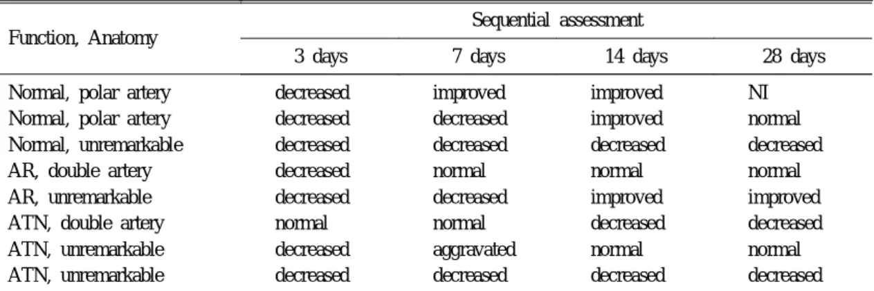
Table 2. Sequential Assessment of Renal Transplants with Focally Decreased Radioactivity on Tc-99m MAG3 SPECT Scans
Function, Anatomy
Sequential assessment
3 days 7 days 14 days 28 days
Normal, polar artery Normal, polar artery Normal, unremarkable AR, double artery AR, unremarkable ATN, double artery ATN, unremarkable ATN, unremarkable
decreased decreased decreased decreased decreased normal decreased decreased
improved decreased decreased normal decreased normal aggravated decreased
improved improved decreased normal improved decreased normal decreased
NI
normal
decreased
normal
improved
decreased
normal
decreased
NI, not interpretable image quality; AR, acute rejection; ATN, acute tubular necrosis.
522 The Korean Journal of Nuclear Medicine : Vol. 33, No. 6, 1999
without any arterial variation. Seven of the 8 pati- ents demonstrated areas of decreased activity on the first SPECT, performed on day 3 following surgery, of which 5 were resolved or reduced in
size and 2 were persistent on follow-up scans. The remaining 1 patient had normal findings on the first postoperative study but demonstrated focally decreased radioactivity on all of the follow-up
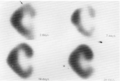
Fig. 1. Coronal tomographs of sequential Tc-99m MAG3 renal SPECTstudies in a 57 years old man with acute rejection of the allograft kidney. There is a focal area of decreased radioactivity (arrow) in the upper pole on the initial scan (3 days after surgery), which continuously improves on follow-up scans.
Fig. 2. Coronal tomographs of sequential Tc-99m MAG3 renal SPECT
studies in a 35 years old woman with acute tubular necrosis of
the allograft kidney. There is focally decreased radioactivity
(arrow) in the upper pole on the initial and first follow-up
scans (3 and 7 days), which were completely resolved in
further follow-up scans.
Ryu JG, et al. Tc-99m MAG3 SPECT on Transplanted Kidney 523
scans.
Of the 30 subjects included in this study, 10 patients (33.3%) had received donor kidneys with arterial variation (Table 3). Four were kidneys with a renal polar artery, and 6 had a double rena artery. Two kidneys with each type of arteria variation demonstrated SPECT findings of focally decreased radioactivity.
Ten of the thirty patients revealed ATN (n=7) o AR (n=3), and among them 5 patients (50%
showed focally decreased radioactivity.
Six patients showed focally increased radioacti vity on SPECT scans; two patients were resolved on follow-up studies; four patients revealed persistent findings (Table 4). These two resolved patients showed normal renal function, one of them had polar artery in the donor kidney. One of th four patients with persistently increased radioac tivity had ATN with double ureter in the dono kidney (Fig. 3). The remaining three of fou
patients showed normal renal function with two of patients having double renal artery.
Dis cus s ion
Because SPECT imaging offers three dimen sional image, which is far superior to planar scan SPECT is considered better than planar scan in evaluating renal parenchymal abnormalities ap pearing on Tc-99m MAG3 planar scan. Neubauer et al7,8) reported that it was possible to evaluate th renal parenchymal abnormality with Tc-99m MAG3 SPECT scan on transplanted kidney.
Tc-99m MAG3 is excreted rapidly via tubular excretion and thus dose not usually allow SPECT acquisition.9,10) However, since it is a high photon flux renal tracer, SPECT can be done with high speed tomographic devices such as multi-head gamma camera, which can shorten the acquisition time. The earlier the starting time of the SPECT
Table 3. Renal Arterial Variation and Tc-99m MAG3 SPECT Scan Findings of Renal Transplant SPECT scan findings
Total Focal decreased Focal increased Normal NI
Polar artery Double arteries Total
2 2 4
1 2 3
0 1 1
1 1 2
4 6 10 NI, not interpretable image quality.
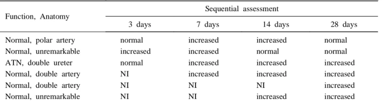
Table 4. Sequential Assessment of Renal Transplants with Focal Increased Radioactivity on SPECT Scans
Function, Anatomy
Sequential assessment
3 days 7 days 14 days 28 days
Normal, polar artery Normal, unremarkable ATN, double ureter Normal, double artery Normal, double artery Normal, unremarkable
normal increased normal NI NI NI
increased increased increased increased NI NI
increased normal increased increased NI increased
normal
normal
increased
increased
increased
increased
ATN, acute tubular necrosis; NI, not interpretable image quality.
524 대한핵의학회지 : 제 33 권 제 6 호 1999
scan, the better the quality image obtained. As all patients void their bladder immediately after the routine planar scan, the SPECT scans could be obtained quickly. As a result, the SPECT studies were initiated at an average of 7.8 minutes fol- lowing the planar scan.
In this study, SPECT scans were performed in patients with normal allograft function as well as in patients with abnormal renal function. Some o the patients had renal arterial variation and it wa expected that sequential SPECT scans could provide information about the clinical significance and course of arterial variation. Subclinical abnor mality could exist even in the presence of norma global renal function, and SPECT scan may be able to demonstrate this abnormality.
SPECT scans showed interpretable image quality in 26 of all 30 patients (86.7%) and in 84 of th total 120 scans (70%), indicating that SPECT scan provides interpretable image quality with relative consistency. Especially, in cases of global excre
tion impairment such as ATN or AR, interpretable quality images were easily acquired due to intense remnant radioactivity in the renal parenchyme.
This finding corresponds to the results reported by Neubauer et al.7,8) Even in cases with normal renal function, SPECT scan showed interpretable image quality at the rate of 61.9%.
Of total 30 patients, 14 showed abnorma findings of focal decreased or increased radioac tivity. But only five of the 14 patients (35.7%
showed abnormality on planar scans, and planar scans demonstrated none of the focal increased radioactivity seen in the SPECT scans. Conse quently, it is obvious that SPECT scan detects much more additional findings than planar scan.
Eight (26.7%) of the thirty patients showed focally decreased radioactivity on SPECT scans of transplanted kidney (Table 2). Two of them had polar artery in the donor kidney, and the area o perfusion from this artery showed decreased radioactivity. The cause of this decreased radioac
Fig. 3. Coronal tomographs of sequential Tc-99m MAG3 renal SPECTstudies in a 29 years old woman with double ureters. There is
normal distribution of radioactivity throughout the transplanted
kidney and good visualization of the double ureters on the
initial scan (3 days), but follow-up scans reveals non-
visualization of upper ureter and persistent focally increased
radioactivity (arrow) in the upper pole.
류종걸 외 2인. 이식 신장에서 시행한 Tc-99m MAG3 SPECT 525
tivity was assumed to be due to a lack of vascular supply.8) Five patients (62.5%) had ATN or AR with (n=2) or without combined arterial variation.
Perfusion abnormality associated with these lesions was assumed to be the cause of decreased radioactivity.7,8,11) No cause was determined in the remaining one case. Five (62.5%) of eight patients with focally decreased radioactivity showed resolved or reduced in size on follow-up scans, which were assumed to be an ischemic area. The other three patients (37.5%) had persistent findings of decreased radioactivity, which were consequently concluded to be an infarcted area.
But we could not rule out the possibility of normal uneven radioactivity distribution in these cases. Further study such as Tc-99m DMSA renal scan maybe necessary to confirm exact nature of focal decreased radioactivity.
Ten (33.3%) of the thirty patients had arteria variation in the donor kidneys (Table 3), similar to the results reported by Zeon et al.12) Four had rena polar artery, and six had double renal artery. Six o the ten patients did not show focally decreased radioactivity in initial and follow-up SPECT scans which suggested that there was at least no ischemic area and further study was not needed. In the remaining four patients, SPECT scan showed focally decreased radioactivity in the transplanted kidney. Three of four cases were resolved on follow-up scans; the area with decreased radioac tivity was assumed to be ischemic area. The remaining one patient showed persistent findings of focally decreased activity, which was believed to be an infarcted area. In these cases with decreased radioactivity, it was thought that further confir matory study was needed to differentiate ischemia or infarction from normal variation of uneven distribution. Sequential SPECT scans could be a guidance for further study that confirmed exac cause of decreased radioactivity.
Among the ten patients with ATN or AR, five showed focally decreased radioactivity on the SPECT scan. Whereas, of the 20 patients with normal allograft function, three showed focally decreased radioactivity. Focally decreased radioac- tivity was more frequent in patients with ATN and AR, but relationship between focally decreased radioactivity and clinical course was not clear (Table 2).
Six (20%) of the thirty patients showed focally increased radioactivity on SPECT scans (Table 4) two (33.3%) of them were resolved on follow-up SPECT scans. In all but four patients (66.7%) areas of increased radioactivity persisted. One of six patients had double ureter in the donor kidney on the first SPECT scan, distribution of radioac tivity was normal with two visible ureters. But on follow-up SPECT scan, one of the two ureters wa not visible and an increased radioactivity was seen in that area. On the next follow-up SPECT scans areas of increased radioactivity persisted. Focal excretion impairment was believed to be the cause of this increased radioactivity (Fig. 3), but othe causes such as venous stasis or an artifact could not be ruled out. Further study is needed define th exact cause.
요 약
목적: 이식 신장에서 시행한 Tc-99m MAG SPECT 스캔의 유용성을 평가하고자 하였다. 대상 및 방법:
신장 이식환자 30명, 120 스캔을 연구대상으로 하였
다(남:여=15:15, 평균연령 35.0세). 수술 후 3일, 7
일, 14일 및 28일에 555~740 MBq의 Tc-99m
MAG3를 순간주사하여 평면스캔을 시행하고, 배뇨
한 후 즉시 SPECT 스캔을 실시하였다(평균 SPECT
스캔 시작 시간: 평면스캔 후 7.8분). 결과: SPECT
스캔은 전체 환자 30명 가운데 26명(86.7%)에서, 전
체 120 스캔 가운데 84 스캔(70%) 에서 판독 가능
한 영상화질을 보여, 비교적 일관성 있게 판독 가능
526 The Korean Journal of Nuclear Medicine : Vol. 33, No. 6, 1999