맥문동 물 추출물의 선천면역 활성과 염증억제 효과
1
경희대학교 대학원 임상한의학과,
2경희대학교 한의과대학 부인과교실 강누리
1, 황덕상
2, 이진무
2, 이창훈
2, 장준복
2ABSTRACT
The Effects of Liriopis Tuber Water Extract on Innate Immune Activation and Anti-Inflammation
Nu-Ri Kang
1, Deok-Sang Hwang
2, Jin-Moo Lee
2, Chang-Hoon Lee
2, Jun-Bock Jang
21
Dept. of Clinical Korean Medicine, Graduate School, Kyung Hee University
2
Dept. of Korean Gynecology, College of Korean Medicine, Kyung Hee University Objectives: This study was designed to examine the anti-cancer activity by innate immunomodulating and anti-inflammatory effects of liriopis tuber water extract (LPE).
Methods: Cell cytotoxicity was tested with 4T1 mouse mammary carcinoma cells, spleen cells, macrophage, and RAW264.7 cells. To investigate innate immunomodulating effects of LPE on macrophage, we measured tumor necrosis factor-alpha (TNF-α), interleukin-12 (IL-12), and interleukin-10 (IL-10). To investigate innate immunomodulating effects of LPE on RAW264.7 cell, we measured TNF-α, interleukin-6 (IL-6). In addition, TNF-α and nitric oxide (NO) induced by lipopolysaccharide (LPS) were measured after treating with LPE to observe innate immunomodulating effect of LPE on RAW264.7 cell. Also, mitogen-activated protein kinase (MAPK) and nuclear factor κB (NF-κB) were examined by western blot analysis.
Results: In an in vitro cytotoxicity analysis, LPE affected tumor cell growth above specific concentration. As compared with the control group, the production of TNF-α, IL-12, and IL-10 were increased in macrophage. As compared with the control group, the production of TNF-α and IL-6 were increased in RAW 264.7 cell. The expression of TNF-α and NO induced by LPS after treating LPE was decreased. In addition, treatment of RAW 264.7 cell with LPE increased the phosphorylation levels of p-extracellular signal-regulated kinase (p-ERK), p-Jun N-terminal kinase (p-JNK), and p-p38.
Conclusions: LPE might have impact on the anti-cancer effect by activation of innate immune system and inflammation control.
Key Words: Liriopis Tuber Water Extract , Cancer, Innate Immune System, Anti-Inflammation
2)
Corresponding author(Jun-Bock Jang) : Kyung-Hee University Korean Medicine Hospital, 23, Kyungheedae-ro, Dongdaemun-gu, Seoul, Republic of Korea
Tel : 02-958-9159 E-mail : [email protected]
Ⅰ. 서 론
여성 질환에서 염증반응은 세균, 바이 러스 등의 다양한 균주에 의한 감염뿐만 아니라 암과 같은 종양 질환에서 세포 증식이 일어날 때 나타난다
1). 암 치료법 은 기존의 수술요법, 방사선요법 및 항 암화학요법 등이 단독 또는 복합으로 시 도되고 있으나 아직 궁극적인 해결책에 이르지 못하고 있다
2). 이러한 기존 치료 법의 한계를 극복할 수 있는 천연물에 대한 관심이 매우 높은 편인데
3), 인체의 방어 기능을 증가시키는 효과가 있어 신 체 내 면역력을 조절하고 방어기전에 효 과적인 한약재에 대한 연구가 최근 들어 활발히 진행되고 있다
4,5).
맥문동은 백합과에 속하는 다년생초인 겨우살이풀의 뿌리로, ≪東醫寶鑑≫의 온 경탕, 자감초탕, 청심연자음, 맥미지황환, 생맥산 등의 방제에 主劑로서 虛勞客熱, 口乾燥渴, 肺痿吐膿, 熱毒身黑目黃 등의 병증에 활용되어
6)養陰潤肺, 凊心除煩, 益胃生津 등의 효능이 알려져 있다
7).
맥문동의 약리작용으로는 혈당강하작 용
8), 항산화작용, 항균작용
9), 항염증작용
10)
, 간 보호 효과
11), 신경 보호 효과
12)및 항암효과가 보고되었으며, 주요 성분 으로는 steroid계 saponin, isoflavonoid, oligosaccharides 및 polysaccharide 등이 함유되어 있는데
13)특히 saponin 단량체 인 Dwarf lilyturf tuber-13(DT-13)의 혈 관 신생 억제를 통한 항암효과
14-8)가 보 고되었으며, 여성호르몬의 천연 대체물 질로 알려져 있는 isoflavone은 에스트로 겐 부족에 효과가 있어 유방암과 난소암 의 예방 및 생체 내 항암 효과
19-21)가 보
고되었다. 맥문동의 항염효과와 암세포 사멸효과에 대한 실험적 연구는 있으나 면역 활성을 통한 항암 효과에 대한 연 구보고는 없었다.
이에 저자는 맥문동 물추출물의 선천 면역 활성과 염증 억제 효과를 연구해 보고자 macrophage와 RAW 264.7 cell을 이용하여 cytokine의 분비를 확인하고, lipopolysaccharide(LPS)자극에 의한 염 증 매개물질의 생산조절, nuclear factor κB (NF-κB), mitogen-activated protein kinase (MAPK) 신호전달경로를 통한 면역활 성을 관찰하여 유의한 효과를 얻었기에 보고하는 바이다.
Ⅱ. 실 험
1. 재 료 1) 약 재
맥문동( Liriopis Tuber )은 백합과(Liliaceae) 에 속하는 다년생 초본인 소엽맥문동 ( Ophiopogon japonicus Ker-Gawl.)의 괴 근이다
7). 경희한약(Wonju, Kangwon)에 서 구입하여 중량의 10배되는 증류수를 첨가한 후 100℃에서 4시간 동안 가열하 였다. 추출물은 원심분리(3000 rpm, 30 min)를 통하여 상등액을 회수한 후 0.2 μm 의 pore size를 가지는 membrane filter (Whatman, Philadelphia, PA, USA)를 이용하여 맥문동의 물 추출물( liriopis tuber water extract , 이하 LPE)를 얻었다. Stock solution의 농도는 phosphate buffered saline (PBS)를 이용하여 건조 중량에 대하여 100 mg/ml의 농도로 준비하였고 4℃에 보관하면서 실험에 적용하였다.
2) 동 물
생후 6-8주령의 자성 BALB/c(20 g±1 g) 를 새론바이오텍에서 분양 받아 사육하 였다. 사육조에 마우스를 5-10 마리씩 넣 어 정수된 물과 실험동물용 펠렛사료 (Samyang Co Ltd, Incheon, Korea)를 자유 공급하였고, 온도 22℃, 습도 50%, 12시간 간격으로 자동 조명되는 상태에 서 스트레스를 받지 않도록 주의하여 사 육하였다.
3) 시 약
종양세포의 배양을 위한 Dulbecco’s modified Eagle’s medium(DMEM)은 WEL GEN (Daegu, Korea)에서, fetal bovine serum (FBS), penicilline과 streptomycin은 Gibco 사(Carlsbad, CA, USA)에서 구입하였고 LPS, thioglycollate은 Sigma-Aldrich(St.
Louis, MO, USA)에서, Griess assay kit는 Promega(WI 53711, USA)에서, PBS는 Gibco사(Grand Island, NY, USA)에서 구입하였다. Cell counting kit(Ez-Cytox) 는 DoGen(Seoul, Korea)에서, ELISA kit 는 R&D system(Inc. MIN 55413, USA) 에서 구입하여 사용하였다.
신호전달을 위한 NF-κB, p65와 β-actin 의 분석을 위한 1차항체는 Santa Cruz Biotechnologies(Santa Cruz, CA, USA)에 서, 인산화된 p65, N-terminal kinase(JNK), extracellular signal-regulated kinase(ERK), p38 protein kinase(p38) 및 beta-actin의 발현을 측정하기 위한 항체는 Cell signaling Technology(Boston, Massachusetts, USA) 에서 구입하였다.
4) 세포주 및 세포 배양
RAW 264.7 cell, 4T1 mammary carcinoma cell은 한국세포주은행(서울, 한국)에서 분 양받았으며, thioglycollate-induced macrophage (이하 macrophage)는 6주령 BALB/c 마
우스 복강으로 3% thioglycollate 1 ml를 주사하고 마우스를 희생시킨 후 복강에 DMEM 배지를 주입하여 얻은 복강 내 세포(peritoneal exudative cells; PEC)를 24 well culture plate에 배양 후 배양액 을 세척하여 얻었다.
각각의 RAW 264.7 cell, 4T1 carcinoma cell 및 macrophage의 배양은 10% FBS 가 함유된 DMEM 배지를 각각 이용하 였고, 5% CO
2, 95% 습도 및 37℃의 배 양기(Thermo Fisher Scientific Inc. Waltham, USA)에서 배양하였다.
2. 방 법
1) 세포 독성 측정
4T1 carcinoma cell을 1×10
4/well의 밀 도로 96 well plate의 각 well에 plating한 후, 3.2 μg/ml, 16 μg/ml, 80 μg/ml, 400 μ g/ml 및 2000 μg/ml 농도의 LPE 추출 물을 첨가하여 3일간 배양하였다. PEC 를 24 well culture plate에 2.5×10
6/well 의 농도로 조정하여 분주하고 2시간 동 안 배양하여 macrophage를 plate에 부착 하고 배양액으로 세척하여 부착되지 않은 세포를 제거한 후, 16 μg/ml, 80 μg/ml, 400 μg/ml, 2000 μg/ml 및 10000 μg/ml 농도의 LPE를 첨가하여 1일간 배양하였 다. RAW 264.7 cell을 4×10
4/well의 밀도 로 96 well plate의 각 well에 plating한 후, 1일간 배양한 후 16 μg/ml, 80 μg/ml, 400 μg/ml, 2000 μg/ml 및 10000 μg/ml 농도의 LPE를 첨가하여 1일간 배양하였다.
각 물질의 세포 독성 효과는 WST를
이용하는 Ez-Cytox를 이용하여 제조사
의 지침에 따라 30분 후에 흡광도를 450 nm
에서 측정하였으며, 시료를 첨가하지 않
은 대조군에 대한 백분율(%)로 표시하
였다.
2) Macrophage의 cytokine 분비 측정 PEC를 24 well culture plate에 2.5×10
6/well 의 농도로 조정하여 분주하였다. 2시간 동안 배양하여 macrophage를 plate에 부 착하고 배양액으로 세척하여 부착되지 않은 세포를 제거하였다. 16 μg/ml, 80 μg/ml, 400 μg/ml 및 2000 μg/ml 농도의 LPE를 첨가하고 24시간 동안 배양한 후, 배양 상등액을 회수하였다. 유도 분비된 tumor necrosis factor-alpha(TNF-α), interleukin-10 (IL-10), interleukin-12(IL-12)의 측정은 각 cytokine에 대한 ELISA kit를 이용하 여 제조사의 지침에 따라 측정하였다.
3) RAW 264.7 cell의 cytokine 분비 측정 RAW 264.7 cell을 24 well culture plate 에 2.0×10
6/well의 농도로 조정하여 분주 하였다. 2시간 동안 배양 후, macrophage 를 plate에 부착하고 배양액으로 세척하 여 부착되지 않은 세포를 제거하였다.
16 μg/ml, 80 μg/ml, 400 μg/ml 및 2000 μ g/ml 농도의 LPE를 첨가하고 24시간 동안 배양한 후, 배양 상등액을 회수하 였다. TNF-α, IL-6의 측정은 각 cytokine 에 대한 ELISA kit를 이용하여 제조사 의 지침에 따라 측정하였다.
4) Macrophage에 LPS 처리 후 염증 억제 활성 측정
RAW 264.7 cell을 24 well plate의 각 well에 2.0×10
6/well이 되도록 분주하고 24시간 배양하여 안정시킨 후, LPE의 최종 농도가 16 μg/ml, 80 μg/ml, 400 μg/ml 및 2000 μg/ml이 되도록 조정하여 2시간 배양하였다. 배양 완료 후 200 ng/ml의 LPS를 첨가하고 24시간 배양한 후 각 배양액에 생산된 염증 매개 물질인 TNF-α 및 nitricoxide(NO)의 양을 측정하였다.
NO의 함량은 Griess assay kit를 이용하 여 제조사의 지침에 따라 측정하였다.
5) Western blotting
RAW 264.7 cell에 100 μg/ml 및 1000 μg/ml 의 시료를 첨가하고 30분 반응 후 4℃의 PBS로 3회 세척 후, protease inhibitor cocktail(Roche Diagnostics Corp., Indianapolis, IN, USA), 1 mM dithiothreitol(Wako, Tokyo, Japan), 1 mM phenylmethylsulfonyl fluoride(Sigma)를 함유하는 RIPA buffer 를 이용하여 30분간 4℃에서 세포를 용 해시켰다. 원심분리(12,000 rpm/20 min) 를 통하여 상등액을 얻은 후, Pierce BCA Protein Assay kit(Thermo Fisher Scientific, Waltham, MA, USA)를 이용하여 단백 질을 정량하였다. 군당 동량의 단백질 (30 μg/well)을 10% 폴리아크릴아마이드 겔 전기영동법(sodium dodecyl sulphate polyacrylamide gel electrophoresis, SDS- PAGE)을 통하여 단백질을 분리한 후에 nitrocellulose membrane(Bio-Rad, USA) 에 전사시켰다. 단백질이 전사된 nitrocellulose membrane은 5% skim milk in tris-buffered saline(TBS) containing 0.1% Tween 20 (TBST)를 이용하여 상온에서 1시간 동안 blocking시켰다. 1차항체가 반응된 membrane 은 TBST를 이용하여 세척단계를 거친 후, HRP가 표지된 2차항체(HRP-conjugated anti-mouse IgG)를 상온에서 1시간 반응 시켰다. 세척단계를 거친 후 membrane 상에서 항체들과 반응한 단백질을 ECL 기질(GE Healthcare Life Sciences)을 적 용시킨 후, X-ray film에 감광시켜 특정 단백질 발현을 분석하였다.
6) 통계 분석
실험 결과의 통계 처리는 SPSS(version
24.0)를 이용하였고, 대조군과 실험군의
비교는 일원분산분석(One-way ANOVA) 또는 독립표본 T 검정(student’s t-test) 를 시행하였다. 분산의 동질성 검정은 Levene 의 등분산 검정을 시행하여 등분산이 가 정될 때 사후 검정은 Tukey’s HSD test 를 이용하였으며, 가정되지 않을 때 사 후검정은 Dunnett T3 test를 시행하였 다. p<0.05일 때 통계적으로 유의한 차이 가 있다고 판정하였다.
Ⅲ. 결 과
1. 세포독성 평가
1) 암세포에 대한 세포독성 평가 4T1 mammary carcinoma cell에 3.2 μg/ml, 16 μg/ml, 80 μg/ml, 400 μg/ml 및 2000 μg/ml 농도의 LPE를 첨가하고 세포독성을 측 정한 결과, 각각 100.4±2.0%, 101.2±5.7%, 100.6±2.4%, 94.3±4.9% 및 79.0±11.8%로 나타났다(Fig. 1).
Fig. 1. Cytotoxicity of LPE on 4T1 mouse mammary.
2) RAW 264.7 cell에 대한 세포독성 평가 Raw 264.7 cell에 16 μg/ml, 80 μg/ml, 400 μg/ml, 2000 μg/ml 및 10000 μg/ml 농도의 LPE를 첨가하고 세포독성을 측정 한 결과, 각각 97.33±6.94%, 113.45±17.56%, 160.12±4.98%, 156.54±17.32% 및 91.39±9.16%
로 나타났다(Fig. 2).
Fig. 2. Cytotoxicity of LPE on RAW 264.7 cell.
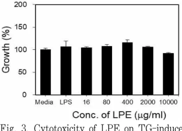
3) 대식세포에 대한 세포독성 평가 Macrophage에 16 μg/ml, 80 μg/ml, 400 μg/ml, 2000 μg/ml 및 10000 μg/ml 농도의 LPE를 첨가하고 세포독성을 측 정한 결과, 각각 104.20±2.64%, 107.84±3.90%, 115.59±6.36%, 105.80±1.51% 및 91.67±1.66%
로 나타났다(Fig. 3).
Fig. 3. Cytotoxicity of LPE on TG-induced macrophage.
2. Macrophage에서의 cytokine 생산 효과 1) TNF-α
Macrophage에 16 μg/ml, 80 μg/ml,
400 μg/ml 및 2000 μg/ml 농도의 LPE를
첨가하고 TNF-α의 분비를 관찰한 결과,
80 μg/ml, 400 μg/ml 및 2000 μg/ml에서
각각 154.2±18.3 pg/ml, 424.9±36.1 pg/ml
및 484.7±20.6 pg/ml로 통계적으로 유의 하게(p<0.05) 증가하였다(Fig. 4).
Fig. 4. Production of TNF-α from TG-induced macrophage stimulated by LPE.
*: p<0.05, **: p<0.01, statistically significant difference compared with media group (one-way ANOVA combined with a Dunnett T3 test).
2) IL-12
Macrophage에 16 μg/ml, 80 μg/ml, 400 μg/ml 및 2000 μg/ml 농도의 LPE를 첨가하고 IL-12의 분비를 관찰한 결과, 16 μg/ml, 80 μg/ml, 400 μg/ml 및 2000 μg/ml 에서 각각 27.0±1.6 pg/ml, 134.6±6.3 pg/ml, 1074.6±33.3 pg/ml 및 1116.9±30.0 pg/ml 로 통계적으로 유의하게(p<0.05) 증가하 였다(Fig. 5).
Fig. 5. Production of IL-12 from TG-induced macrophage stimulated by LPE.
**: p<0.01, statistically significant difference compared with media group (one-way ANOVA combined with a Dunnett T3 test).
3) IL-10
Macrophage에 16 μg/ml, 80 μg/ml, 400 μg/ml 및 2000 μg/ml 농도의 LPE를 첨가하고 IL-10의 분비를 관찰한 결과, 80 μg/ml, 400 μg/ml 및 2000 μg/ml에서 각각 74.2±6.4 pg/ml, 1833.9±37.0 pg/ml 및 2662.9±82.2 pg/ml로 통계적으로 유의 하게(p<0.05) 증가하였다(Fig. 6).
Fig. 6. Production of IL-10 from TG-induced macrophage stimulated by LPE.
**: p<0.01, statistically significant difference compared with media group (one-way ANOVA combined with a Dunnett T3 test).
3. RAW 264.7 cell에서의 cytokine 생 산 효과
1) TNF-α
RAW 264.7cell에 16 μg/ml, 80 μg/ml,
400 μg/ml 및 2000 μg/ml 농도의 LPE를
첨가하고 TNF-α의 분비를 관찰한 결과,
16 μg/ml, 80 μg/ml, 400 μg/ml 및 2000 μg/ml
에서 각각 57.89±3.28 pg/ml, 215.49±18.81
pg/ml, 451.16±12.17 pg/ml, 557.05±23.50
pg/ml로 통계적으로 유의하게(p<0.05) 증
가하였다(Fig. 7).
Fig. 7. Production of TNF-α from RAW 264.7cell stimulated by LPE.
*: p<0.05, **: p<0.01, statistically significant difference compared with media group (one-way ANOVA combined with a Dunnett T3 test)
2) IL-6
RAW 264.7cell에 16 μg/ml, 80 μg/ml, 400 μg/ml 및 2000 μg/ml 농도의 LPE를 첨가하고 IL-6의 분비를 관찰한 결과, 400 μg/ml 및 2000 μg/ml에서 각각 674.23
±25.70 pg/ml, 2503.21±25.40 pg/ml로 통 계적으로 유의하게(p<0.05) 증가하였다 (Fig. 8).
Fig. 8. Production of IL-6 from RAW 264.7cell stimulated by LPE.
**: p<0.01, ***: p<0.001, statistically significant difference compared with media group (one-way ANOVA combined with a Dunnett T3 test)
4. Macrophage에 LPS 처리 후 염증 매개물질 생산 억제 효과
1) TNF-α
Macrophage에 LPS와 함께 LPE를 16 μ g/ml, 80 μg/ml, 400 μg/ml 및 2000 μg/ml 농도의 LPE를 첨가하고 TNF-α의 분비 를 관찰한 결과, LPS만 처리한 경우 469.19
±52.35 pg/ml 에 비하여 2000 μg/ml에서 349.36±37.22 pg/ml로 통계적으로 유의하 게(p<0.05) 억제되었다(Fig. 9).
Fig. 9. Production of TNF-α from macrophage stimulated by LPE and LPS.
**: p<0.01, statistically significant difference compared with LPS group (one-way ANOVA combined with a Tukey HSD test)
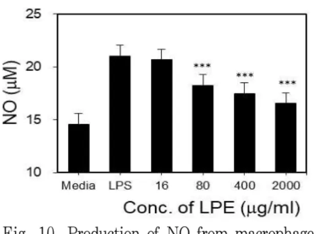
2) NO
Macrophage에 LPS와 함께 LPE를 16 μg/ml,
80 μg/ml, 400 μg/ml 및 2000 μg/ml 농
도의 LPE를 첨가하고 NO의 분비를 관찰
한 결과, LPS만 처리한 경우 21.02±0.29 μM
에 비하여 80 μg/ml, 400 μg/ml 및 2000
μ g/ml에서 각각 18.25±0.45 μM, 17.46±0.74
μ M, 16.52±0.33 μM로 통계적으로 유의
하게(p<0.01) 억제되었다(Fig. 10).
Fig. 10. Production of NO from macrophage stimulated by LPE and LPS.
***: p<0.001, statistically significant difference compared with LPS group (one-way ANOVA combined with a Tukey HSD test)