HaCaT 세포의 산화 스트레스로 인한 세포자멸사에서 정향의 보호효과
박숙자#*
대구한의대학교 제약공학과
Protective effect of Caryophylli Flos on apoptosis caused by oxidative stress in HaCaT cells
Sook Jahr Park
#*Department of Pharmaceutical Engineering, Daegu Haany University
ABSTRACT
Objective : Caryophylli Flos has been used in Korean medicine to relieve vomiting and pains caused by chills that make fluid circulation difficult. This study was designed to investigate the protective effect of ethanol extract of Caryophylli Flos (CF) in hydrogen peroxide (H
2O
2)-induced apoptotic cell death in human keratinocyte HaCaT cells.
Methods : CF was prepared by extracting 200 g of Caryophylli Flos in 2 L of ethanol for 48 h. Cell viability was measured by MTT assay, and the protein expression was monitored by Western blot analysis. Apoptosis was determined by terminal deoxynucleotidyl transferase dUTP nick end labeling (TUNEL) assay. Reactive oxygen species (ROS) was measured using fluorescent dye, and reduced glutathione (GSH) was determined with a colorimetric commercial kit.
Results : CF protected HaCaT cells from cell death caused by oxidative stress after H
2O
2treatment. H
2O
2amplified generation of ROS and induced depletion of GSH, whereas these changes in ROS and GSH were inhibited by GF treatment. In addition, H
2O
2resulted in apoptosis as assessed by TUNEL assay and the expression of apoptosis regulator proteins. However, cells treated with CF showed a decrease in TUNEL-positive cells and restored the reduced expression of procaspase-9, -3 and PARP.
Conclusion : This study showed cytoprotective effects of CF by anti-apoptotic activity while exerting antioxidative activity in H
2O
2-treated HaCaT cells. These results suggest that CF could be beneficial in skin damage caused by oxidative stress.
1)Key words : Caryophylli Flos, H
2O
2, ROS, GSH, apoptosis, caspase
Ⅰ. 서 론
정향(丁香)은 도금양과(Myrtaceae) 식물인 정향나무 (
Eugenia caryophyllataThunberg)의 꽃봉오리로 녹색에서 붉은색으로 변하는 시기에 채취하여 사용한다
1). 특이하고 강한 향기가 있어 식품의 향신료로 사용되며 한의학에서는 약성이 따뜻하고 매운 맛을 지니고 있어 비위(脾胃)를 따뜻하게 하고 냉기로 인한 복통, 구토, 소화불량 등을 치료하는 약재로 사용 되었다
2). 주요성분으로는 eugenol, acetyleugenol, beta-
caryophyllene 등의 정유 성분을 가지고 있으며
3)플라보노이 드인 rhamnetin, keampferol과 oleanolic acid, eugeniin, eugenitin 등을 함유한다
4). 정향의 약리작용으로는 항산화
5), 항균
6)및 항진균
7), 항염증
8), 항당뇨
9), 항혈전
10)등이 보고되 었다.
피부는 몸의 가장 바깥층을 이루고 있기 때문에 물리화학 적인 측면에서 외부 환경 자극에 대한 장벽 역할을 하여 피부 항상성을 유지하고 인체를 보호한다
11). 하지만 외부 유해인자 들에 오랜 시간 노출되게 되면 산화 스트레스가 유발되고 여러
#*Corresponding and First author : Sook Jahr Park, Department of Pharmaceutical Engineering, Daegu Haany University, Gyeongsan 38610, Republic of Korea.
·Tel : +82-53-819-1298 ·Fax : +82-53-819-1406 ·E-mail : [email protected] ·Received : 11 August 2021 ·Revised : 02 September 2021 ·Accepted : 25 September 2021
피부 문제가 발생한다. 태양광과 같은 환경 자극은 피부 세포를 손상시키고 주름, 처침, 색소 침착이 특징인 피부 노화의 진 행을 가속화한다. 피부는 표피, 진피, 피하지방으로 구성되어 있으며, 자외선은 피부의 표피뿐만 아니라 진피까지 침투하여 NADPH oxidase, xanthine oxidase를 포함하는 활성산소종 생성효소의 활성화를 통해 superoxide anion radicals를 생 성한다
12). 샴푸 등에 사용되는 계면활성제인 sodium lauryl sulfate는 피부 각질세포주인 HaCaT cell에서 superoxide anion radicals의 생성을 촉진한다는 것이 보고되었다
13). 대기 오염 물질도 피부의 산화 스트레스를 유발하게 되는데, 실제로 대기 오염 지역에서 비오염 지역과 비교하여 피부 주름과 색소 반점이 빈번하게 발생하는 것으로 나타났다
14).
산화 스트레스는 세포 내에서 막지질의 산화를 유발하여 세포막의 구조를 파괴하고 단백질, 핵산과 같은 생체 분자들의 손상을 유발한다
15). 활성산소종 (reactive oxygen species, ROS)은 정상적인 산소 대사의 부산물이며 신호전달과 항상성 유지에 중요한 역할을 하지만 세포의 항산화 용량을 넘어 과도 하게 축적되면 세포 손상을 유발하는 산화 스트레스가 나타나게 된다
16). 피부 각질세포를 포함한 대부분의 세포에서 ROS의 축적에 의한 미토콘드리아 기능 장애와 관련된 DNA 손상이 세포자멸사를 유도하는 것으로 보고되고 있다
17,18). 따라서 세포 내 산화 방어 시스템은 산화 스트레스와 관련된 질병의 예방 및 치료에 필수적이라 할 수 있다. 본 연구에서는 H
2O
2로 산화 스트레스를 유도한 HaCaT 세포 손상 모델에서 정향 추출물이 항산화 활성을 통한 세포보호 효과를 나타내는지 살펴보고자 하였다.
Ⅱ. 재료 및 방법
1. 시약
Dulbecco’s modified Eagle’s medium/F12 (DMEM/F12), penicillin-streptomycin, fetal bovine serum (FBS)은 Gibco (Rockville, MD, USA)제품을 사용하였고 dimethyl sulfoxide (DMSO), 2,7’-dichlorofluorescein diacetate (DCF-DA), 3-(4,5-dimethylthiazol-2-yl)-2,5-diphenyltetrazoleu m (MTT)는 Sigma (St. Louis, MO, USA)에서 구입하였다.
GSH determination kit는 Oxis International Inc (Tampa, FL, USA)에서 구입하였고
in situapoptosis detection kit는 Roche (Mannheim, Germany)에서 구입하였다. β-actin 항체는 Santa Cruz Biotechnology (Bergheimer, Germany) 에서 구입하였고, anti-poly (ADP-ribose) polymerase (PARP), anti-procaspase-3, anti-procaspase-9 항체는 Cell Signalling Technology (Beverly, MA, USA)에서 구입 하였다.
2. 정향 에탄올 추출물(CF)의 제조
정향(원산지: 인도네시아산)은 삼홍건재약업사(Seoul, Korea) 에서 시판되는 것을 구입하여 품질 검증을 실시한 후에 실험에 사용하였다. 건조 중량으로 200 g의 정향을 에탄올 2 L에 넣고 상온에서 48시간 동안 추출하여 300 ㎜ filter paper (Toyo
Roshi Kaisha Ltd, Tokyo, Japan)로 여과하였다. 이 여과 액을 회전농축기(EYELA, Tokyo, Japan)로 감압농축한 후에 ultra-low temperature freezer(FDU-1100; EYELA, Tokyo, Japan)에 12시간동안 넣어 동결건조시켰다. 정향 에 탄올 추출물(CF)의 최종 수율은 10.2%였으며, -20℃에 보관 하여 사용하였다.
3. 세포 배양
HaCaT cell은 American Type Culture Collection (ATCC, Rockville, MD, USA)에서 구입하였으며 10% FBS, 100 units/㎖ penicillin이 포함된 DMEM/F12 배지를 이용하여 37℃, 5% CO
2조건의 incubator에서 배양하였다. 모든 실험 과정에서 세포는 80~90%의 confluence 범위에 도달하도록 배양하여 사용하였다.
4. MTT assay
세포 생존율을 측정하기 위해 HaCaT cell을 24 well plate 에 1×10
5cells/well로 분주하여 24 시간 배양한 후에, CF를 농도별(0, 10, 30, 100 ㎍/㎖)로 한 시간 전처치하고 500 μM H
2O
2를 첨가하여 18 시간 더 배양하였다. 배양 배지는 걷어 내고 phosphate-buffered saline (PBS)로 세척한 세포에 0.1 g/㎖ MTT 용액을 첨가하여 37℃에서 2 시간 반응시킨 후 생성된 formazan을 DMSO로 녹여 microplate reader (Tecan, Männedorf, Switzerland)를 사용하여 570 ㎚의 파장에서 흡광도를 측정하였다. 세포 생존율은 대조세포에 대한 백분율 로 다음과 같은 수식에 의해 계산하였다 [cell viability (%) = 100 × (absorbance of treated sample)/(absorbance of control)].
5. Reactive oxygen species (ROS) 측정
세포 내 ROS 수준은 DCF-DA를 이용하여 형광 흡광도로 측정하여 조사하였다. 처치가 완료된 세포에 10 μM DCF- DA를 30분 동안 반응시키고, trypsin으로 세포를 분리하여 black plate에 100 ㎕씩 옮겨준 다음, excitation 485 ㎚, emission 530 ㎚ 파장에서 형광강도를 측정하였다.
6. Glutathione (GSH) 함량 측정
GSH의 함량을 측정하기 위해서 500 ㎕의 metaphosphoric acid를 처치가 완료된 세포에 첨가하여 용해한 후, 원심분리 (3,000 ×g, 4℃, 10 min)하여 상층액을 얻었다. 이 상층액은 GSH determination kit를 사용하여 400 ㎚ 파장에서 흡광 도를 측정하였다.
7. TUNEL assay
4 well chamber slide의 well당 1×10
5개의 농도로 세포를
배양하여 CF를 한 시간 전처치한 후에 H
2O
2를 18 시간 더 처치
하였다. PBS로 2회 washing한 후에 4% paraformaldehyde로
고정하고
in situapoptosis detection kit (ab206386)로 apoptotic cell을 염색하여 light microscope (eclipse Ti-E, Nikon, Japan)로 관찰하였다.
8. Western blot analysis
처치가 끝난 세포는 radioimmunoprecipitation assay (RIPA) buffer (1% NP-40, 1% sodium deoxycholate, 0.1% SDS, 25 mM Tris-HCl pH 7.6, 150 mM NaCl)를 첨가하여 4℃에서 30 분동안 lysis시켜 전세포 추출액 (whole cell lysates)을 획득하였다. 전세포 추출액은 BCA protein assay kit로 단백질 정량을 하였고 10%의 SDS- polyacrylamide gel에서 전기영동하였다. Gel 상의 단백질을 nitrocellulose membrane으로 전이하고, 일차항체(1:1000) 및 이차항체(1:20000)를 반응시켜 준 다음 enhanced chemiluminescense detection kit로 발광시켜준 후에 Amersharm Imager 600 (GE Healthcare, Freiburg, Germany)에서 단백질 밴드를 확인하였다. 각 단백질의 발현 정도는 Image J (version 1.50i, National Institutes of Health)를 이용하여 densitometric analysis로 조사하였다.
9. 통계적 검정
모든 실험은 3회 이상 반복 실시하였으며 실험 결과는 평균
± 표준편차로 나타내었다. 유의성 검정은 윈도우용 SPSS ver. 23 프로그램을 사용하여 one way analysis of variance (ANOVA) 분석을 실시하였다. 사후 검증은 Tukey HSD를 사 용하였으며,
p값이 0.05 미만일 때 통계적으로 유의하다고 판정하였다.
III. 실험결과
1. H
2O
2로 유도된 HaCaT 세포 독성에 대한 CF의 보호 효과
H
2O
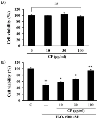
2로 유도된 세포 독성에 대해 CF가 보호 효과를 나타 내는지 관찰하기 위하여 MTT assay를 실시하였다. 먼저 MTT assay로 CF의 단독 처치에 의한 세포 독성을 조사한 결과, 10-100 ㎍/㎖ 농도에서 세포 독성이 나타나지 않아 최적 실험 농도로 결정하였다 (Fig. 1A). 500 μM의 H
2O
2는 세포 독성 을 유도하여 HaCaT 세포의 생존율을 급격하게 감소시켰다.
하지만 H
2O
2에 의해 감소한 세포 생존율은 CF에 의해 농도 의존적으로 증가하는 경향을 나타내었으며, 100 ㎍/㎖의 농도 에서 가장 효과적인 세포 보호 효과가 관찰되었다 (Fig. 1B).
2. CF가 H
2O
2로 과다 생성된 ROS에 미치는 영향
CF가 H
2O
2로 유도된 산화 스트레스에 대해 어떻게 작용하 는지 조사하기 위해 DCF-DA로 염색하여 세포 내 ROS 수준을 형광 흡광도를 측정하여 조사하였다. H
2O
2를 단독으로 처치 하였을 때, control과 비교하여 ROS가 192.4 ± 4.7% 증가
하였다. CF의 경우 H
2O
2로 매개된 ROS의 비정상적인 증가가 유의하게 감소했는데, 100 ㎍/㎖ CF에서는 control 대비 60.7
± 2.5%를 나타내었다 (Fig 2). 따라서 CF가 H
2O
2로 유도된 ROS의 과다 생성을 저해함으로써 산화 스트레스를 낮추는 효과를 확인할 수 있었다.
(A)
(B)
Fig. 1. Effect of CF on H2O2-induced cytotoxicity in HaCaT cells (A) Cells were treated with various concentrations (10, 30, 100
㎍/㎖) of CF for 24 h to determine the optimal experimental concentration. (B) HaCaT cells were pre-treated with CF for 1 h and then exposed with 500 μM of H2O2 for an additional 18 h.
C, control; ns, not significant; ##p < 0.01, significant as compared to control; **p < 0.01, *p < 0.05, significant as compared to H2O2 alone.
Fig. 2. Effect of CF on H2O2-induced ROS production
HaCaT cells were pre-treated with CF for 1 h and then exposed with H2O2 for an additional 18 h. ROS level was measured after DCF-DA staining for 15 min and the fluorescence intensity was expressed as percentage of control. C, control; ##p < 0.01, significant as compared to control; **p < 0.01, significant as compared to H2O2 alone.
3. CF가 H
2O
2로 유도된 GSH 고갈에 미치는 영향
GSH는 세포의 비효소적 항산화 체계에서 자신의 thiol group 에서 일어나는 산화환원반응을 활용하여 산화물을 제거하는 항산화 물질이다. H
2O
2를 처리하였을 때 나타나는 GSH의 함 량 변화를 관찰한 결과, control과 비교하여 72.9 ± 8.9%로 감소하였다. 하지만 100 ㎍/㎖ CF 전처치에 의한 GSH 함량은 124.1 ± 12.5%로 통계적으로 유의하게 증가하였다 (Fig. 3).
이 결과를 통해 CF는 H
2O
2로 고갈된 GSH의 함량을 증가시 킴으로써 세포 내 산화 방어 시스템에 관여함을 알 수 있다.
Fig. 3. Effect of CF on GSH levels altered by H2O2 treatment HaCaT cells were pre-treated with CF for 1 h and then exposed with H2O2 for an additional 18 h. GSH contents were measured in cell homogenates as described in material and method section.
Data were expressed as percentage of control. C, control; ##p <
0.01, significant as compared to control; **p < 0.01, significant as compared to H2O2 alone.
4. H
2O
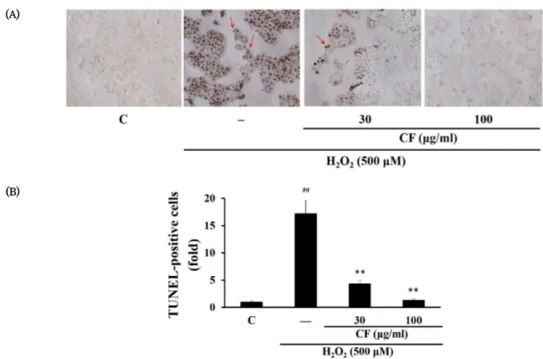
2로 유도된 세포자멸사에 대한 CF의 보호 효과
HaCaT cell에서 H
2O
2에 의한 세포자멸사를 확인하고 CF의 세포 보호 효과를 살펴보기 위해 TUNEL assay를 시행하였다.
Fig. 4에 나타난 바와 같이 DNA 분절 (fragmentation)이 일 어난 핵이 염색된 TUNEL-양성 세포 (TUNEL-positive cell)의 수가 H
2O
2의 첨가에 의해 유의하게 증가되었고, 이는 세포자멸사가 유도되었음을 나타낸다. CF를 처리한 경우 H
2O
2단독으로 자극된 세포와 비교하여 TUNEL 양성 세포의 수가 감소하였다 (Fig. 4B). 이러한 결과는 CF 처리가 농도 의존 적으로 H
2O
2에 의한 세포자멸사를 유의하게 억제하였음을 보 여준다.
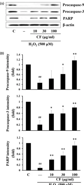
5. CF가 세포자멸사 관련 단백질의 발현에 미치는 영향
Caspase-3, caspase-9, PARP는 세포자멸사에서 중요한 역할을 담당하는 매개 단백질이다. CF가 HaCaT cell의 세포 자멸사에서 이들 단백질 발현에 어떤 영향을 미치는지 살펴보 기 위해 Western blot을 실시하였다. HaCaT 세포에 CF를 농도별로 처리하고 H
2O
2에 의해 변화된 procaspase-9, procaspase-3, PARP의 단백질 발현을 측정한 결과, H
2O
2에 의한 비활성 형태의 procaspase-9와 –3 및 PARP의 단백질 발현 감소가 확인되었으나 CF의 처리 농도에 따라 증가된 것 으로 나타났다 (Fig. 5).
(A)
(B)
Fig. 4. Effect of CF on H2O2-induced apoptosis in HaCaT cells
(A) Apoptotic cells were detected by TUNEL assay. Arrows indicate TUNEL-positive cells with dark brown color. (B) TUNEL-positive cells were counted, and the results were expressed as the fold value of control. C, control; ##p < 0.01, significant as compared to control;
**p < 0.01, significant as compared to H2O2 alone.
(A)
(B)
Fig. 5. Effect of CF on the expression of apoptosis-associated proteins
(A) HaCaT cells were pre-treated with CF for 1 h and then exposed with H2O2 for an additional 18 h. The level of expression of the apoptosis marker protein was monitored by Western blot analysis. β-actin was used as a loading control. (B) The relative intensity of protein band was measured by scanning densitometry and quantified as a fold value of control. C, control; ##p < 0.01, significant as compared to control; **p < 0.01, *p < 0.05, significant as compared to H2O2 alone.
Ⅳ. 고 찰
피부는 신체의 가장 바깥쪽을 구성하는 조직으로 자외선, 중금속, 미세먼지 등과 같은 환경에서 발견되는 독성 성분들에 지속적으로 노출되어 공격을 받는다. 이러한 환경적 노출과 세포의 항상성을 유지하려는 능력 사이의 균형이 교란되면 산화
스트레스 상황이 발생하여 심각한 손상을 일으키고 세포 사멸을 촉발할 수 있다
11,12). 본 연구에서는 과산화수소 (H
2O
2)로 산화 스트레스를 유발한 인체 각질형성세포주 HaCaT cell에서 항 산화 활성을 통한 정향 에탄올 추출물 (CF)의 세포 보호 효과 를 확인하였다.
과산화수소는 주로 피부의 표피층에 축적되고 세포와 조직 안팎으로 자유롭게 확산될 수 있기 때문에 산화 스트레스 모 델에 사용되는 가장 일반적인 산화제 중 하나이다
20,21). 세포 내 H
2O
2수준의 증가는 다양한 산화촉진 반응으로 인해 ROS를 과도하게 증폭 생성하게 되고 각질세포에 산화 스트레스를 심화 하여 막지질 과산화, 세포 손상 및 사멸을 유도하게 된다
22). 본 연구에서도 각질세포주에서 H
2O
2에 의한 비정상적인 ROS 생성과 세포 생존율 감소를 확인할 수 있었으나, CF는 이러한 변화에 대항하여 세포 내 ROS 수준을 감소시키고 세포 생존 율을 회복시켰다 (Fig.1, 2). CF는 H
2O
2와 함께 18시간 동안 HaCaT 세포 배양에 노출되었기 때문에 배양액 속에서 직접 적으로 H
2O
2를 제거하였을 가능성도 배제할 수 없다. 이와 관련하여, CF는 세포 내외에서 H
2O
2를 비롯한 ROS를 제거 함으로써 세포생존율을 회복시키는데 관여하였을 것으로 생 각된다.
세포 내에서 ROS의 증가로 산화 스트레스가 생기는 이유는 항산화 시스템의 작동 오류에 기인한다. 낮은 농도에서 ROS는 세포의 증식, 분화 및 사멸을 포함하여 세포의 정상적인 생리 기능을 유지하는 데 필수적이며 세포의 효소 및 비효소적 항 산화 체계가 정상 수준의 ROS를 유지하기 위해 작동하고 있다.
하지만 과도한 ROS의 생성에 의해 항산화 방어력이 압도되면 산화 스트레스가 유발되고 세포 손상이 나타난다
15,16). 산화적 스트레스에 의한 각질세포의 손상은 각종 피부질환의 유발과 밀접한 관련이 있는 것으로 알려져 있다
23,24). 따라서 ROS 생 성과 항산화 활성 사이의 균형을 유지함으로써 피부노화와 질 환 발생을 억제할 수 있으며, 잘 알려진 항노화 물질인 α- tocopherol은 HaCaT 각질세포에서 GSH 수준을 증가시키는 것으로 보고되었다
25). 본 연구에서는 CF가 H
2O
2로 고갈된 GSH 수준을 농도 의존적으로 향상시킬 수 있음을 확인했다 (Fig. 3).
산화 스트레스에 의한 ROS의 비정상적인 축적은 DNA 손
상에 따른 세포자멸사를 유도하는 메커니즘 중 하나이다
17,18).
TUNEL은 손상된 DNA의 3'-hydroxyl 말단을 표지함으로써
광범위한 DNA 분해로 인한 세포자멸사를 감지할 수 있게 한
다
26). 본 연구에서는 TUNEL 시스템을 통해 H
2O
2에 의한 세
포자멸사를 관찰하였고 CF의 처리에 의해 세포자멸사가 억제
됨을 확인하였다 (Fig. 4). 산화 스트레스로 인한 세포자멸사
과정에서 caspase의 활성화는 중추적인 역할을 담당한다
27).
우선, 막지질의 산화로 손상된 미토콘드리아로부터 세포질로
방출된 cytochrome
c는 apoptosome을 형성하여
procaspase-9를 절단하고 활성화한다. Caspase-9의 활성
화는 세포자멸사 경로에서 주요한 초기 단계로 caspase-3를
순차적으로 활성화하여 결국 세포자멸사를 초래한다. 이 과정
에서 caspase-3에 의한 PARP 단백질의 절단이 손상된
DNA의 복구를 불가능하게 한다. H
2O
2에 의한 HaCaT의 세
포자멸사에서도 caspase의 활성이 증가하고 PARP의 분해가
일어남이 보고되어 있다
28,29). 본 연구에서는 비활성형의
procaspase-9, -3과 caspase의 기질로 DNA 복구에 관여 하는 PARP의 단백질 발현을 Western blot을 통해 조사한 결 과, H
2O
2에 의해 감소된 procaspase-9, -3의 발현이 CF에 의해 다시 증가됨을 확인하였다 (Fig. 5). 이러한 결과는 procaspase가 절단되어 활성형태로 전환되는데 H
2O
2가 관여 하였으며 CF는 procaspase의 절단을 억제하여 caspase의 활성화를 저해하였음을 간접적으로 보여준다. CF가 caspase 의 활성화에 미치는 직접적인 영향을 설명하기 위해서는 cleaved caspase의 발현양상과 caspase의 효소활성을 더 살 펴볼 필요가 있다. PARP의 발현은 H
2O
2에 의해 감소했으나 CF에 의해 증가되어, CF가 PARP의 분해를 막아 세포자멸사 를 억제하는데 기여하였을 것으로 사료된다.
Ⅴ. 결 론
본 연구에서는 H
2O
2로 유도된 HaCaT 각질형성세포의 산 화 스트레스와 세포자멸사에 대하여 정향(丁香) 에탄올 추출 물 (CF)이 나타내는 효과를 조사하여 다음과 같은 결과를 얻 었다.
1. H
2O
2처리로 인해 감소된 세포 생존율이 CF에 의해 유 의하게 회복되었다.
2. H
2O
2는 ROS의 수준을 증폭시키고 GSH의 감소로 인한 산화 스트레스를 유도하였으며, CF는 세포 내 GSH 수 준을 향상시키고 ROS를 감소시키는 항산화 효과를 나 타내었다.
3. H
2O
2에 의해 DNA 분절을 통한 각질세포의 세포자멸사 가 확인되었으며, CF는 농도 의존적으로 세포자멸사를 억제하였다.
4. CF는 H
2O
2에 의해 감소된 procaspase-9, procaspase-3, PARP의 발현을 증가시켰다.
이러한 결과들은 CF가 산화 스트레스 및 세포자멸사를 차 단함으로써 H
2O
2로 유도된 세포 독성으로부터 HaCaT 각질 세포를 보호할 수 있음을 보여준다. 본 연구는 CF가 산화 스 트레스로 인한 피부 질환을 치료하고 피부 노화를 방지할 수 있는 제약 및 화장품 원료로서의 가능성이 있음을 시사한다.
References