빈랑자와 천련자 복합물의 만성 역류성 식도염에서 보호 효과
신미래1#, 이진아1, 김민주1, 안효진2, 노성수1*
1:대구한의대학교 한의과대학 본초약리학교실, 2:상지대학교 한의과대학 약리학교실
The protective effect of Areca Semen and Toosendan Fructus mixture in a chronic model of reflux esophagitis
Mi-Rae Shin
1#, Jin A Lee
1, Min Ju Kim
1, Hyo-Jin An
2, Seong-Soo Roh
1*1:Department of Herbology, College of Korean Medicine, Daegu Haany University, Republic of Korea 2:Department of Pharmacology, College of Korean Medicine, Sangji University, Republic of Korea.
ABSTRACT
Objective:The aim of present study was to clarify the effect of Areca Semen and Toosendan Fructus Mixture (AT-mix) on chronic reflux esophagitis (CRE) in rats.
Methods:The antioxidant activity of AT-mix was measured through DPPH and ABTS radical scavenging activities
in vitro. CRE was induced in SD rats (5 weeks, male) by ligating the border forestomach and granular portion with 2-0 silk and the duodenum near the pyloric portion was covered with 2-mm wide piece of 18-Fr Nélaton catheter.
And then rats were treated AT-mix 200 mg/kg one daily for 14 days. The anti-oxidant and inflammatory protein levels were evaluated using western blotting.
Results:Gross lesion of esophageal mucosa after AT-mix treatment showed a superior enhancement compared with that of CRE control rats. AT-mix treatment strongly reduced both DPPH and ABTS radical scavenging activities (DPPH, IC
508.15±0.14 g/mL; ABTS, IC
5024.69±0.03 g/mL, repspectively). Levels of the NADPH oxidase subunit including NOX4 and p22
phoxincreased in CRE control rats. Otherwise, AT-mix treatment significantly reduced. The activation of Nuclear factor-erythroid 2-related factor 2 (Nrf2) led to significantly the up-regulation of HO-1. The inhibition of IB phosphorylation led to NF-B inactivation. Subsequently, NF-B inactivation significantly induced the decrease of COX-2, iNOS, TNF-, and IL-6 protein expressions.
Conclusion:Taken together, these results suggest that AT-mix treatment can attenuate the esophageal mucosal ulcer though inhibiting NF-B pathway and enhancing Nrf2/HO-1 pathway. Thus, the additional mechanism study about AT-mix would need for the development as a safe herbal therapy for CRE.
1)Key words : Areca Semen and Toosendan Fructus Mixture, chronic reflux esophagitis, esophageal mucosal ulcer, anti-oxidant, anti-inflammatory
Ⅰ. Introduction
Gastroesophageal reflux disease (GERD), which is characterized by distinguishing symptoms such as
dysphagia and heartburn, develops when the reflux of gastric contents toward the esophagus
1). GERD is recently the most popular gastrointestinal disorder encountered from various ages in gastroenterology practice
2). GERD
*Corresponding author:Seong-Soo Roh, College of Korean Medicine, Daegu Haany University, 136, Sincheondong-ro, Suseong-gu, Daegu, 42158, Republic of Korea.
·Tel : +82-53-770-2258 ·Fax : +82-53-768-6340 ·E-mail : [email protected]
#First author:Mi-Rae Shin, College of Korean Medicine, Daegu Haany University, 136, Sincheondong-ro, Suseong-gu, Daegu, 42158, Republic of Korea.
· Tel : +82-53-770-2258 ·Fax : +82-53-768-6340 ·E-mail : [email protected]
·Received : 10 Dec 2019 ·Revised : 21 Jan 2020 ·Accepted : 25 Jan 2020
is more highly represented in USA and Europe than in Asia in the past
3). However, the prevalence of Korea among Asia has been recently increased in parallel with the remarkable growth of obesity associated with westernized or irregular eating habits, lack of exercise, stress, smoking, alcohol consumption, and usage status of health-care utilization
4-6). The increased prevalence of Korea caused by the economic burden because of requiring long-term use of proton pump inhibitors (PPIs)
7). Since the introduction of omeprazole in 1989, PPIs for acid suppression are the cornerstone of therapeutic strategy for GERD treatment
8). As of 2015, there are six PPIs (omeprazone, esomerazole, lansoprazole, dexlansoprazole, pantoprazole, and rabeprazole) approved by the United States Food and Drug Administration (FDA)
9). Despite their excellent efficacy, PPIs have some limitations related to meal timing (preprandial dosing), an slow expression of medical effect, and their short plasma half-lives
10,11). Long-term use of high-dose PPIs led to possible risk of fractures of the hip, wrist, and spine. Moreover, it associated with hypomagnesemia, acute interstitial nephritis, and vitamin B12 deficiency
12,13). Accordingly, the current researches are focused on a new and rational approach about medical herbs or their mixture which can satisfy both the great drug efficacy and safe use of long term
14-16).
Oxidative stress (OS) harmfully affects esophageal mucous membrane through overproduction of the reactive oxygen species (ROS) including superoxide anions, hydrogen peroxide and induces chronic reflux esophagitis
17,18). OS deteriorates when there is an excessive generation and inefficient scavenging of ROS
19). Therefore, a redox balance is referred to as the cellular biochemical process for a maintenance of homeostasis. Nuclear factor erythroid 2‐related factor 2 (Nrf2), is essential modulator of redox status and cellular detoxification responses is reported to bind promoters of antioxidant genes to promote antioxidant enzymes expression
20-22).Furthermore, Nrf2 also plays a key role in improving inflammatory response, which is associated with the activation of nuclear factor kappa B (NF-B) pathway23
). Hence, Nrf2 has been revealed to be one of possible therapeutic targets for various disorders
24).
Arecae Semen, the fruits of
Areca catechuL.
(Arecaceae), widely distributed in South and Southeast Asia. It has been used as the treatment agent for the improvement of gastrointestinal motor function including functional dyspepsia
25,26). In addition, it has various pharmacological activities including anti-oxidant, anti- inflammatory, antibacterial, anti-parasitic, antifungal,
analgesic, and anti-allergic effects
27). However, health disorders the association with repeated and continuous chewing habit of areca nut is a serious issue at present28). Toosendan Fructus, the mature fruit of
Melia toosendanSieb. et Zucc, has been used as medicinal plant in China and Korea to treat gastritis, stomach ache, and cholecystitis
29,30). Toosendan Fructus was primally recorded in the Chinese pharmacology BEN CAO ZHENG and subsequently included in the People's Republic of China Pharmacopoeia
31). Modern pharmacological studies indicate that Toosendan Fructus has an insecticidal effect and can treat coccidiosis
32). Toosendan Fructus has anti-oxidant, anti-inflammatory, antibacterial, anti-H1N1 virus, and analgesic effects
33,34). In addition, Toosendan Fructus can reduce the pain sensitivity
35). As mentioned above, major effects of Arecae Semen and Toosendan Fructus are considered with the anti-oxidant, anti-inflammatory, and analgesic activities.
Accordingly, Areca Semen and Toosendan Fructus Mixture (AT-mix) may alleviate effectively esophageal mucosal ulcer on chronic reflux esophagitis caused by oxidative stress. However, the protective mechanism of AT-mix treatment in esophageal mucosal ulcer by chronic reflux has not yet been reported. Therefore, we investigated whether AT-mix will exert the protective effect through Nrf2–mediated anti-oxidative function in a chronic model of reflux esophagitis.
Ⅱ. Materials and Methods
1. Experimental materials
The protease inhibitor mixture solution and ethylene diamine tetra acetic acid (EDTA) were purchased from Wako Pure Chemical Industries, Ltd. (Osaka, Japan).
Phenyl methyl sulfonyl fluoride (PMSF) was purchased
from Sigma-Aldrich (St. Louis, MO, USA). The pierce
bicin choninic acid (BCA) protein assay kit was
obtained from Thermo Fisher Scientific (Waltham, MA,
USA). ECL Western Blotting Detection Reagents and
pure nitrocellulose membranes were supplied by GE
Healthcare (Chicago, IL, USA). Rabbit polyclonal
antibodies against nuclear factor-erythroid 2-related
factor 2 (Nrf2), heme oxygenase-1 (HO-1), superoxide
dismutase (SOD), glutathione peroxidase-1/2 (GPx-
1/2), p22
phox; Goat polyclonal antibodies against tumor
necrosis factor- (TNF-), interleukin-6 (IL-6),
interleukin-1 (IL-1); mouse monoclonal antibodies
against Nuclear factor-kappa B p65 (NF-Bp65),
phosphorylated inhibitor of B (p-IB),
cyclooxygenase-2 (COX-2), inducible nitric oxide synthase (iNOS), histone, and -actin were purchased from Santa Cruz Biotechnology, Inc. (Santa Cruz, CA, USA). Rabbit polyclonal anti-reduced nicotinamide adenine dinucleotide phosphate oxidase 4 (NOX4) was purchased from LifeSpan BioSciences (Seattle, WA, United States). Rabbit anti-goat, goat anti-rabbit, and goat anti-mouse immunoglobulin G (IgG) horseradish peroxidase (HRP)-conjugated secondary antibodies were acquired from Santa Cruz Biotechnology, Inc.
(Santa Cruz, CA, USA). All other chemicals and reagents were purchased from Sigma-Aldrich (St Louis, MO, USA).
2. Test material
Areca Semen and Toosendan Fructus were purchased from Ominherb Co. (Youngcheon, Korea). A voucher herbarium specimen has been deposited at the Herbarium of Daegu Haany University and was identified by Seong-Soo Roh, the herbarium leader of Daegu Haany University. Dried Areca Semen (15 g) and Toosendan Fructus (15 g) mixture (AT-mix) boiled with distilled water (300 mL) at room temperature for 2 h and the solvent was evaporated
in vacuoto give an extract with a yield of 9.7% by weight, of the original AT-mix.
3. DPPH radical scavenging activity
Antioxidant activity determination of AT-mix was performed by the DPPH radical scavenging according to the method of Hatano
et al.
36). In microwells, 100 ㎕ of an ethanolic solution of the sample (blank: 100 ㎕ of ethanol) was added to an ethanolic solution of DPPH (60 ㎛). After mixing gently and leaving to stand for 30 min at room temperature, the optical density was determined using a Microplate Reader, model infinite M200 PRO (Tecan, Switzerland). The mixture was measured spectrophotometrically at 540 nm. The antioxidant activity of each sample was expressed in terms of the IC
50(micromolar concentration required to inhibit DPPH radical formation by 50%) calculated from the log-dose inhibition curve. The radical scavenging activity was calculated as a percentage using the following equation:
DPPH radical scavenging activity (%) = [1 − (A
sample/A
blank)] × 100
4. ABTS radical scavenging activity
ABTS radical scavenging activity of the different
extracts was measured according to the modified method of Re
et al.
37). ABTS stock solution was dissolved in water to a 7.4 mM concentration. The ABTS radical cation (ABTS) was produced by reacting ABTS stock solution with 2.45 mM potassium persulfate and allowing the mixture to stand for 14 h at room temperature in the dark. The ABTS solution was diluted with ethanol to obtain an absorbance of 0.70 ± 0.02 at 415 nm. After adding 1.0 mL of diluted ABTS solution (A415 nm = 0.70 ± 0.02) to 0.01 mL of sample, the mixture was left at room temperature for 30 min in the dark. The absorbance at 415 nm was measured using a microplate reader. The blank was prepared in the same manner, except distilled water was used instead of the sample. The radical scavenging activity was calculated as a percentage using the following equation:
ABTS radical scavenging activity (%) = [1 − (A
sample/A
blank)] × 100
5. Experimental animals and treatment
Animal experiments were carried out according to the “Guidelines for Animal Experimentation” approved by the Ethics Committee of the Daegu Haany University with certificate number DHU2019-129.
Four-week-old male Sprague-Dawley rats (B.W. 135
- 145 g) were purchased from Daehan Bio Link
(Eumseong, Korea). Rats were maintained under a
12-h light/dark cycle, housed at a controlled
temperature (24±2
oC) and humidity (about 60%). After
adaptation (1 week), the rats were fasted for 18 h prior
to surgical procedures and kept in raised mesh-bottom
cages to prevent coprophagy. And then rats were
anaesthetized with an injection of Zoletil at 0.75
mg/kg (Virbac S.A. France). The CRE model was
developed by following the methods proposed by
Omura
et al.
38). A midline laparotomy was performed
to expose the stomach and the transitional region (i.e.,
limiting ridge) between the fundus and the glandular
portion of the stomach was ligated with 2–0 silk thread
in order to restrict the compliance of the stomach,
which led to the reflux of gastric contents into the
esophagus. Additionally, a latex ring (2 mm in
thickness; ID, 4 mm, made from 18-Fr Nélaton
catheter) was placed around the pyloric sphincter so as
to restrict the emptying of gastric contents. Rats were
injected with gentamicin sulfate (antibiotic, subcutaneously
injection) and dexamethasone (anti-inflammatory
agent, subcutaneously injection) for 3 days to prevent
infection. After surgery, the rats fasted for a further
48 h but water was provided 24 h after surgery. All animals had an operation adjustment for 7 days after surgery. At 21 days (an operation adjustment period; 7 days + drug treatment period; 14 days) after surgery, rats were sacrificed and the esophageal tissues were obtained for further processing and analysis. Rats divided into three groups. The normal and CRE control groups were given water, while the drug group was orally administered AT-mix at a dose 200 mg/kg body weight daily using a stomach tube for 14 days (
n=8 in each group). The entire esophagus was removed immediately and examined for gross mucosal injury.
The esophageal tissue was immediately frozen in liquid nitrogen and blood samples were collected by vena cava puncture from anesthetized rats. Subsequently, the esophagus and serum were kept at -80
oC until analysis.
6. Preparation of cytosol and nuclear fractions
Protein extraction was performed according to the method of Komatsu with minor modifications
39). Esophageal tissues for cytosol fraction were homogenized with ice-cold lysis buffer A (250 mL) containing 10 mM HEPES (pH 7.8), 10 mM KCl, 2 mM MgCl
2, 1 mM DTT, 0.1 mM EDTA, 0.1 mM PMSF, and 1,250 L protease inhibitor mixture solution. The homogenate incubated at 4
oC for 20 min. And then 10% NP-40 was added and mixed well. After centrifugation (13,400×g for 2 min at 4
oC) using Eppendorf 5415R (Hamburg, Germany), the supernatant liquid (cytosol fraction) was seperated new e-tube. The left pellets were washed twice by buffer A and discard the supernatant. Next, the pellets were suspended with lysis buffer C (20 mL) containing 50 mM HEPES (pH 7.8), 50 mM KCl, 300 mM NaCl, 1 mM DTT, 0.1 mM EDTA, 0.1 mM PMSF, 1% (v/v) glycerol, and 100 L protease inhibitor mixture solution suspended and incubated at 4
oC for 30 min.
After centrifugation (13,400 ×g for 10 min at 4
oC), the nuclear fraction was prepared to collect the supernatant. Both cytosol and nuclear fractions were kept at -80
oC before the analysis.
7. Immunoblotting analyses
For the estimation of Nrf2, NF-Bp65, and histone, 12 g of protein from each nuclear fraction was electrophoresed through 8-10% sodium dodecylsulfate polyacrylamide gel (SDS-PAGE). Separated proteins were transferred to a nitrocellulose membrane, blocked with 5% (w/v) skim milk solution for 1 h, then incubated with primary antibodies (Nrf2, NF-Bp65,
and histone) and overnight at 4℃. After the blots were washed, they were incubated with anti-rabbit or anti-mouse IgG HRP-conjugated secondary antibody for 1 h at room temperature. In addition, 8 g proteins of each cytosol fraction of SOD, GPx-1/2, HO-1, COX-2, iNOS, TNF-, IL-6, IL-1β , NOX4, p22
phox, and β -actin were electrophoresed through 8-15% SDS-PAGE. Each antigen-antibody complex was visualized using ECL Western Blotting Detection Reagents and detected by chemiluminescence with Sensi-Q 2000 Chemidoc (Lugen Sci Co., Ltd., Gyeonggi-do, Korea). Band densities were measured using ATTO Densitograph Software (ATTO Corporation, Tokyo, Japan) and quantified as the ratio to histone or β -actin. The protein levels of the groups are expressed relative to those of the normal rat (represented as 1).
8. Statistical analysis
The data are expressed as the mean ± SD (or SEM).
Significance was assessed by one-way analysis of variance (ANOVA) followed by Least Significant Difference (LSD) method using SPSS version 22.0 software (SPSS Inc., Chicago, IL, USA). Values of
p<
0.05 were considered significant.
Ⅲ. Results
1. DPPH radical scavenging activity and ABTS radical scavenging activity
Two analysis methods (such as DPPH and ABTS methods) were performed to confirm antioxidant activity of AT-mix. IC
50(g/ mL) represent half maximal concentration of tested compounds to scavenge DPPH and ABTS radical. As shown in Figure 1, the IC
50of DPPH radical scavenging activity of AT-mix was found at 8.15±0.14 g/ mL and the IC
50value of ascorbic acid as a positive control was 1.11±0.03 g/
mL. The calculated IC
50value of AT-mix against the ABTS radical was determined to be 24.69±0.03 g/
mL and the IC
50value of ascorbic acid as a positive control was 3.34±0.04 g/mL.
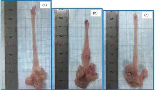
2. Gross mucosal damage in the esophagus.
Normal rats had no detectable esophageal mucosa lesions, whereas esophageal ulcers were readily apparent in the middle or distal esophagus of CRE control rats.
As compared to the normal rats, CRE control rats
exhibited basal layer thickening and esophagus length
decreased and enlarged. Moreover, esophagus mucosal
Figure 2. Representative image in each group of esophageal mucosal ulcer after surgical induction of CRE. A: Normal rat; B: CRE control rat; C: AT-mix-treated rat.
Figure 1. DPPH and ABTS radical scavenging activities.
AT-mix. AT-mix, water extract of Areca Semen and Toosendan Fructus Mixture.
Data are mean ± SEM. Each experiment was run in triplicate.
ulcer is severe. On the other hand, the AT-mix rats remarkably alleviated an extensive damage (Figure 2).
Figure 3. Esophageal NOX4 and p22phox protein expressions. N, normal rats; Con, CRE control rats; AT-mix, AT-mix 200 mg/kg body weight/day-treated CRE rats.
Data are mean ± SD. (n=8) Significance: *P < 0.05 vs. CRE control rats.
Figure 4. Esophageal Nrf2, HO-1, SOD, Catalase, and GPx-1/2 protein expressions. N, normal rats; Con, CRE control rats;
AT-mix, AT-mix 200 mg/kg body weight/day-treated CRE rats. Data are mean ± SD. (n=8) Significance: #P <0.05, ##P <0.01 vs. normal rats and *P <0.05, **P <0.01 vs. CRE control rats.
3. Esophageal NOX4 and p22
phoxprotein expressions.
An important cellular source of ROS is the NADPH oxidase family. The protein expressions of both NOX4 and p22
phox(the markers of NADPH oxidase activity in the esophageal tissues) were augmented in CRE control rats. However, its change didn’t show a significant
increase. Otherwise, AT-mix treatment had significantly down-regulated NADPH oxidase (Figure 3).
4. Esophageal Nrf2, HO-1, SOD, Catalase, and GPx-1/2 protein expressions.
As shown in Figure 4, the Nrf2 protein expression
Figure 5. Esophageal p-I
B
and NF-
Bp65 protein expressions. N, normal rats; Con, CRE control rats; AT-mix, AT-mix 200 mg/kg body weight/day- treated CRE rats. Data are mean ± SD. (n=8) Significance: ##P <0.01, ###P <0.001 vs. normal rats and *P <0.05 vs. CRE control rats.was significantly decreased in CRE control rats compared with normal rats, whereas AT-mix treatment significantly up-regulated Nrf2 expression. CRE control rats significantly showed decreased expressions of HO-1, SOD, Catalase, and GPx-1/2 in esophagus compared with normal rats; however AT-mix administration up-regulated all four antioxidant enzymes compared with CRE control rats. Especially, HO-1 showed a significant increase (Figure 4).
5. Esophageal p-Iκ Bα and NF-κ Bp65 protein expressions.
The protein levels of the oxidative stress-related proteins, such as p-IκBα and NF-κ Bp65, were examined.
As shown in Figure 5, the protein levels of p-Iκ Bα and NF-κ Bp65 increased in the esophagus of CRE control rats, whereas these elevated levels significantly reduced in AT-mix treated CRE rats.
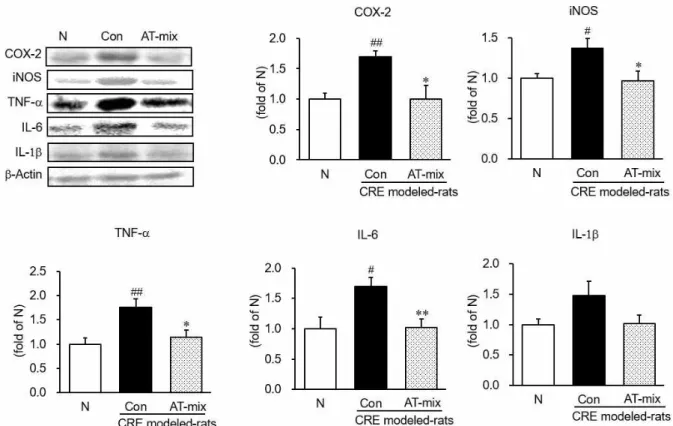
6. Esophageal COX-2, iNOS, TNF-α , IL-6, and IL-1β protein expressions.
We quantified COX-2, iNOS, TNF-α , IL-6, and IL-1β protein expressions (Figure 6). The inflammation- related protein expressions in CRE control rats were significantly augmented in the esophagus compared with normal rats. However, treatment with AT-mix suppressed these proteins in the esophagus. Herein, AT-mix supplementation significantly decreased COX-2,
iNOS, TNF-α , and IL-6. In addition, AT-mix administration reduced nearly to normal levels or below in levels of COX-2, iNOS, and IL-6. However, IL-1 didn’t exhibit a significant change.
Ⅳ. Discussions
GERD is rapidly increasing worldwide and causing a
considerable economic impact. Reflux esophagitis (RE),
which is known as the early stage of GERD can be
principally diagnosed using esophageal pH value and
gastroscopy when the abnormalities of upper
gastrointestinal motility or the esophageal mucosa is
found
40,41). Inflammatory mediators and other factors
can break down the esophageal mucosal barrier, lower
the esophageal peristalsis, and reduce the lower
esophageal sphincter pressure, ultimately leading to
RE
42,43). The pathogenesis of RE results from an
imbalance between a numerous natural defense
mechanisms and aggressive factors damaging the
esophagus. The fundermental aggressive components
of gastric refluxate include acid, pepsin, pancreatic
enzymes, and bile cause inflammation, destruction of
the normal squamous epithelium of the esophagus,
and ulceration. In recent years, a number of studies
have detected that the occurrence of esophageal
mucosal injury in RE patients are closely associated
with oxidative stress factors
44). Excessive reactive oxygen
species (ROS) are produced by mucosal epithelial cells
Figure 6. Esophageal COX-2, iNOS, TNF-α, IL-6, and IL-1β protein expressions. N, normal rats; Con, CRE control rats; AT-mix, AT-mix 200 mg/kg body weight/day-treated CRE rats. Data are mean ± SD. (n=8) Significance: #P <0.05, ##P <0.01 vs. normal rats and *P <0.05, **P <0.01 vs. CRE control rats.