online©ML Comm
-146- 대한두경부종양학회지
제 25 권 제 2 호 2009
Ceruminous Adenoma of the External Auditory Canal
- Report of Two Cases -
Na Rae Kim, MD1, Kyu Cheol Han, MD2, Hee Young Hwang, MD3, Hyun Yee Cho, MD1 Departments of Pathology,1 Otolaryngology2 and Radiology,3 Gachon University Gil Hospital, Incheon, Korea
외이도의 귀지샘종
- 2예 보고 -
가천의과학대학교 길병원 병리과,1 이비인후과,2 영상의학과3 김나래1·한규철2·황희영3·조현이1
=
국 문 초 록 =외이도의 종양은 드물며, 귀지샘에서 기원한 종양은 더욱 흔하지 않다. 저자들은 이루를 동반한 2예의 귀지샘종을 보 고하고자 한다. 현미경적으로, 2예 모두 중층 혹은 단층으로 둘러싸인 세관 혹은 샘으로 이루어진 경계가 좋은 종양이었 다. 종양세포는 과립성의 풍부한 호산성 세포질을 가졌고, 세포질의 관내 돌출이 관찰되어 아포크린화생을 보였다. 완전 절제후 재발은 관찰되지 않았다. 귀지샘종은 경계가 좋은 양성종양이며, 광범위 절제 치료하지만, 높은 재발율을 보인다.
여기에서 외이도에서 발생한 귀지샘종의 임상적 소견과 함께 병리 소견에 대해 기술하였다.
중심 단어:귀지샘종·외이도.
Introduction
The external auditory canal is divided into the inner osse- ous(2/3) and the outer cartilaginous portions(1/3). The skin of the bony portion contains few appendages, and the skin of the cartilaginous portion shows numerous hair follicles, sebaceous glands and a modified apocrine sweat gland, i.e., the ceruminous gland. This gland is found in the deep der- mis of the overlying skin lining the cartilaginous portion of the external auditory canal. Together with sebaceous glands, they produce cerumen(the ear wax). Cerumen plays an im- portant role to protect the external auditory canal against physical damage and microbial invasion. Ceruminous ade- noma is an infrequent tumor and it should be distinguished from other ceruminous gland tumors.1) There have been only two reports of ceruminous adenomas in Korea.2,3)
Here, we report on two cases of ceruminous adenoma that originated in the external auditory canal, and briefly describe the clinical features and surgical treatment.
Case Report
Case 1 was is a 53-year-old male who suffered from inter- mittent otorrhea in the right ear for 1 year. A protruding mass was detected at the cartilagenous portion of the right exter- nal auditory canal. The right tympanic membrane couldn’t be checked due to a canal mass. The left tympanic membrane was intact. He had no hearing loss, tinnitus or vertigo. Tem- poral bone computed tomography(CT) showed a tumor shad- ow of soft-tissue density on the posterior wall of the right external auditory canal, but there was no sign of bone de- struction(Fig. 1A). Endoscopic examination showed an oval mass at the right external auditory canal(Fig. 1B). Dissec- tion and mass removal were done under local anesthesia. A bulging mass was seen at the posterior wall of the right ex- ternal auditory canal. Sharp incision was done at the midpor- tion of the mass, and turbid discharge was expelled. A lobu- 교신저자:조현이, 405-760 인천광역시 남동구 구월1동 1198
가천의과학대학교 길병원 병리과
전화:(032) 460-3865·전송:(032) 460-3073 E-mail:hicho@gilhospital.com
-147- lated mass on the posterior wall measured 1.5×1.5×1.3cm.
There has been no evidence of recurrence or ear trouble during 1 year of follow up. He was then lost to follow up.
Case 2 was a 71-year old male who presented with a 2 week history of left otorrhea. Head and neck temporal bone CT showed inflammatory exudates in the left middle ear cavity and accumulation of fluid at the left mastoid area. A space-occupying soft tissue density was observed in the left external auditory canal. The ear drum was intact. A poly- poid mass, measuring 0.8×0.7×0.5cm, was found at the left external auditory canal. He had no otalgia, hearing loss, tinnitus or vertigo. Polypectomy was performed, and the pa- tient’s postoperative course was uneventful. He was lost to follow up.
1. Pathological findings
In case 1, a gray tan colored, irregular shaped mass, meas- uring 1.5×1.5×1.3cm, was well delineated. Microscopi- cally, ceruminous glands and small cysts had proliferated in
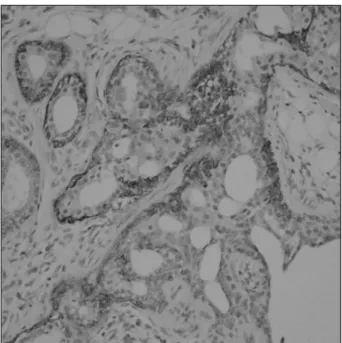
a hyalinized stroma with an infiltrative pattern of growth (Fig. 2A). The tumor cells formed tubuloglandular patterns of inner ceruminous epithelial cells that were decapitating, and these cells were subtended by a layer of spindled to cu- boidal myoepithelial cells(Fig. 2B). Intracytoplasmic and intraluminal yellow to brown pigments were found. Neither mitosis nor atypism was seen. Case 2 showed a polypoid oval shaped mass that measured 0.8×0.7×0.5cm. The mass showed tubuloglandular patterns of inner ceruminous epi- thelial cells with decapitating, and these cells were sub- tended by a layer of spindled cuboidal myoepithelial cells.
The ceruminous tumor cells were cuboidal to columnar cells with eosinophilic cytoplasm and apical snouts(Fig. 2C).
Intracytoplasmic and intraluminal yellow to brown pig- ments were found. Neither mitosis nor atypism was seen.
Immunohistochemically, the tumor cells of both cases were immunopositive for pancytokeratin(AE1/AE3;Dako, Glo- strup, Denmark, prediluted), and they were focally positive for S-100 protein(Polyclonal, 1:1200 dilution, Zymed, San
Fig. 1. Case 1. A:A coronal image of case 1 shows an irregular soft tissue density in the right external ear canal. B:Endoscopy shows an oval mass of smooth surface.
A B
Fig. 2. A:Case 1. The glandular and small cysts are seen at low magnification(H-E, ×100). B:Case 1. Ceruminous glandular prolifera- tion in tubular or papillary patterns is noted in a hyalinized stroma(H-E, ×200). Inset indicating ceruminous decapitation secretion in the luminal cells(H-E, ×400). C:Case 2. Ceruminous glandular proliferation of tubular or papillary patterns is noted in a hyalinized stroma(H-E, ×200). Arrow indicates brown colored pigmented secretion in the lumen(H-E, ×200).
A B C
-148- Francisco, CA, USA), and they were negative for GFAP (Dako, 1:350 dilution). The myoepithelial cells lining the glands were positive for smooth muscle actin(1A4;Dako, 1:100 dilution, Fig. 3). Ceruminous adenoma was diagnosed.
Discussion
The pathologic differential diagnosis of ceruminous ade- noma in the external auditory canal includes pleomorphic adenoma(chondroid syringoma), ceruminous adenocarcino- ma and cylindroma of the ceruminous gland.1) Eccrine cy- lindroma is most likely a very primitive sweat gland tumor differentiating toward either the eccrine or apocrine line.
Thus, its histologic features show compact nests of tumor cells surrounded by a thick basement membrane.4) Eccrine cylindroma is an uncommon tumor in the external auditory canal. Ceruminous adenocarcinoma shows infiltrating growth or perineural invasion, and ceruminous adenoma does not show significant cytologic atypism or mitotic activity with well-circumscribed borders.5) Pleomorphic adenoma of the external auditory canal is extremely rare.6) Pleomorphic ade- oma shows the pathology of irregular islands and cordons of epithelial cells forming ducts and tubule-like structures in a myxoid matrix with occasional chondroid areas. Ceruminous adenoma demonstrates a dual cell population of basal myo- pithelial-type cells and luminal ceruminous cells. Both the diffuse and strong immunoreactivity for CK7 of the luminal cells and the immunonegativity for smooth muscle actin and S-100 protein of the basal myoepithelial-type cell help us to
distinguish this tumor from other neoplasms that occur in this site.
There is still debate concerning the nomenclature and classification of ceruminous gland tumors. The term “ceru- minoma” has long been used as a collective denomination for all ceruminous gland tumors, which causes confusion re- garding differentiating between benignness and malignancy.
So, the term was excluded from the World Health Organiza- tion classification in 1991.7) Unfortunately, it is currently being used. Ceruminous adenoma is defined as a well-dif- ferentiated, localized, benign neoplasm, it is occasionally cystic and it shows papillary proliferation of glands that are histologically similar to normal ceruminous glands;irregular glandular structures lined by cuboidal epithelium with lu- minal blebbing or snouting, i.e., apocrine differentiation. Im- munohistochemically, the tumor cells of ceruminous ade- noma are positive for pancytokeratin and S-100 protein, which supports a ceruminous gland origin.1)
Ceruminous gland tumors are infrequent lesions of the ex- ternal auditory canal. Ceruminous tumors mainly develop in the cartilaginous canal and rarely in the osseous canal be- cause of the above mentioned anatomical distribution of the glands.8) Ceruminous adenomas cause very few symptoms and in general they are nonspecific. The symptoms associ- ated with ceruminous adenoma, such as conductive hearing loss, otorrhea and otalgia, are usually related to the size of the tumor and the degree of canal obstruction.
Ceruminous adenoma is a benign and well-delineated tu- mor, and it is best treated by wide local excision, yet ceru- minous adenomas have a high recurrence rate. It is difficult to make a distinction whether or not gross total resection is complete because of the anatomic restrictions of the area.1) Albeit its rare occurrence, a ceruminous adenoma must be taken into consideration in the differential diagnosis of tu- mors that are found in the external auditory canal.
References
1) Thompson LD, Nelson BL, Barnes EL. Ceruminous adenomas: A clinicopathologic study of 41 cases with a review of the literature.
Am J Surg Pathol. 2004;28(3):308-318.
2) Kang YK, Chi JG. Ceruminous gland tumors: 5 cases report. Ko- rean J Pathol. 1994;28(4):414-419.
3) Kim SC, Lee HK, Ahn SY, Lee JH. Two cases of ceruminous ade- noma arising from the external auditory canal. Korean J Otolar- yngol - Head Neck Surg. 1997;40(7):1059-1062.
4) Sharma HS, Meorkamal MZ, Zainol H, Dharap AS. Eccrine cylin- droma of the ear canal--report of a case. J Laryngol Otol. 1994; 108(8):706-709.
Fig. 3. The basal cells of the tumor glands are stained with smooth muscle actin(smooth muscle actin, ×200).
-149-
5) Kim CW, Rho YS, Cho SJ, Lee CH, Bae WJ. A case of cerumin- ous adenocarcinoma of the external auditory canal presenting as an aural polyp. Am J Otolaryngol. 2008;29(3):205-208.
6) Koyuncu M, Karagoz F, Kiliacarslan H. Pleomorphic adenoma of the external auditory canal. Eur Arch Otorhinolaryngol. 2005;262 (12):969-971.
7) Lassaletta L, Patron M, Oloriz J, Perez R, Gavilan J. Avoiding misdiagnosis in ceruminous gland tumours. Auris Nasus Larynx.
2003;30(3):287-290.
8) Yamamoto E, Tabuchi K, Mori K. Ceruminous adenoma in the osseous external auditory canal(a case report). J Laryngol Otol.
1987;101(9):940-945.