• 검색 결과가 없습니다.

Identification of the microstructural components of crumb rubber modified asphalt binder (CRMA) and the feasibility of using environmental scanning electron microscopy (ESEM) coupled with energy dispersive X-Ray spectroscopy (EDX)

N/A
N/A
Protected

Academic year: 2021

Share "Identification of the microstructural components of crumb rubber modified asphalt binder (CRMA) and the feasibility of using environmental scanning electron microscopy (ESEM) coupled with energy dispersive X-Ray spectroscopy (EDX)"

Copied!
10
0
0

로드 중.... (전체 텍스트 보기)

전체 글

(1)

Kim, Hyun Hwan 김`현`환 Member·Texas State University·Graduate Assistant (E-mail : [email protected]) Mithil Mazumder Texas State University ·Graduate Assistant (E-mail : [email protected])

Lee, Moon Sup 이`문`섭 Member·Korea Institute of Civil Engineering and Building Technology·Senior Researcher·

Corresponding Author (E-mail : [email protected])

Lee, Soon Jae 이`순`제 Member·Texas State University·Associate Professor (E-mail : [email protected])

1. INTRODUCTION

The use of unmodified petroleum asphalt has several

disadvantages such as crack prone in low temperature, poor aging and fatigue resistance, premature distress like rutting

Int. J. Highw. Eng. Vol. 18 No. 6 : 41-50 DECEMBER 2016 https://doi.org/10.7855/IJHE.2016.18.6.041

ABSTRACT

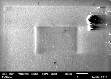
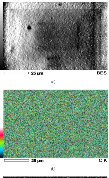
OBJECTIVES : In this study, microstructural components of crumb rubber modified asphalt (CRMA) binder were investigated using environmental scanning electron microscope (ESEM). To clearly understand the elemental composition of the CRMA binder, energy dispersive X-ray spectroscopy (EDX) was employed on the ESEM samples.

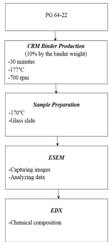
METHODS : CRMA binders were produced using open blade mixers at 177 ℃ for 30 min. The binders were artificially aged through a series of accelerated aging processes. Sample preparation was done by making a mold shape on the glass slide. Thereafter, the morphology of the CRMA binder was observed using the ESEM coupled with the EDX.

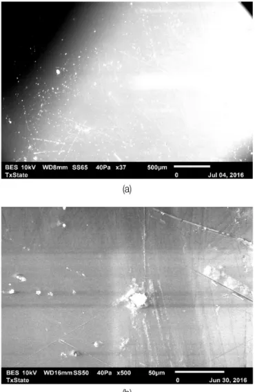
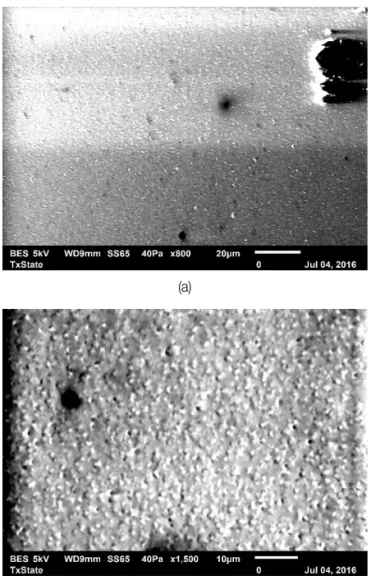
RESULTS : The images captured from the ESEM indicate that the unaged CRMA binder appears to have a single-phase continuous nonuniform structure after the addition of crumb rubber particles, whereas the artificially aged CRMA binder was observed to have two different phases. ESEM coupled with EDX shows detailed internal structure of the modified binders compared to other technologies (i.e., optical microscopy, atomic force microscopy, and conventional scanning electron microscope).

CONCLUSIONS : The captured images resemble the internal structures such as the viscous properties of the unaged CRMA binder and the interaction between the rubber particles and the base binder at aged condition. ESEM is a powerful instrument and with the introduction of EDX, it provided more details of the network microstructure of the asphalt binder. ESEM coupled with EDX is recommended for use in future investigation of microstructure of asphalt binders.

Keywords

Environmental scanning electron microscopy, Energy dispersive X-ray spectroscopy, Crumb rubber modified asphalt

Corresponding Author : Lee, Moon Sup, Senior Researcher Korea Institute of Civil Engineering and Building Technology, 283, Goyangdae-ro, Ilsanseo-gu, Goyang-si, Gyeonggi-do, 10223, Korea.

Tel : +82.31.9100.690 Fax : +82.31.9100.161 E-mail : [email protected]

International Journal of Highway Engineering http://www.ksre.or.kr/

ISSN 1738-7159 (print) ISSN 2287-3678 (Online)

Received Aug. 18. 2016 Revised Nov. 16. 2016 Accepted Nov. 16. 2016

Identification of the microstructural components of crumb rubber modified asphalt binder (CRMA) and the feasibility of using environmental scanning electron

microscopy (ESEM) coupled with energy dispersive X-Ray spectroscopy (EDX)

ESEM과 EDX를 사용한 CRM 바인더의 미세구조 성분 분석

(2)

and moisture induced damage. As a result the addition of polymer additives (i.e., SBS, SBR), domestic waste polymers (i.e., crumb rubber from scrap tires and waste plastic) and chemical admixtures into the asphalt binder have become an effective solution not only because of their resistant to the failures but also due to their environmental and economical advantages (Bahia et al. 2001; Sun and Lu 2003; Polacco et al. 2006; Garcia-Morales et al. 2006; Yildrim 2007; Yu et al.

2007). Using crumb rubber in the asphalt binder as a modifier, on the one hand, can enhance the mechanical properties and riding quality, asphalt stability at high temperature, improve the rutting and cracking resistance of asphalt pavements (Bahia and Davis 1994; Ruth et al. 1995;

Liang et al. 1996; Way 1998; Huang et al. 2002; Palit et al.

2004; Shen and Amirkhanian 2005; Xiao et al., 2009; Wang et al. 2012a; Kim et al. 2013); on the other hand, can recycle waste rubber to reduce the burden on the environment (Adhikari et al. 2000; Azizian et al. 2003; Xiang et al. 2009;

Karakurt 2015). In recent years crumb rubber modified asphalt (CRMA) has attracted a close attention of many countries in the world due to its cost effectiveness and environmental issues.

Crumb rubber is produced by grinding whole rubber to approximately 4.75-0.075 mm at ambient or cryogenic temperature (Siddique and Naik 2004). Ambient crumb rubber modifier (CRM) is manufactured by grinding rubber to the required sizes at room temperature and cryogenic CRM is produced by freezing rubber by using liquid nitrogen, and the embrittled rubber is then shattered to the required sizes by using impact device (Blumenthal 1994).

CRM can be incorporated into the asphalt binder in two ways either a dry or wet process. In the dry process, crumb rubber is added to the aggregates before adding bitumen whereas in the wet process, crumb rubber is added to the binder at elevated temperature which is approximately 190℃

and the interaction conditions are maintained for 1-4 h (Visser and Verhaegle 2000). This investigation is concerned with CRMA produced with wet process. The rubber particles and bitumen interaction is very complex.

This interaction mechanism can be classified into three categories which are swelling of rubber molecules, dissolution of rubber in bitumen and devolatilization and cross-linking in rubber (Shen et al. 2009). When the rubber particle is added to the bitumen it absorbs the lighter

aromatic fraction and rubber particle swells, leads to a gel like structure which is the reason behind the increase of viscosity (Heitzman 1992; Bahia and Davis 1994).

In order to understand the interaction between the rubber particles and the bitumen, it requires the understanding of the microstructural properties of such mechanical behavior.

Development of modern technology has allowed to examine asphalt binder at smaller scales using several equipment like optical microscopy, scanning electron microscopy (SEM), and atomic force microscopy (AFM).

However, direct observation of the change in the binder morphology due to modifier-binder interaction is quite difficult. Although the optimal microscopy can provide good images, the magnification is not enough to take accurate roughness data. Loeber et al. (1996) investigated the microstructural properties of asphalt using AFM and SEM. They concluded that AFM can identify the structure of asphalt without any sample preparation with low resolution. On the other hand using SEM the same results are observed with better resolution. The nanoscale surface images and roughness of asphalt binder can be measured using AFM, but there are several limitations like additional work for fixing tip and scanning time by indirect observation. Apart from that Masson et al. (2006) mentioned that AFM can differentiate the binder based upon the morphology and the different phases but it cannot identify the change in network structure after the modifier interaction. SEM is considered to provide better microscopic characterization for asphalt binder including accurate surface image. However, measuring visible image for oil based material like asphalt is quite challenging under high vacuum mode of conventional SEM. Also, it requires a good sample preparation with artificial coating (Liu et al.

2014). Some studies have been performed using SEM to observe the morphology of the CRMA (Shen and Amirkhanian 2005; Xiang et al. 2009; Liu et al. 2014).

Of all the microscopic techniques, the environmental

scanning electron microscope (ESEM) which has little

different to conventional SEM technique is considered as

one of the best techniques for oil bearing materials like

asphalt. The primary advantage of the ESEM over

conventional SEM is that the ESEM does not require the

test sample to be under high vacuum. Thus, wet, oily, dirty,

or nonconductive samples can be examined in their natural

(3)

state without modification or preparation (Kimseng et al.

2001). At first Rozeveld et al. (1997) performed the ESEM techniques to understand the internal structure of unmodified and polymer-modified binders. They reported that ESEM can help to characterize the morphology of the network structure of asphaltenes and resins. Later Williams et al. (1998) used ESEM to understand the asphalt water interactions in terms of water stripping and water penetration in asphalts. They concluded that ESEM has potential applications to study the microscopic properties of asphalt binder and can be used as a practical method for studying roadway failure mechanisms. Although ESEM introduced very early, limited studies have been performed using ESEM techniques to understand the morphology of the asphalt binder with CRM (Xin et al. 2012; Divya et al.

2013; Yu et al. 2014).

Xin et al. (2012) investigated the difference between the microstructural properties of CRMA and CRMA binder with Sasobit using ESEM. They reported that the addition of Sasobit changes the uneven state of the CRMA and improve the surface microstructure by fully embedded into the binder and more evenly distributed. Divya et al. (2013) investigated the morphology of 12 binders using ESEM and carried out the energy dispersive X-ray spectroscopy (EDX) to identify the chemical composition of the network structure obtained from ESEM. They have used wet process to produce CRMA and analyzed the ESEM images to differentiate the surface morphology in between blended and blown bitumen. Yu et al. (2014) have conducted the experiment to investigate the performance and microstructural properties of CRMA with and without Evotherm-DAT additives. They differentiated the surface morphology between CRMA binder and CRMA binder with Evotherm-DAT using ESEM. By observing the images through ESEM they have concluded that the additive can change the aggregation of the rubber particles.

Although previous studies have demonstrated the difference between the microstructural properties of CRMA and CRMA with warm-mix additives, the morphology of long term aged (LTA) CRMA binder has not yet been performed using ESEM and EDX techniques. The main objective of this study is to investigate the difference between the microstructural properties of unaged CRMA and aged CRMA binder using ESEM. Furthermore, to understand the

chemical composition of the surface morphology obtained from ESEM, EDX was carried out on the aged CRMA binder. Also, the feasibility of these two techniques to characterize the internal structure of the binder needs to be investigated for future use. Figure 1 shows a flow chart of the experimental design used in this study.

2. EXPERIMENTAL DESIGN

2.1. Crumb-Rubber Modifier and Asphalt binder Performance grade (PG) 64-22 asphalt binder produced by NuStar was used as a base binder for this study. Table 1 summarizes the results through three ageing process. The crumb rubber produced by mechanical shredding at ambient temperature was obtained from one source. Table 2 shows a gradation of crumb rubber. The CRM binder was produced in the laboratory at 177℃ for 30 minutes by an open blade mixer at a blending speed of 700 rpm (Putman 2005; Lee et al. 2006; Lee et al. 2007). The percentages of crumb rubber added for rubberized binder were 10%, by weight of the base binder. To ensure that the consistency of the CRM was maintained throughout the study, only one batch of crumb rubber was used in this study.

Fig. 1 Flow Chart of Experimental Design Procedures

(4)

2.2. Ageing Process

The unaged CRMA binder was artificially aged through a series of accelerated aging processes. The short term ageing was conducted in the rolling thin film oven (RTFO) for 85 min at 163℃. The RTFO aged binder was further aged using the pressure aging vessel (PAV) system, which simulated the long-term aging after setting at 2.10 MPa of pressure for 20 h at 100℃ [Asphalt Institute 2003].

2.3. Environmental Scanning Electron Microscope The ESEM used in this study is manufactured by JEOL

(Model #: JSM-6010PLUS/LA) to examine the surface microstructure of unaged and aged CRMA. The degree of magnification was chosen to be 200, 400, 500, 800, 1500 and 2000. The scan sizes used were 10, 20, 50, and 100 µm. The equipment settings used for scanning were as follows, 5-10kV;

pressure, 40Pa. Figure 2 shows the JEOL SEM used in this study.

2.4. Energy Dispersive X-ray Spectroscopy

EDX is normally used to find the chemical composition of the materials. It can create the elemental composition maps. It used a software called EDX-Genesis to gather and analyze the energy spectra. The software works to find out the peaks based on the gathered elemental spectrum and makes it a comprehensive survey tools to do the quantitative analysis of the chemical compositions.

2.5. Sample Preparation

In order to investigate the morphology of CRMA binders, sample preparation is considered as one of the most important task. A sample was prepared by pouring melted binder on the surface of a glass slide. All binders were preconditioned by controlled heating at 170℃ in an oven. No outside medium like spatula was used to spread the sample because it may manipulate the flow properties of CRMA. Using thin tape a mold shape was formed on the glass slide with a 1 mm thickness and pored the melted binder on the mold. The binder flowed smoothly into the mold and glass slides were placed on heating plate to produce a well-dispersed sample. The samples were examined immediately after they cooled down and after 24 hours or more.

At least three different regions of the surface were scanned. The CRMA binder sample was shown in Figure 3.

Table 1. Properties of Base Asphalt Binder (PG 64-22) Aging states Test properties Test result

Unaged binder

Viscosity @135℃ (cP) 531 G*/sin δ@ 64℃ (kPa) 1.415 G*/sin δ@ 64℃ (kPa) 2.531 RTFO aged residual G*/sin δ@ 25℃ (kPa) 2558

RTFO+PAV aged residual

Stiffness @ -12℃ (kPa) 287 m-value @ -12℃ 0.307

Table 2. Gradation of CRM Used in This Study Sieve number (㎛) % Cumulative passed

30 (600) 100.0

40 (425) 91.0

50 (300) 59.1

80 (180) 26.2

100 (150) 18.3

200 (75) 0.0

Fig. 2 JEOL SEM Fig. 3 CRMA Binder Sample for ESEM

(5)

3. RESULTS and DISCUSSION

3.1. ESEM Investigations on Morphology of CRMA Binder

As mentioned earlier samples were preconditioned by controlled heating to 170℃ in an oven. The samples were placed in the ESEM chamber. Figure 4 (a) shows the initial exposure of the sample in the ESEM chamber with relatively low magnification (37×). It seems like a plain surface.

Figure 4 (b) presents the image of unaged CRMA binder with a scan size of 50 µm and a magnification level of 500×.

Although it shows detailed surface shape, no noticeable changes in the surface morphology has been observed. The reason might be the scanning done very adjacent to the tape surface. While making samples in this procedure one needs to be careful about the placement of the binder sample in order to avoid such situation.

Figure 5 (a) shows the liquid state of the CRMA binder at 200× with the scan size of 100 µ m. The image was

captured as soon as the sample was exposed in the ESEM chamber. The microstructure of CRMA binder appears to be a single-phase continuous non-uniform structure after adding crumb rubber particles. The rubber particles are densely packed and enveloped by the base binder. It represents the state of high viscous CRMA binder after the addition of rubber particles in the base binder. This visual effect is similar to the mechanism explained in the article of Xin et al. (2012) and Liu et al. (2013). Figure 5 (b) represents more detailed shape of the microstructural surface of the CRMA binder with higher magnification of 500×. The image captured the network structure between the asphalt binder and crumb rubber. As a whole the rubber particles are not uniformly distributed in the asphalt binder.

Figure 6 (a) shows a microscopic image of a uniform view in between rubber particles and asphalt binder after 24 hours with a magnification degree of 800× at a certain part of the sample. The more detailed image of this internal structure is shown in Figure 6 (b).

(a)

(b)

Fig. 4 ESEM Image of CRM Asphalt Binder at the Border Side of the Sample

(a)

(b)

Fig. 5 ESEM Image of Liquid State of CRMA Binder

(6)

ESEM images were capture for the aged CRMA binder after a series of artificial aging. Figure 7 (a) presents the image of aged CRMA binder with a scan size of 10 µm and a high magnification level of 2000×. The reason behind using high magnification is to capture the liquid state of the aged CRMA binder as soon as it placed in the ESEM chamber after taking it from the oven. It seems like the image indicates the presence of rubber particles in the asphalt binder but due to high magnification it is quite challenging to observe the actual morphology. This is because the density of electrons coming from the electron guns increases with the magnification of image (Danilatos, 1990). Therefore, the sample surface becomes unstable and produces a blurred image. As a result it was decided to observe the development of microstructure of the aged CRMA binder after 24 hours. Figure 7 (b) shows the morphology of the aged CRMA binder after 24 hours. As it is obvious that the microstructure of the CRMA binder

should be changed due to the molecular mobility of the binder at room temperature and the oxidation of the surface of the binder. In order to observe this phenomenon in the aged CRMA binder the same spot was observed at a high magnification degree of 800×. Figure 7 (c) illustrates the molecular changes in the aged CRMA binder after experiencing the artificial aging.

(a)

(b)

Fig. 6 ESEM Image of Interaction between Rubber Particles and Asphalt

(a)

(b)

Fig. 7 ESEM Image for Aged CRMA Binder

(c)

(7)

The image appears to have two different phases. At the lower phase there is a broad dark black region and the upper phase quite light compared to that bottom region. Those two phases resemble the interaction between the rubber particles and base binder at aged condition. High resolution showed more detailed shape of some small spot (red circles) which represent the property of aged asphalt binder. Masson et al.

(2006) reported that this shape is formed due to the existence of asphaltene which is considered as primary material observed in aged asphalt binder.

In general, the enlarged images were taken from lower resolution to higher resolution. When decreasing the resolution from highest resolution, rectangle shapes matching with visual size at each magnification were detected on sample surface. Figure 8 shows those damages on the surface of the binder. Although it is possible to take the images of asphalt binder using ESEM, the operation of ESEM needs to be careful considering sample usefulness

and research plan. It is recommended to use ESEM at the end of all micro-characterizing process regardless the sample damages.

It is worth noting that this study also tried to take images using conventional SEM. The purpose is to observe if this sample preparation is suitable to take the image using Helios 400 SEM. It is very difficult to take the visible image of the CRMA binder at liquid state just after taking the sample from the oven. Due to that reason the sample was dried in room temperature for 24 hours. Then the sample was loaded in the FEI Helios 400 SEM. Imaging was done in field free mode and with ETD detector for secondary electron. Since the sample contained organic compound, the imaging was started at lowest possible accelerating voltage and current (i.e., 2.0kV and 5.3Pa). The detector could not detect any image at this voltage. Then, voltage was increased to 5kV keeping the current at 5.3Pa which did not produce any image. Voltage and current was further raised to 8kV and 86Pa. This time, sample was visible but the sample appeared to be melting. The detector was paused immediately and voltage was reduced to 5kV. Another attempt was made to take images and similar phenomenon of sample damaging was observed once again. The imaging was stopped at this point. The main reason for such a behavior was found to be the chamber pressure. The chamber pressure in the FEI Helios 400 was very low because it usually works in ultra-high vacuum (<0.1torr).

Based on such behavior of SEM it was confirmed that the SEM imaging of the present sample is only possible in ESEM where the pressure is not very low i.e. 1-50 Torr or 0.1-6.7 kPa.

3.2. Chemical Composition Analysis with EDX One of the benefit of ESEM is elemental analysis by EDX mode. Using spot mode within a very short time the energy spectrum can be recorded and analyzed by the EDX-Genesis software in order to get the chemical composition. The peak high ratios are considered as the measurement of the chemical composition. Figure 9 shows the EDX spectrum as a plot of X-ray counts versus energy (keV) for aged CRMA binder. The energy peaks corresponding to the various elements present in the aged CRMA binder are also shown.

The analysis helps to understand the chemical composition of (a)

(b)

Fig. 8 Sample Damage by Electron Beam and Vacuum Pressure

(8)

the aged CRMA binder. The peak high ratio of CRMA binder indicates the primary composition of binder is hydro carbon. As shown on the graph, most part of the material is consisted of hydro carbon and trace amount of oxygen and sulfur were detected. It is considered that the oxygen is generated through the aging process of CRMA binder, and the sulfur is produced by the addition of crumb rubber. All expected constituents were detected through elemental analysis using ESEM. Currently, there are lots of research including different kinds of additives to produce better asphalt binder for paving roads. Therefore, it is expected that the application of elemental analysis using EDX will be beneficial for identifying the elemental property of different kinds of asphalt binder.

4. CONCLUSIONS

The objective of this study is to investigate the microstructural components of CRMA binder. Performance grade (PG) 64-22 asphalt binder was used to produce the crumb rubber modified (CRM) binder. Control CRMA binders were artificially aged through a series of accelerated aging process. A good sample preparation has been introduced by making a mold shape on the glass slide without interrupting the natural flow of the CRMA binder. To accomplish the objective, microscopic morphology and (a)

(b)

(c)

(d)

(e)

Fig. 9 EDX Spectrum for Aged CRMA Binder Acquisition Conditon

Volt : 5.00kV Process Time : T2 Real Time : 196.60 sec.

Count Rate : 2148.00 CPS

Instrument : 6010LA Current : --- Live time : 195.10 sec.

Dead time : 1.00%

(9)

chemical composition of the CRMA binder was investigated by using ESEM and EDX techniques. On the basis of the observation following conclusions and recommendations can be drawn

The morphological feature of CRMA binder with and without ageing has been observed through ESEM.

Unaged CRMA binder appears to have a single-phase continuous non-uniform structure after adding crumb rubber particles. The captured images resemble the viscous properties of the CRMA binder due to the addition of rubber particles in the base binder.

The artificially aged CRMA binder is observed to have two different phases. The lower phase consists of the broad dark region and the upper phase relatively light which resembles the interaction between the rubber and asphalt binder due to oxidation.

Chemical composition of aged CRMA binder obtained through EDX indicates that this technology can be considered as an effective and time consuming technique.

ESEM is a powerful instrument and with the addition of EDX, it can provide more details of the network microstructure of asphalt binder.

While taking images using ESEM with high resolution (i.e., magnification degree of 2000×) caution needs to be taken for the sample.

Previous studies have observed the morphology of the unaged asphalt binder with and without wax additives. It is recommended to observe the microstructure of the long-term aged binder containing various wax additives using ESEM coupled with EDX.

Through this study, it can be expected that this equipment has the potential to understand the microstructural properties of asphalt binders relating to their engineering properties. It is also recommended to use other characterizing equipments along with ESEM to validate the observational method of morphology images.

Acknowledgement

This study was conducted under research project (Development of SAP Concrete for Bridge Deck Overlay) funded by the Ministry of Land, Infrastructure and Transport (MOLIT) and the Korea

Agency for Infrastructure Technology Advancement (KAIA). The authors would like to thank the members of research team, MOLIT and KAIA for their guidances and supports throughout the project.

REFERENCES

The Asphalt Institute (2003). Performance graded asphlt binder specification and testing, SP-1. The Asphalt Institute, Lexington.

Adhikari, B., De D, Maiti, S. (2000). Reclamation and recycling of waste rubber. Prog Polym Sci, 25(7), 909-48.

Azizian, F., & Nelson, O., Thayumanavan, P. & Williamson, J.

(2003). Environmental impact of highway construction and repair materials on surface and ground waters: case study:

crumb rubber asphalt concrete. Waste Manage, 22(8), 719-28.

Bahia, H. U., & Davis, R. (1994). Effect of crumb rubber modifiers (CRM) on performance-related properties of asphalt binders. J.

Assoc. Asphalt Paving Technol., 63, 414-449.

Bahia, H. U., Hanson, D. I., Zeng, M., Zhai, H., Khatri, M. A., &

Anderson, R. M. (2001). Characterization of modified asphalt binders in superpave mix design. Rep. No. 459, Transportation Research Board, National Research Council, Washington, DC.

Blumenthal, M. (1994). Producing ground scrap tire rubber: A comparison between ambient and cryogenic technologies. Proc., 17th Biennial Waste Processing Conf., ASME, New York.

Danilatos, G.D., (1990). Theory of the gaseous detector device in the environmental scanning electron microscope. Advances in Electronics and Electron Physics, 78, 1-102.

Divya, P. S., Gideon, C. S., & Krishnan, M. (2013). Influence of the type of binder and crumb rubber on the creep and recovery of crumb rubber modified bitumen. Journal of Materials in Civil Engineering, 25(4), 438-449.

Garci、 a-Morales M, Partal P, Navarro FJ, Gallegos C. (2006). Effect of waste polymer addition on the rheology of modified bitumen, Fuel, 85, 936-945.

Heitzman, M. (1992). “State of the practice design and construction of asphalt paving materials with crumb rubber modifier.”Rep.

No. FHWA A-SA-92-022, Federal Highway Administration, Washington, DC.

Huang B, Mohammad LN, Graves PS, Abadie C. (2002). Louisiana experience with crumb rubber-modified hot-mix asphalt pavement. Transport Res Rec: J Transport Res Board, 1789, 1- 13.

Karakurt, C. (2014). Microstructure properties of waste tire rubber composites: an overview. J Mater Cycles Waste Mang, 17, 422- 433.

Kimseng, K. & Meissel, M. (2001). Short overview about the ESEM: The Environmental Scanning Electron Microscope.

Maryland: Produced by CALCE Electronic Products and Systems Centre, University of Maryland.

Kim, H. & Lee, S.-J. (2013). Laboratory investigation of different

(10)

standards of phase separation in crumb rubber modified asphalt binders. Journal of Materials in Civil Engineering, 25(12), 1975-1978.

Lee, S.-J., Amirkhanian, S., & Shatanawki, K. (2006). Effects of crumb rubber on aging of asphlt binders. In: Asphalt rubber 2006, 3, California: Palm Springs, 779-95.

Lee, S.-J. (2007). Characterization of recycled aged CRMA binders.

Dissertation (Ph.D.), Clemson University.

Liu, H., Chen, Z., Wang, W., Wang, H., & Hao, P. (2014).

Investigation of the rheological modification mechanism of crumb rubber modified asphalt (CRMA) containing TOR additive. Construction and Building Materials, 67, 225-233.

Liang, RY., & Lee S. (1996). Short-term and long-term aging behavior of rubber modified asphalt paving mixtures. Transport Res Rec: J Transpor Res Board,1530(1), 1-7.

Loeber, L., Sutton, O., Morel, J.V.J.M., Valleton, J.M. & Muller, G.

(1996). New direct observations of asphalts and asphalt binders by scanning electron microscopy and atomic force microscopy.

Journal of Microscopy, 182(1), 32-39.

Masson, J.F., Leblond, V. & Margeson, J. (2006). Bitumen morphologies by phase

detection atomic force microscopy.

Journal of Microscopy, 221(1), 17-29.

Palit, S. K., Sudhakar Reddy, K., & Pandey, BB. (2004). Laboratory evaluation of crumb rubber modified asphalt mixes. J Mater Civ Eng, 16(1), 45-53.

Polacco, G., Stastna, J., Biondi, D., & Zanzotto, L. (2006). Relation between Polymer architecture and nonlinear viscoelastic behavior of modified asphalts, Current Opinion in Colloid &

Interface Science, 11(4), 230-245.

Putman, B.J. (2005). Qualification of the Effects of Crumb Rubber in CRM Binder, Dissertation (Ph.D.), Clemson University.

Rozeveld, S. J., Shin, E. E., Bhurke, A., France, L., & Drzal, L.

T.(1997). Network morphology of straight and polymer modified asphalt cements. Microsc. Res. Tech., 38(5), 529-543.

Ruth, B.E., & Roque, R. (1995). Crumb rubber modifier (CRM) in asphalt pavements. In: Proceedings of the transportation

congress, 768-85.

Shen, J., & Amirkhanian, S.N. (2005). The influence of crumb rubber modifier (CRM) microstructures on the high temperature properties of CRM binders, The International Journal of Pavement Engineering, 6(4), 265-271.

Sun, D., & Lu W. (2003). Investigation and Improvement of storage stability of SBS modified asphalt, Petroleum Science and Technology, 21, 901-910.

Shen, J., Amirkhanian, S.N., Xiao, F., &Tang, B. (2009). “Influence of surface area and size of crumb rubber on high temperature properties of crumb rubber modified binders.”Constr. Build.

Mater., 23(1), 304-310.

Siddique, R., & Naik, T. R. (2004). Properties of concrete containing scrap-tire rubber-An overview. Waste Manage., 24(6), 563-569.

Visser, A., and Verhaegle, B. (2000). “Bitumen rubber: Lessons learned in South Africa. ”Proc., Asphalt Rubber, Faro, Portugal (Nov. 14-17, 2000).

Wang, H., You, Z., Mills-Beale, J., & Hao, P. (2012a). Laboratory evaluation on high temperature viscosity and low temperature stiffness of asphalt binder with high percent scrap tire rubber.

Constr. Build. Mater., 26(1), 583-590.

Williams, T., & Miknis, F. (1998). Use of Environmental SEM to study asphalt-water interactions. Journal of materials in Civil Engineering, 10(2), 121-124.

Way GB. (1998). OGFC Meets CRM-Where the Rubber Meets the Rubber-12 Years of the durable success. The Asphalt Conference, Atlanta, Georgia.

Xiao, F., Zhao, W., & Amirkhanian, S. N. (2009). Fatigue behavior of rubberized asphalt concrete mixtures containing warm asphlt additives. Construction and Building Materials, 23(10), 3144- 3151.

Xiao, F., Punith, V. S., & Amirkhanian, S. N. (2012). Effects of

non-foaming WMA additives on asphalt binders at high

performance temperatures. Fuel, 94, 144-155.

수치

Fig. 1 Flow Chart of Experimental Design Procedures
Table 1. Properties of Base Asphalt Binder (PG 64-22) Aging states Test properties Test result
Figure 4 (b) presents the image of unaged CRMA binder with a scan size of 50 µm and a magnification level of 500×.
Fig. 6 ESEM Image of Interaction between Rubber Particles and  Asphalt
+3

참조

관련 문서

Surface morphology and chemical composition of samples were characterized by field emission scanning electron microscopy (FE-SEM), contact angle measurement and

The purpose of this study is to evaluate the marginal and internal fit of coping made by CAD/CAM using different scanning methods.. Zirconia coping was made

Therefore, the execution of Eco-STEAM program using Arduino can expect the students’ changes in perception of environmental problems, motivation for them

Excitation Detection X-ray photoelectron spectroscopy (XPS) Photons(X-ray) Electrons UV photoelectron spectroscopy (UPS) Photons (UV) Electrons

In this regard, to overcome the limitations associated with quantitative evaluation of ECT using the bobbin probe and to consider the field requirements in terms of scanning

The structure and film optical properties were investigated by X-ray diffraction(XRD), the particle size and thickness were investigated by scanning

Scanning electron micrographs showing sheathed Leptothrix ochraceas(L) and helical stalks of Gallionellas (G fi.: Gallionella filamenta, G fe.:

In this study, paper purposes to evaluate characteristics of machined surface using Atomic Force Microscope in the precision cutting of aluminum( Al 6061 )