http://dx.doi.org/10.14316/pmp.2016.27.1.14
We would like to thank Yongnam Kang, Ph.D. and Hong-Seok Jang, M.D. in the Department of the Radiation Oncology, Seoul St. Mary’s Hospital for a valuable advice of treatment planning and IRB process.
This research was supported by the Mid-career Researcher Program through the National Research Foundation (NRF) funded by the Ministry of Science, ICT & Future Planning (MSIP) of Korea (NRF- 2014R1A2A1A10050270) and by the Radiation Technology R&D program through the National Research Foundation of Korea funded by the Ministry of Science, ICT & Future Planning (No. 2013M2A2A 7038291).
Received 29 February 2016, Revised 15 March 2016, Accepted 18 March 2016
Correspondence: Tae-Suk Suh ([email protected]) Tel: 82-2-2258-7232, Fax: 82-2-2258-7506 cc This is an Open-Access article distributed under the terms of the Creative Commons Attribution Non-Commercial License (http://creativecommons.org/licenses/by-nc/4.0) which permits unrestricted non-commercial use, distribution, and reproduction in any medium, provided the original work is properly cited.
Evaluating the Dosimetric Characteristics of
Radiation Therapies according to Head Elevation Angle for Head and Neck Tumors
Geum-Seong Cheon* † , Seong-Hee Kang † , Dong-Su Kim † , Tae-Ho Kim † , Tae-Suk Suh †
*Department of Radiation Oncology, Seoul St. Mary’s Hospital, The Catholic University of Korea,
† Department of Biomedical Engineering and Research Institute of Biomedical Engineering, College of Medicine, The Catholic University of Korea, Seoul, Korea
Since the head and neck region is densely located with organs at risk (OAR), OAR-sparing is an important issue in the treatment of head and neck cancers. This study—in which different treatment plans were performed varying the head tilt angle on brain tumor patients—investigates the optimal head elevation angle for sparing normal organs (e.g. the hippocampus) and further compares the dosimetric characteristics of different types of radiation equipment. we performed 3D conformal radiation therapy (3D-CRT), intensity-modulated radiation therapy (IMRT), and tomotherapy on 10 patients with brain tumors in the frontal lobe while varying the head tilt angle of patients to analyze the dosimetric characteristics of different therapy methods. In each treatment plan, 95%
of the tumor volume was irradiated with a dose of 40 Gy in 10 fractions. The step and shoot technique with nine beams was used for IMRT, and the same prescription dose was delivered to the tumor volume for the 3D-CRT and tomotherapy plans. The homogeneity index, conformity index, and normal tissue complication probability (NTCP) were calculated. At a head elevation angle of 30 o , conformity of the isodose curve to the target increased on average by 53%, 8%, and 5.4%. In 3D-CRT, the maximum dose received by the brain stem decreased at 15 o , 30 o , and 40 o , compared to that observed at 0 o . The NTCP value of the hippocampus observed in each modality was the highest at a head and neck angle of 0 o and the lowest at 30 o . This study demonstrates that the elevation of the patients’ head tilt angle in radiation therapy improves the target region’s homogeneity of dose distribution by increasing the tumor control rate and conformity of the isodose curve to the target.
Moreover, the study shows that the elevation of the head tilt angle lowers the NTCP by separating the tumor volume from the normal tissues, which helps spare OARs and reduce the delivered dose to the hippocampus.
Key Words: Head&Neck Cancer, Hippocampus, Head elevation angle, Treatment planning
서 론
Brain tumor의 방사선 치료는 환자의 병기(Stage)정도에 따라 Grade III∼IV인 종양의 경우에는 55∼60 Gy, Grade I∼
II 인 경우에는 적어도 45∼55 Gy 등의 정상장기의 임계선 량을 넘는 선량을 조사 해야 한다. 5) 특히 두 경부에 고 선 량의 방사선이 조사될 때 1,12,13) Primary target의 위치에 따라 서 두부 안의 결정장기(organ at risk, OAR)들의 한계선량 (tolerance dose) 4) 이 초과되는 경우, 시신경(시력결손), 뇌간 (호흡곤란, 어지럼증), 내이(청력손상), 수정체(백내장)등 부 위별로 심각한 신체적 장애와 부작용이 발생될 수 있기 때 문에 두 경부 암 치료에 있어서 OAR-sparing은 매우 중요 한 issue이다.
최근 두 경부 암 치료 시 선량학적 이점을 갖기 위하여
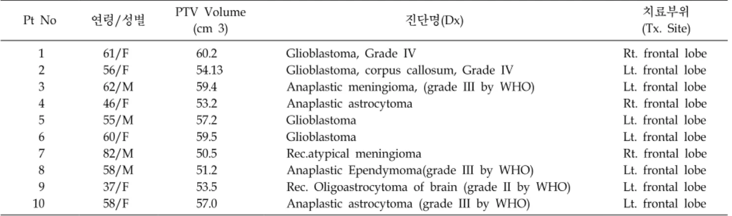
Table 1. Characteristics of 10 patients participated in the study.
Pt No 연령/성별 PTV Volume
(cm 3) 진단명(Dx) 치료부위
(Tx. Site)
1 61/F 60.2 Glioblastoma, Grade IV Rt. frontal lobe
2 56/F 54.13 Glioblastoma, corpus callosum, Grade IV Lt. frontal lobe
3 62/M 59.4 Anaplastic meningioma, (grade III by WHO) Lt. frontal lobe
4 46/F 53.2 Anaplastic astrocytoma Rt. frontal lobe
5 55/M 57.2 Glioblastoma Lt. frontal lobe
6 60/F 59.5 Glioblastoma Lt. frontal lobe
7 82/M 50.5 Rec.atypical meningioma Rt. frontal lobe
8 58/M 51.2 Anaplastic Ependymoma(grade III by WHO) Lt. frontal lobe
9 37/F 53.5 Rec. Oligoastrocytoma of brain (grade II by WHO) Lt. frontal lobe
10 58/F 57.0 Anaplastic astrocytoma (grade III by WHO) Lt. frontal lobe
종양체적과 정상조직간의 선량 경사도(dose gradient)를 높 여 종양체적에는 고 선량을 조사하면서 결정장기에는 최소 의 선량을 전달하는 치료 기법인 최적화된 비 균일 플루언 스(nonuniform fluence)를 만들어 선량분포를 형성하는 세기 조절방사선치료(Intensity modulation radiation therapy, IMRT) 를 이용하거나, 8,16,18,20)
64개의 binary MLC를 이용하여 최적 화된 beamlet을 생성하고, 환자 주변으로 360도 회전하며 치료 선량을 전달하는 토모테라피(Hi-Art helical tomotherpay, TOMO)를 이용 6,9,19) 하여 치료를 수행하고 있다.
이러한 최신 치료 기술을 이용함에도 불구하고 여전히 복잡한 구조를 지닌 두 경부 암 치료에서 OAR-sparing을 충분히 하지 못할 수 있다. 예를 들어, 뇌의 관자엽 안쪽, 대뇌 겉 질 밑에 위치하는 해마(Hippocampus)는 학습과 기 억에 관여하고, 감정행동 조절, 신경학적 인지 기능 등을 담당한다고 알려져 국내외학회 등에서 활발히 연구가 진행 중에 있다. 그렇지만 종양이 해마 인근에 위치한 환자의 경 우 주변선량을 제한해야 하기 때문에 종양체적에 선량 전 달이 부 정확해 질 수 있으며 이에 따라 치료효과 저하 및 국소재발을 일으킬 수 있다고 알려져 있다. 14,15)
Joshua 등에 의해 17) Whole brain 방사선 치료 시 두 경부 각도를 주어 방사선을 조사하여 Hippocampus Sparing을 한 연구 10) 내용이 보고되었으며, Head angle을 기울인 경우 OAR-sparing이 향상되고 뇌간(Brain stem)과 렌즈(lens)등의 최대선량을 줄여 주었다는 보고가 있었다. 본 연구에서는 이러한 연구를 기반으로 두 경부 종양(Brain tumor)환자를 대상으로 방사선치료의 3가지 기법 즉, 3차원 입체조형치 료(Three-dimensional conformal radiation therapy, 3D-CRT), 11) IMRT, TOMO를 이용, 치료계획을 수립하여 해마를 비롯한 정상장기를 보호하기 위한 두 경부 거상각도(head elevation
angle)에 의한 장비 별 선량특성을 비교 분석해 보고자 한다.
재료 및 방법
1. Patients and image acquisition
2012년 3월에서 2014년 12월까지 두 경부 종양의 확진 을 받고 방사선치료를 받은 환자 중 전두엽에(frontal lobe) 종양이 위치하고 중요 장기인 해마와 인접된 10명의 환자 를 대상으로 연구를 수행하였다. 환자군 의 나이는 37세에 서 82세 사이였으며 성별은 남자 4명, 여자 6명으로 구성되 었다. 환자들의 병소 위치는 우 전두엽(Rt. frontal lobe) 3명, 좌 전두엽(Lt. frontal lobe) 7명이었고 환자의 특성은 Table 1과 같다. 10명의 환자들 모두 Treatment Planning을 위해 Aquaplast RT mask fixation (Aquafast, Avondale, PA)를 착용 하고, SOMATOM Sensation Open (Simens, Munich, Germany) 을 이용하여 슬라이스 두께 3 mm와 해상도 1024×1024 pixels 의 전산화 단층영상을 얻었다. 전산화 단층영상만으로는 교모세포종(Glioblastoma), 악성뇌교종(Anaplastic astrocytoma) 등의 주변 악성 부종(malignant edema)과 주변 정상장기의 (e.g., 해마, 시신경, 뇌간 등) 구분이 어려워 정확한 종양체 적의 설정이 어렵다. 따라서 정확한 종양체적 설정을 위하 여 VERIO 3T (Simens, Erlangen, Germany)를 이용하여 T1 GD (가돌리늄)가중영상을 획득하였다.
2. Head angle position and delineation
각도 별 환자의 CT영상을 획득하고 Treatment Planning
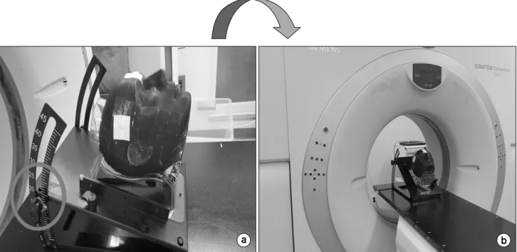
및 structure delineation을 수행하기 위하여 Fig. 1과 같이
Alderson-Rando phantom과 Head Tilting Base plate를 이용하
여 0 o , 15 o , 30 o , 그리고 40 o 로 변화시켜가며 전산화 단층촬
Fig. 1. (a) RANDO Phantom and head-board, (b) RANDO Phantom and head-board scan with CT simulator.
Fig. 2. Delineation of the hippocampus in MRI (Orange Line: Hippocampus, Light green Line: Hippocampus 3 mm expansion).
영영상을 획득하였다. 이후 Alderson Rando anthropomorphic phantom과 환자의 CT영상을 fusion program을 이용하여 영 상융합을 하였다. 각도 별로 새롭게 획득된 환자의 CT영상
과 MR영상은 전산화 치료계획장치(Pinnacle v8.0, Phi-lips
Medical System, Cleveland, USA)의 Normalized Mutual Infor-
mation algorithm을 이용하여 영상융합(image fusion)을 수행
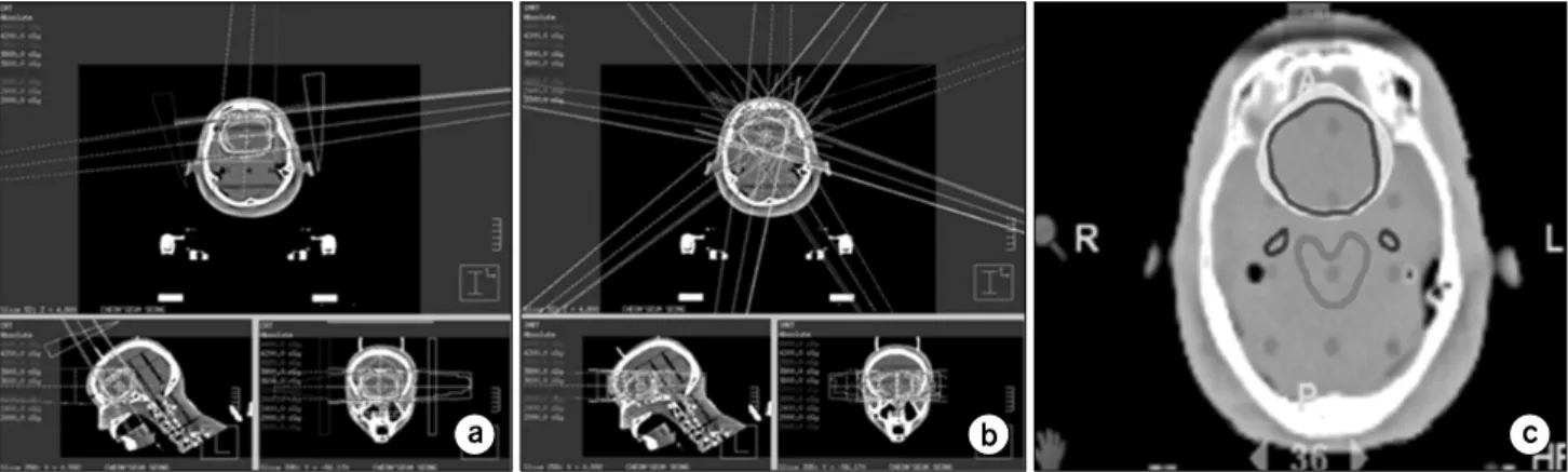
Fig. 3. Dose distribution among the 3 different treatment techniques (a) 3D-CRT, (b) Linac-IMRT and (c) Tomotherapy.
하였다. 21) Fig. 2와 같이 Image fusion된 자기 공명영상에 방 사선 종양학 전문의가 T1 GD 영상에서 보이는 종양을 육 안적 종양체적(Gross tumor volume, GTV)으로 설정하였고, 종양체적에 처방선량 전달을 위해 GTV에 5 mm margin을 추가로 설정 2) 하여 치료계획용적(Planning Target Volume, PTV)을 만들어 치료 계획에 이용하였다. 종양체적 주변의 결정장기를 평가하기 위하여 해마(Hippo campus), 안구 (Eyeball), 뇌간(Brain stem), 수정체(lens), 내이(Cochlea) 등의 OAR을 묘사 하였다.
3. 치료 계획 및 선량 분포 획득
두 경부 거상각도(head elevation angle)에 의한 장비 별 선량특성을 비교분석 하기 위해 3가지 다른 Modality를 사 용하여 치료계획을 수립하였다. 3D-CRT와 IMRT기법의 치 료계획 수립을 위해 전산화 치료계획장치(Pinnacle v8.0, Philips Medical System, Cleveland, USA)를 이용하였다.
3D-CRT의 치료계획은 종양체적(PTV)에 400 cGy/10 fx, 총 40 Gy를 처방하여 조사하였으며, 3) 6 MV의 광자선을 3 o , 82 o , 262 o , 그리고 300 o 에서는 Table을 90도 회전시켜 총 4개 의 비 동일평면(non Coplanar-plane) 조사 빔을 기본으로 대 상 환자의 종양체적 위치와 정상장기의 보호 정도를 고려 하여 최적의 선 속 방향과 가중치를 설정하였다. IMRT기 법의 경우 3D-CRT의 치료계획과 동일한 에너지와 처방선 량으로 종양체적의 95% 이상이 조사받도록 치료계획을 수 립하였다. 치료계획의 최적화를 위하여 DMPO (Direct Machine Parameter Optimization algorithm)알고리즘을 이용 하였으며, 3 o , 35 o , 70 o , 110 o , 150 o , 190 o , 230 o , 280 o , 320 o 로 구성된 9개 동일평면(Coplanar-plane) 조사 빔을 단계별조사 기법(Step-and-shoot)을 이용하여 선량분포(dose distribution)
를 획득하였다(Fig. 3).
TOMO 치료계획은 Hi-Art helical tomotherpay, TOMO Software version (4.2.3)의 이용하여 기존 치료계획들과 동 일한 선량을 처방하였고 부가인자(parameter)로는 Dose Cal- culation Grid는 Fine, Field Width는 1.05 cm Jaw, Modulation Factor는 2.0, Pitch 0.3의 Parameters를 적용하여 효율적으로 delivery될 수 있도록 지속적으로 변수들을 변화시키고 조 절하였다(Fig. 3).
4. 평가 및 분석
수립된 세 가지 치료계획기법(3D-CRT, IMRT, TOMO)으 로부터 획득한 선량-체적 히스토그램을 통하여 두 경부 거 상각도에 따른 종양체적 과 주변 OAR의 선량 4) 을 비교 분 석하였다(Fig. 3). 7)
종양체적은 선량 일치 성지수(conformity index, CI), 선량 균질 성 지수(homogeneous index, HI) 종양평균선량(PTV mean dose) 등을 비교하였으며, 선량 일치 성 지수와 선량 균일도 지수는 아래 식(1)을 이용하여 획득하였다.
×
(1)
위 수식에서 V TV 는 처방선량에 들어간 볼륨이고, V PTV 는 PTV의 볼륨, TV PV 는 처방선량에 들어간 볼륨 안의 PTV 볼 륨을 나타내며, CI 값이 1에 가까울수록 선량 일치성이 좋 음을 의미한다.
여기서(식 2), D 2% 는 PTV 2% 선량이고, D 98% 는 PTV 98%
선량, D median 은 PTV의 중간 선량이며, 그 수치가 0에 가까
울수록 선량 균질성이 좋음을 의미한다.
(2)
정상조직의 경우 QUANTEC 25,26) 을 참고하여 뇌간, 수정 체의 maximum dose를 비교하였으며, 안구의 경우 mean dose, 그리고 해마의 경우 Dmax dose와 D100%를 비교하였 다. D100 % 는 PTV의 100% 선량을 의미한다. 추가적으로 정 상조직 부작용 발병률(normal tissue compli cation proba- bility, NTCP)을 비교하기 위하여 Lyman-Kutcher-Burman model 22,23,24) 을 이용하여 비교분석을 수행하였다.
해마에 대한 m, n, D50이 문헌 22,23) 을 통해 지정되어 있으 며, 다음의 수식을 이용하여 NTCP값을 획득하였다.
∞
×
(3)
여기서, d ref 는 reference dose로 DVH에서 가장 큰 선량 값 으로 정의되며, v eff 는 patial effective volume의 합으로 LKB 모델에서 방사선조사를 받은 체적 v j (j=1,...,k), 조사된 선량 d j 와 d ref 를 통해 계산된다.
결 과
해마 및 정상장기 보호를 위해 최적화된 두 경부 거상각 도(head elevation angle)를 확인하고 평가하기 위해 3가지 서로 다른 Modality를 비교 한 정량적 결과는 다음과 같다.
1. 종양표적 선량
치료계획 평가 변수 비교에서 3D-CRT, IMRT, 그리고 TOMO의 CI 및 선량 HI를 계산하여 Table 2에 나타내었다.
CI의 경우, 두 경부 각도 0 o 와 비교하여 30 o 에서 가장 좋은 결과를 나타내었다. 두 경부 거상각도 0 o 에서 각 modality의 CI값은 1.7±0.12, 1.21.1±0.14, 그리고 1.13±0.04로 계산되었 고, 두 경부 각도를 30 o 거상 시 1.11.1±0.02, 1.12±0.04, 그리 고 1.072±0.061로 계산되어 종양표적과 isodose curve의 일 치성이 평균적으로 53%, 8%, 그리고 5.4% 향상됨을 확인 할 수 있었다. HI 역시 두 경부 각도 0 o 와 비교하여 30 o 에서 가장 좋은 결과를 나타내었다. 각 Modality의 HI값은 0 o 에
서 0.11±0.05, 0.05±0.45, 그리고 0.04±0.32로 계산되었으나, T a b
le 2 . C o m p a ri so n o f th e d o se re ce iv e d b y tu m o r in d iffe re n t ra d ia tio n t h e ra p ie s w it h v a rie d h e a d e le va tio n a n g le s. 3D -CR T Li nac -IM RT To motherapy 0 o 15 o 30 o 40 o 0 o 15 o 30 o 40 o 0 o 15 o 30 o 40 o CI 1. 70 ± 0. 12 1.6 1± 0. 22 1.1 1± 0. 02 1. 51 ± 0.0 4 1.2 1± 0. 14 1. 18 ± 0.2 3 1. 12 ± 0. 04 1.1 9± 0. 21 1. 13 ± 0. 04 1. 08 ± 0.0 5 1. 07 ± 0.0 6 1.0 9± 0.0 1 H I 0. 11 ± 0. 05 0.0 9± 0. 09 0.0 8± 0. 10 0. 09 ± 0.1 6 0.0 5± 0. 45 0. 04 ± 0.3 2 0. 03 ± 0. 28 0.0 3± 0. 48 0. 04 ± 0. 32 0. 04 ± 0.4 4 0. 03 ± 0.5 8 0.0 9± 0.3 5 PTV M ean d o se ( G y ) 40. 80 ± 0.8 0 41 .20 ± 1. 01 40 .30 ± 0.9 3 41. 20 ± 0.7 1 40 .60 ± 0.5 2 42. 90 ± 0.8 2 40. 60 ± 0.5 1 41 .10 ± 0. 61 40 .80 ± 0. 72 41. 40 ± 0.6 3 40. 90 ± 0.8 2 40 .90 ± 1.0 2 CI: Conformity index, HI: Ho mogeneity index.
30 o 거상 시 0.08±0.1, 0.03±0.28, 그리고 0.03±0.58로 계산되 어 종양의 선량 균질성이 더욱 향상됨을 확인하였다. 거상 각도 15 o , 40 o 에서도 0 o 와 비교하여 CI, HI 값이 향상된 결 과를 보였지만, 30 o 에 비해서는 상대적으로 낮은 결과값을 보였다. 두 경부 거상각도 30 o 에서 각 Modality의 종양평균 선량은 40.3±0.9 Gy, 40.6±0.5 Gy, 40.9±0.82 Gy 결과를 보 였으나 두 경부 각도 0 o 에서는 40.8±0.8 Gy, 40.6±0.5 Gy, 40.8±0.7 Gy로 종양평균선량은 각도와 Modality에 따른 차 이가 크지 않음을 Table 2에서 확인 할 수 있다.
2. 정상장기 선량비교
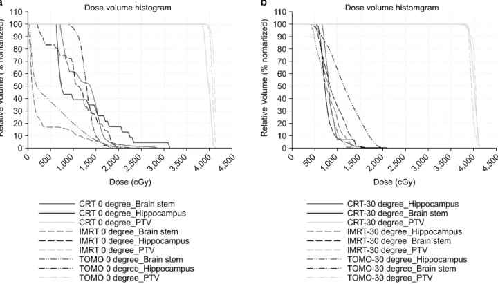
두 경부 거상에 따른 치료계획을 비교하기 위하여 3D-CRT, IMRT, 그리고 TOMO로부터 획득한 정상장기 흡 수선량은 Table 3에 나타내었다. 뇌간의 경우 각 Modality에 서 모두 35 Gy 이하를 나타내도록 치료계획을 수립하였으 며, 특히 30 o 거상 시 각 modality 별 최대선량이 23.7±1.8 Gy, 19.1.1±3.2 Gy, 21.4±1.2 Gy로 다른 각도에 비교해서 가 장 낮은 선량을 나타내는 것을 Table 3에서 확인할 수 있 다. 수정체의 최대선량은 30 o 거상 시 좌측 수정체는 4±1 Gy, 2.5±0.8 Gy, 2.5±0.7 Gy, 우측 수정체는 5.2±2.5 Gy, 3.5±1.8 Gy, 1.8±0.4 Gy로 가장 낮은 선량을 나타냈으며, 안 구의 평균선량 역시 30 o 거상 시 좌측 안구는 8.9±0.8 Gy, 4.1±0.7 Gy, 4.5±0.4 Gy, 우측 안구는 6.5±0.7 Gy, 3.2±0.4 Gy, 4.3±0.8 Gy로 가장 낮은 선량을 나타냈다. Fig. 3은 case‘2'의 뇌간과 해마의 DVH curve를 보여주고 있다. Fig.
4는 case 2의 두 경부 거상각도 0 o 와 30 o 의 DVH curve를 보 여주고 있다. Table 4는 각 modality에서 해마의 최대선량과 D100%의 선량을 보여주고 있다.
Table 4는 각 modality에서 해마의 최대선량과 D100%의 선량을 보여주고 있다. 해마의 흡수선량은 두 경부 각도 0 o 와 비교하여 30 o 에서 가장 좋은 결과를 나타냈다. 두 경부 거상각도 0 o 에서 각 modality에 따른 해마의 최대선량 (Maximum dose)은 평균 28.4 Gy, 20.4 Gy, 그리고 18.7 Gy 를 나타냈으며, 두 경부 각도를 30 o 거상 시 14.8 Gy, 14.1 Gy, 그리고 13.3 Gy를 나타내어 평균적으로 48%, 31%, 그 리고 29% 낮은 선량을 나타냈다.
해마의 D100% 선량은 두 경부 거상각도 0 o 에서 각 mo- dality에서 평균 8.09 Gy, 6.89 Gy, 그리고 6.31 Gy를 나타내 었으나, 두 경부 각도를 30 o 거상 시 7.64 Gy, 6.76 Gy, 그리 고 5.98 Gy로 0 o 와 비교하여 낮은 선량을 나타냈다.
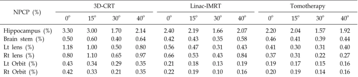
해마, 뇌간, 수정체 그리고 안구의 NTCP값은 Table 5에
나타내었다. 두 경부 각도 0 o 와 비교하여 30 o 에서 가장 좋 T a b
le 3 . C o m p a ri so n o f th e d o se re ce iv e d b y n o rm a l o rg a n s in d if fe re n t r a d ia ti o n t h e ra p ie s w it h v a ri e d h e a d e le va ti o n a n g le s. 3D -CR T Li na c-IMR T Tomot h er apy 0 o 15 o 30 o 40 o 0 o 15 o 30 o 40 o 0 o 15 o 30 o 40 o Max ( B ra in stem ) (G y ) 32 .00 ± 2. 50 28. 90 ± 3. 10 23 .70 ± 1. 80 24. 80 ± 3.2 0 33 .00 ± 2. 20 23 .30 ± 2. 70 19. 10 ± 3.2 0 31. 60 ± 2.0 0 25. 20 ± 3.0 0 22. 50 ± 2.1 0 21 .40 ± 1. 20 24 .20 ± 3. 40 Max ( L t le ns) (G y ) 9.5 0± 3.9 0 8.7 0± 2. 50 4.1 0± 1.4 0 7.2 0± 1. 80 4.5 0± 1.5 0 3.8 0± 1. 20 2. 50 ± 0. 80 3. 50 ± 0. 10 3.3 0± 0. 20 2.7 0± 1. 70 2.5 0± 0. 70 3. 20 ± 0.9 0 Max ( R t le ns) (G y ) 6.5 0± 1.2 0 8.9 0± 2. 20 5.2 0± 2.5 0 7.8 0± 1. 80 5.3 0± 1.2 0 4.3 0± 2. 10 3. 50 ± 1. 80 4. 20 ± 0. 70 3.0 0± 0. 50 2.5 0± 0. 70 1.8 0± 0. 40 2. 20 ± 0.7 0 M ea n ( L t O rb it ) (G y ) 13 .10 ± 1. 20 10 .20 ± 1. 10 8.9 0± 0. 80 10 .50 ± 0. 70 6.5 0± 0.5 0 5.5 0± 0. 40 4. 10 ± 0. 70 5. 90 ± 0. 50 5.7 0± 0. 60 5.1 0± 0. 50 4.5 0± 0. 40 5. 10 ± 0.3 0 M ea n ( R t O rb it ) (G y ) 12 .80 ± 0.4 0 9. 90 ± 0.4 0 6.5 0± 0.7 0 10. 50 ± 0.8 0 6.6 0± 0. 90 5.8 0± 0. 20 3. 20 ± 0.4 0 4. 80 ± 0.5 0 6. 20 ± 0.7 0 5. 80 ± 0.6 0 4.3 0± 0. 80 4.8 0± 0. 50
Fig. 4. DVH (Dose Volume Histogram) among the 3 different treatment techniques. (a) 0 degree, (b) 30 degree.
은 결과를 나타내었으며, 해마의 경우 각 Modality에서 두 경부 각도 0 o 의 NTCP 값은 3.3%, 2.4%, 그리고 2.2%로 계 산되었고, 두 경부 각도를 30 o 거상 시 1.7%, 1.66%, 그리고 1.57%로 계산되어 30 o 에서 부작용 발병률이 가장 낮은 결 과를 확인할 수 있었다. 해마 이외의 정상장기 역시 TOMO 의 좌측 수정체를 제외한 뇌간, 우측 수정체 그리고 양쪽 안구에서 환자의 두 경부를 30 o 거상한 경우 모든 modality 에서 부작용 발병률이 가장 낮은 결과를 확인 할 수 있었다.
고 찰
본 연구에서는 Joshua 등의 연구방법을 사용하여 전두엽 (frontal lobe)에 종양이 위치하고 해마와 인접한 환자를 대 상으로 두 경부 거상각도와 장비 별 선량의 특성을 관찰해 보고자 하였다. Table 2에서와 같이 세 가지 방법(modality) 을 이용한 모든 각도의 치료계획에서 치료계획용적에 흡수 되는 종양평균선량이 처방선량(prescription dose)의 95%이 상을 만족하는 것을 확인할 수 있다.
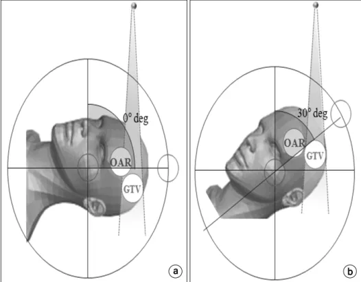
두 경부 각도를 거상 하면(Fig. 5)의 (b)와 같이 인접 정 상장기의 체적(OAR volume)과 종양체적의 조사 면을 분리 시킬 수 있기 때문에 인접정상장기(OAR) 주변에 방사선이
입사되지 않도록 조사야를 조절하여 방사선 조사 시 정상 조직에 흡수된 전체 흡수선량 즉, 용적선량(Integral volume dose)을 감소시킬 수 있었다.
이로 인해 치료계획 시 정상조직을 보호하기 위해서 과 도한 선 속 제한(Constrains)으로 인해 발생하는 정상조직과 인접된 종양체적의 과 선량(Hot dose)이 발생하는 현상을 최소화 할 수 있어 종양의 선량분포곡선(isodose curve)의 일치 성이 향상 될 수 있다. 또 다른 효과로는 정상조직과 종양체적이 분리되면서 기존 0 o 와 비교하여 뚜렷한 선량감 소(fall-off)를 얻을 수 있어, 치료 범위 내에서 선량 분포를 개선하여 종양 제어 율을 향상시키고 선량의 균일 성을 확 보할 수 있었다.
정상조직 측면에서도 두 경부 각도를 거상하면 종양 체
적과 인접한 정상조직이 분리되어 OAR과 해마의 흡수선
량을 줄여 주고, NTCP가 낮아지는 효과를 확인 할 수 있었
다. 3D-CRT에서 뇌간의 경우 0 o 와 비교하여 15 o , 30 o , 그리
고 40 o 에서 최대선량(Max dose)이 각각 10.7%, 35.8%, 그리
고 29% 감소하였다. 양쪽 수정체와 안구의 경우에서도 두
경부를 거상한 경우, 0 o 에 비해 흡수선량이 감소함을 확인
할 수 있었으며, 그 중 30 o 에서 가장 좋은 결과를 보여주었
다. IMRT, TOMO의 경우 역시 두 경부를 30 o 거상한 경우
Table 4. Comparison of the dose received by the hippocampus with varied head elevation angles.
Hippocampus dose (Gy)
Patient 0 o 15 o 30 o 40 o 0 o 15 o 30 o 40 o 0 o 15 o 30 o 40 o
3D-CRT Linac-IMRT Tomotherapy
1 Max dose 34.0 30.0 17.4 27.2 22.0 19.6 14.7 19.3 20.0 16.0 12.0 18.0
D100% 7.0 8.0 7.5 8.2 3.0 6.5 6.5 7.0 6.0 5.0 6.2 8.0
2 Max dose 37.0 33.0 15.2 23.2 22.5 20.1 15.2 19.8 21.2 16.5 13.0 18.9
D100% 8.2 7.0 6.8 7.8 6.5 6.0 5.8 7.0 5.8 5.4 5.1 6.0
3 Max dose 33.0 32.0 16.0 17.2 21.7 20.5 15.0 17.8 20.2 19.4 14.2 16.6
D100% 7.2 7.1 6.8 7.3 5.7 5.3 5.1 6.0 5.2 5.3 5.0 5.8
4 Max dose 32.0 27.0 15.1 17.2 22.2 20.2 14.1 17.8 19.2 18.2 13.2 15.8
D100% 7.6 7.4 7.0 7.7 6.8 6.6 6.3 6.8 5.9 5.7 5.3 5.7
5 Max dose 28.2 24.0 13.0 15.3 21.7 19.2 13.4 16.7 18.5 18.2 14.2 16.9
D100% 8.3 8.5 8.2 8.4 7.8 7.7 7.3 7.6 6.7 6.4 6.2 7.0
6 Max dose 26.2 23.0 12.4 15.7 20.4 19.0 15.2 17.2 17.6 18.1 14.0 16.4
D100% 9.0 8.5 8.2 8.0 8.8 8.2 7.8 7.9 7.2 7.0 6.8 6.9
7 Max dose 24.5 22.6 16 17.2 19.2 17.2 14.5 17.2 17.8 16.2 13.6 16.5
D100% 8.9 8.7 8.3 8.4 7.8 7.6 7.2 7.7 6.2 6.1 6.0 6.3
8 Max dose 23.5 20.2 14.4 16.2 18.7 17.5 12.6 16.7 17.2 16.8 12.8 14.5
D100% 8.1 7.9 8.0 8.1 7.8 7.4 7.1 7.3 6.5 6.3 6.0 6.4
9 Max dose 25.0 22.4 15.1 16.8 19.4 18.2 13.4 17.2 18.4 17.3 13.5 15.2
D100% 7.7 7.6 7.2 7.4 7.2 7.1 7.3 7.4 6.4 6.3 6.3 6.5
10 Max dose 21.0 21.1 14.2 15.9 16.4 15.4 13.1 16.8 17.4 16.8 13.1 14.9
D100% 8.9 8.7 8.4 8.3 7.5 7.3 7.2 7.3 7.2 7.1 6.9 7.0
AVG Max dose 28.4 25.5 14.8 18.1 20.4 18.6 14.1 17.6 18.7 17.3 13.3 16.3
D100% 8.1 7.9 7.6 7.9 6.8 6.9 6.7 7.2 6.3 6.1 5.9 6.5
AVG: average.
Table 5. The NTCP value of normal organs in three different radiation therapies.
NPCP (%) 3D-CRT Linac-IMRT Tomotherapy
0 o 15 o 30 o 40 o 0 o 15 o 30 o 40 o 0 o 15 o 30 o 40 o
Hippocampus (%) 3.30 3.00 1.70 2.14 2.40 2.19 1.66 2.07 2.20 2.04 1.57 1.92
Brain stem (%) 0.50 0.60 0.40 0.64 0.42 0.43 0.35 0.58 0.46 0.41 0.39 0.44
Lt lens (%) 1.18 1.00 0.50 0.80 0.56 0.47 0.31 0.43 0.41 0.30 0.31 0.40
Rt lens (%) 0.80 1.10 0.65 0.97 0.66 0.53 0.43 0.84 0.37 0.31 0.22 0.27
Lt Orbit (%) 0.43 0.34 0.29 0.35 0.21 0.18 0.13 0.19 0.19 0.17 0.15 0.16
Rt Orbit (%) 0.42 0.33 0.21 0.35 0.22 0.19 0.10 0.16 0.20 0.19 0.14 0.16
15 o 와 40 o 에 비해 좋은 결과를 확인할 수 있었다.
이와 같은 결과는 두 경부 기울기 30 o 에서 OAR과 종양 체적의 분리가 가장 잘 되었으나, 15 o 에서는 30 o 에 비교하 여 분리 정도가 부족하였고 40 o 에서는 오히려 OAR과 종양 체적이 더 겹쳐지는(Overlap) 차이가 발생한 것으로 보인다.
RTOG 0933 Protocol에 의하면 해마의 유의한 선량으로 100% 체적의 선량은 9 Gy 이하(D100%≤9 Gy)로 정하며 최대선량은 16 Gy 이하(maximum dose≤16 Gy)를 권고하고
있다. 본 연구에서 두 경부를 거상하여 서로 다른 치료법 (즉, 3D-CRT, IMRT, TOMO)에 동일하게 적용했을 때 두 경부를 30 o 거상 시 두 경부 각도 0 o 와 비교하여 해마의 최 대선량은 평균적으로 48%, 44.6%, 그리고 29% 낮은 선량 을 나타냈다.
MLC를 이용해 intensity를 변화시켜 빔을 전달하는 IMRT,
TOMO와 비교하여 3D-CRT 치료계획은 종양체적의 선량과
정상장기의 보호를 위한 선량의 최적화나 Segments 등의
Fig. 5. Difference of received dose of OAR among the tilting angle. (a) 0 degree, (b) 30 degree.
조절이 어려워 두 경부 종양에서 정상조직의 흡수선량 조 절에 어려움이 있다. 그러나 두 경부 각도를 거상한 3D-CRT 치료계획의 경우 정상장기에 선 속을 제한하거나 다엽콜리 메이터를 이용한 차폐가 용이하였다. 또한 IMRT 치료계획 이나 TOMO 치료계획과 비교하여 임상적으로 적절한 선량 분포를 구현하고 정상조직의 흡수선량과 부작용 발병률도 비교적 대등하게 낮아지는 것을 확인 할 수 있었다.
본 연구에서 두 경부를 일정 각도(30 o )로 거상하면 환자 의 전신 상태에 따라 굽힘이 어려워 물리적인 제약이 따를 수 있는 제한 점이 있었다. 향후에는 이러한 제한 점을 극 복하는 연구를 진행 할 예정이며, 실제 환자를 대상으로 연 구를 진행하여 임상적 데이터를 확보하고자 한다.
결 론
본 연구에서는 두 경부 각도를 거상하여 치료 범위 내에 서 선량 분포를 개선하여 종양의 선량분포곡선(isodose curve) 의 일치성 과 종양 제어율을 향상시켜 선량의 균일성을 확 보할 수 있었으며 정상조직 측면에서도 두 경부 각도를 거 상하면 종양 체적과 인접한 정상조직이 분리되어 OAR과 해마의 흡수선량을 줄여 주고, NTCP값이 낮아지는 효과를 확인 할 수 있었다.
References
1. Lee SW, Back GM, Yi BY, et al: Preliminary results of a phase I-II study of simultaneous modulated accelerated radio- therapy for nondisseminated nasopharyngeal carcinoma. Int. J.
Radiat. Oncol. Biol. Phys. 65:152-160 (2006)
2. Shiau AC, Lai PL, Liang JA, et al: Dosimetric verification of surface and superficial doses for head and neck IMRT with different PTV shrinkage margins. Med. Phys. 38:1435-1443 (2011)
3. Pezner RD, Archambeau JO: Brain tolerance unit: A meth- od to estimate risk of radiation brain injury for various dose schedules. Int. J. Radiat. Oncol. Biol. Phys. 7:397-402 (1981) 4. Milano MT, Constine LS, et al: Normal Tissue Tolerance
Dose Metrics for Radiation Therapy of Major Organs. Semin.
Radiat. Oncol. 17: 131-140 (2007)
5. Sahgal A, Barani lJ, Jr JN, et al: Prescription Dose Guideline Based on Physical Criterion for Multiple Metastatic Brain Tumors Treated With Stereotactic Radiosurgery. Int. J.
Radiat. Oncol. Biol. Phys. 78: 605-608 (2010)
6. Jeraj R, Mackie TR, Balog J, et al: Radiation character- istics of helical tomotherapy. Med. Phys. 31:396-404 (2004) 7. Kan MW, Cheung JY, Leung LH, et al: The accuracy of
dose calculations by anisotropic analytical algorithms for stereo- tactic radiotherapy in nasopharyngeal carcinoma. Phys. Med.
Biol. 56:397-413 (2011)
8. Park SH, Park HC, Oh DH, et al: Multi-institutional
Comparison of Intensity modulated radiation Therapy (IMRT)
Planning strategies and Planning Results for Nasopharyngeal Cancer. J. Korean. Med. Sci, 24, pp. 248-255, 2009.
9. Mackie TR, Balog J, Ruchala K, et al: Tomotherapy.
Semin. Radiat. Oncol. 9, pp. 108-117, (1999)
10. Marsh JC, Godbole R, Diaz AZ, et al: Sparing of the hip- pocampus, limbic circuit and neural stem cell compartment dur- ing partial brain radiotherapy for glioma: A dosimetric feasibility study. J. Med. Imag. Radiat. On. 55, 442-449, (2011) 11. Huang D, Xia P, Akazawa P, et al: Comparison of treat-
ment using intensity-modulated radiotherapy and three-dimen- sional conformal radiotherapy for paranasal sinus carcinoma, Int.
J. Radiat. Oncol. Biol. Phys. 56, No. 1, pp. 158-168, (2003) 12. Niroomand RA, Razavi R, Thobejane S, et al: Radiation
dose perturbation at tissue-titanium dental interfaces in head and neck cancer patients. Int. J. Radiat. Oncol. Biol. Phys.
34:475-480 (1996)
13. Choi JY, Won YJ, Park JY, et al: Development of a Thermoplastic Oral Compensator for Improving Dose Uniformity in Radiation Therapy for Head and Neck Cancer. Progress in Medical Physics. 23: 269-278 (2012)
14. Prokic Vesna, Wiedenmann Nicole, Fels F, et al: Whole Brain Irradiation With Hippocampal Sparing and Dose Escalation on Multiple Brain Metastases: A Planning Study on Treatment Concepts. Int. J. Radiat. Oncol. Biol. Phys. 85, No. 1, pp.
264-270, (2013)
15. Gondi Vinai, Tolakanahalli R, Mehta MP, et al: Hippo- campal-Sparing Whole-Brain Radiotherapy: A “How-To”
Technique Using Helical Tomotherapy and Linear Accelerator- Based Intensity-Modulated Radiotherapy. Int. J. Radiat. Oncol.
Biol. Phys. 78, No. 4, pp. 1244-1252, (2010)
16. Wang X, Zhang X, Dong L, et al: Effectiveness of non- coplanar IMRT planning using a parallelized multiresolution beam angle optimization method for paranasal sinus carcinoma.
Int. J. Radiat. Oncol. Biol. Phys. 63, 594-601 (2005)
17. Siglin J, Champ CE, Vakhnenko Y, et al: Optimizing pa-
tient positioning for intensity modulated radiation therapy in hip- pocampal-sparing whole brain radiation therapy. Pract. Radiat.
Oncol. 4, 378-383 (2014)
18. Joseph KJ, Syme A , Small C, et al: A treatment planning study comparing helical tomotherapy with intensity-modulated radiotherapy for the treatment of anal cancer. Radiother. Oncol.
94, 60-66, (2010)
19. Lee FK, Yip CW, nkie Chun-hung Cheung, et al:
Dosimetric difference amongst 3 techniques: TomoTherapy, sliding-window intensity-modulated radiotherapy (IMRT), and RapidArc radiotherapy in the treatment of late-stage nasophar- yngeal carcinoma (NPC). Med. Dosim. 39 , 44-49, (2014) 20. Alber M, Nusslin F: Optimization of intensity modulated ra-
diotherapy under constraints for static and dynamic MLC delivery. Phys. Med. Biol. 46:3229-3239 (2001)
21. Chui H, Rangarajan A: A new point matching algorithm for non-rigid registration. Comp. Vis. Image Und. 89, 114-141, (2003)
22. Semenenko VA, Li: Lyman-Kutcher-Burman NTCP model parameters for radiation pneumonitis and xerostomia based on combined analysis of published clinical data. Phys. Med. Biol.
53, 737-55 (2008)
23. G Luxton G, Keall PJ, King CR: A new formula for normal tissue complication probability (NTCP) as a function of equiv- alent uniform dose (EUD). Phys. Med. Biol. 53:23-36 (2008) 24. Kutcher GJ, Burman C, Brewster L, et al: Histogram re-
duction method for calculating complication probabilities for three-dimensional treatment planning evaluations. Int. J. Radiat.
Oncol. Biol. Phys. 21:137-146 (1991)
25. Makrs LB, Yorke ED, Jackson A, et al: Use of Normal Tissue Complication Probability Models in the Clinic. Int. J.
Radiat. Oncol. Biol. Phys. 76, S10-S19 (2010)
26. Emami B, Lyman J, Brown A, et al: Tolerance of normal tissue to therapeutic irradiation. Int. J. Radiat. Oncol. Biol. Phys.
21: 109-122 (1991)
두 경부 종양 치료 시 거상각도에 따른 치료기법 별 선량특성 평가