유방암 환자의 3D-CRT, TOMO 방법에 따른 선량 분포 평가
국립암센터 양성자치료센터
김대웅ㆍ김종원ㆍ최윤경ㆍ김정수ㆍ황재웅ㆍ정경식ㆍ최계숙
목 적: 방사선치료계획에 있어서 정상조직과 치료부위의 선량 분포는 매우 중요하다. 이에 본원에서는 유방암 환자를 대상으 로 Three-dimensional conformal radiation therapy (3D-CRT), Helical tomotherapy (TOMO)의 방법으로 방사선치료계획을 세웠으 며 이에 선량분포를 분석하여 실제 임상에서의 적용여부를 알아보고자 한다.
대상 및 방법: 20명의(좌측: 10명, 우측: 10명) 유방보존절제술 환자를 대상으로 시행하였으며 방법으로는 같은 조건에서 3D-CRT는 Philips사의 Pinnacle을, TOMO는 TomoTherapy사의 TOMO Planning System을 이용해 치료계획을 세웠다. Dose- Volume Histogram (DVH)의 prescribed dose (PD)에 대한 PTV의 Homogeneity index (HI)와 Conformity index (CI)를 구하였고, 정 상조직의 dose- volume 관계를 비교하였다.
결 과: Homogeneity index (HI)와 Conformity, index (CI)는 TOMO에서 우수한 결과를 나타났다. V_50_IB-NPTV (the percentage ipsilateral non-PTV breast volume that was delivered 50% of the prescribed dose)는 3D-CRT: 40.4%, TOMO: 18.3%, V20_IL (the average ipsilateral lung volume percentage receiving 20% of the prescribed dose)는 3D-CRT: 4.8%, TOMO: 14.2%, V20_10H (the average heart volume percentage delivered 20% and 10% of the prescribed dose in left breast cancer)는 3D-CRT: 1.6%, 3%
TOMO: 9.7%, 26.3%의 결과를 보여준다.
결 론: 유방암 환자의 방사선치료계획 방법들은 PTV에서 원하는 선량분포를 보여줬다. 그러나 TOMO는 좋은 Homogeneity index (HI), Conformity index (CI)와 Breast를 보호하는 장점이 있는 반면에 Lung과 Heart에서는 많은 피폭선량이 있음을 알 수 있기에 TOMO의 방사선치료계획시 주의해야 할 점으로 사료된다.
핵심용어: 유방암, 3D-CRT, TOMO
이 논문은 2007년 10월 5일 접수하여 2007년 11월 27일 채택되었음.
책임저자:김대웅, 국립암센터 양성자치료센터 Tel: 031)920-0132, Fax: 031)920-0149 E-mail: [email protected]
서 론
유방암은 전 세계적으로 여성암 중 1위를 차지하는 대표적 인 질병이다. 우리나라 여성의 유방암 발병은 서구국가에 비 해서 낮은 편이지만 생활양식이 서구화되어 가고 지방섭취 의 증가와 이로 인한 비만의 증가, 출산율 및 수유의 감소, 만혼, 조기 초경 및 폐경기 지연 등의 사회적인 현상으로 유 방암은 점차 증가할 것으로 예상된다. 한국유방암학회에서 발표된 한국인 유방암 환자의 연령별 분포 특징을 살펴보면 미국은 환자 대다수가 폐경 이후의 환자들이 많은 반면에 한 국인은 60% 이상의 환자가 50세 이하의 젊은 환자라는 특징 이 있어 서구보다 15∼20년 정도는 더 젊은 나이에 유방암이 많이 발생하여 유방암에 대한 관심이 증가하고 있다.
유방암 환자의 평균 연령이 낮아짐에 따라 유방의 미용에
도 치료의 초점이 맞추어 지고 있다. 방사선 치료 역시 주변 장기에는 방사선을 최소화하고 표적에는 더 많은 선량을 줄 수 있는 형태로 발전해 가고 있다. 2차원 방사선치료→3차원 방사선치료(3-Dimensional Radiation Therapy)→세기조절 방 사선치료(Intensity Modulated Radiation Therapy)형태로 발전 해 왔고, 최근에는 영상 유도 방사선치료(Imaged Guided Radiation therapy)와 나선형으로 방사선 세기가 조절 가능한 Tomotherapy가 도입되면서 방사선 치료 기법은 빠르게 변화 하고 있다.1-3)
본 연구에서는 현재 가장 많이 시행되고 있는 방사선치료 의 형태인 3D-CRT와 가장 최근에 개발된 방사선 치료인 Tomotherapy를 이용하여 치료계획을 세웠으며, 각각의 치료 계획에 따른 선량분포, 선량-체적 히스토그램을 이용하여 산 출된 값들을 비교ㆍ분석하고자 한다.
대상 및 방법
본원에 내원한 유방암 보존절제술 20명(우측: 10명, 좌측:
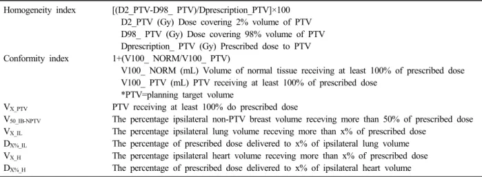
Homogeneity index [(D2_PTV-D98_ PTV)/Dprescription_PTV]×100 D2_PTV (Gy) Dose covering 2% volume of PTV D98_ PTV (Gy) Dose covering 98% volume of PTV Dprescription_ PTV (Gy) Prescribed dose to PTV Conformity index 1+(V100_ NORM/V100_ PTV)
V100_ NORM (mL) Volume of normal tissue receiving at least 100% of prescribed dose V100_ PTV (mL) PTV receiving at least 100% of prescribed dose
*PTV=planning target volume
VX_PTV PTV receiving at least 100% do prescribed dose
V50_IB-NPTV The percentage ipsilateral non-PTV breast volume receving more than 50% of prescribed dose VX_IL The percentage ipsilateral lung volume receving more than x% of prescribed dose
DX%_IL The percentage of prescribed dose delivered to x% of ipsilateral lung volume VX_H The percentage ipsilateral heart volume receving more than x% of prescribed dose DX%_H The percentage of prescribed dose delivered to x% of ipsilateral heart volume Table 1. Dosimetric parameters
HI, CI 3D-CRT TOMO
HI (Homogeneity index) 4.7∼13.1 (8.25) 4.85∼8.5 (7.16) CI (Conformity index) 1.9∼8.88 (3.25) 1.05∼1.19 (1.11) HI: Homogeneity Index/CI: Conformity Index
Table 2. Comparison of HI, CI
PTV 3D-CRT TOMO
V100_PTV 87.3∼98.8 (94.8) 94.4∼95.9 (95.1)
V95 PTV 99.5∼100 (99.9) 99.2∼99.9 (99.6)
V90_PTV 100 (100) 100 (100)
VX_PTV: PTV receiving at least 100% do prescribed dose Table 3. Comparison of VX_PTV
Non-PTV breast 3D-CRT TOMO
V50_IB-NPTV 25∼64.7 (40.4) 11.7∼24.2 (18.3) V50_IB-NPTV: the percentage ipsilateral non-PTV breast volume receving more than 50% of prescribed dose
Table 4. Comparison of V50_IB-NPTV
10명)을 대상으로 하였다. 각각의 환자는 방사선치료용 자세 고정 장치 Breast board에 누운 자세로 일정한 환자 자세를 유지하였다. 전산화 단층촬영장치(Computed Tomography, Ultra-Z, Philips)를 이용해 CT모의치료를 시행하여 영상을 획득하였다. 치료계획실에서는 이미지에 관심영역 표시는 종 양은 계획용 표적체적(Planning target volume)으로 정상장기 는 폐, 심장, 유방으로 나타냈다. 3D-CRT 치료계획(Pinnacle Ver 7.6 c, Philips)은 non-coplanar beam으로 표적에는 충분 한 선량을 정상장기에는 선량이 최소화 되도록 치료계획을 세웠다. Tomotherapy 치료계획(Tomotherapy planning sta- tion)은 field width: 2.5 cm, pitch: 0.25, modulation factor: 2 의 값을 동일하게 적용하여 최적의 치료계획을 수립되도록 하였다. 치료계획에 의해 얻어진 선량-체적 히스토그램을 바 탕으로 Homogeneity index, Conformity index, VX_PTV, V50_
IB-NPTV, VX_IL, DX%_IL, VX_H, DX%_H값을 산출하여 비교하였고 정의는 다음과 같다(Table 1).4,5)
결 과
3D-CRT와 Tomotherapy의 치료계획을 수립한 후 Dosimet- ric parameters에 의한 선량-체적 히스토그램을 비교하였다.
Homogeneity index와 Conformity index의 선량 비교는 3D-CRT는 Homogeneity index: 8.25, Conformity index: 3.25 의 값을 TOMO는 Homogeneity index: 7.16, Conformity in- dex: 1.11로 나타났다(Table 2).
VX_PTV의 선량 비교는 3D-CRT는 V100_PTV: 94.8, V95_PTV: 99.9, V90_PTV: 100의 값을 TOMO는 V100_PTV: 95.1, V95_PTV: 99.6, V90_PTV: 100로 나타났다(Table 3).
V50_IB-NPTV의 선량 비교는 3D-CRT는 V50_IB_-NPTV: 40.4의 값
을 TOMO: V50_IB_-NPTV: 18.3로 나타났다(Table 4).
VX_IL의 선량 비교는 3D-CRT는 V20_IL: 4.8, V10_IL: 8.1, V5_IL: 13.8의 값을 TOMO는 V20_IL: 14.2, V10_IL: 35.4, V5_IL: 55.9로 나타났다(Table 5, Fig. 1).
DX%_IL의 선량 비교는 3D-CRT는 D20%_IL: 3.1, D10%_ IL: 8.6, D5%_IL: 19.4의 값을 TOMO는 D20%_IL: 15.5, D10%_IL: 23.3, D5%_IL: 31로 나타났다(Table 6, Fig. 2).
VX_H의 선량 비교는 좌측 유방암을 대상으로 3D-CRT는
Ipsilateral lung 3D-CRT TOMO
D20%_IL 1.5∼5.5 (3.1) 7.6∼28.2 (15.5)
D10% IL 3.6∼19 (8.6) 13.6∼34.9 (23.3)
D5%_IL 9.8∼40.7 (19.4) 21.8∼39.8 (31)
*DX%_IL: the percentage of prescribed dose delivered to x% of ipsilateral lung volue
Table 6. Comparison of DX%_IL
Fig. 3. Comparison of VX_H.
Heart 3D-CRT TOMO
D20%_H 0.7∼3.7 (2.1) 7.8∼24.8 (14.8)
D10% H 1.1∼9.8 (4) 9.8∼30 (19.3)
D5%_H 1.3∼33.8 (8.8) 11.4∼34.3 (23.4) DX%_H: the percentage of prescribed dose delivered to x% of ipsilateral heart volume
Table 8. Comparison of DX%_H
Ipsilateral lung 3D-CRT TOMO
V20_IL 1.8∼9.4 (4.8) 5.6∼33.2 (14.2)
V10 IL 3.7∼15.4 (8.1) 14.3∼67.6 (35.4) V5_IL 7.4∼26.7 (13.8) 28.3∼87.6 (55.9) VX_IL: the percentage ipsilateral lung volume receving more than x% of prescribed dose
Table 5. Comparison of VX_IL
Heart 3D-CRT TOMO
V20_H 0∼7.1 (1.6) 0∼25.8 (9.7) V10 H 0∼9.9 (3) 8.9∼49.6 (26.3) V5_H 0∼15 (6.3) 39.5∼90.9 (69.9) VX_H: the percentage ipsilateral heart volume receving more than x% of prescribed dose
Table 7. Comparison of VX_H
Fig. 1. Comparison of VX_IL.
Fig. 2. Comparison of DX%_IL.
V20_H: 1.6, V10_H: 3, V5_H: 6.3의 값을 TOMO는 V20_H: 9.7, V10_H: 26.3, V5_H: 69.9로 나타났다(Table 7, Fig. 3).
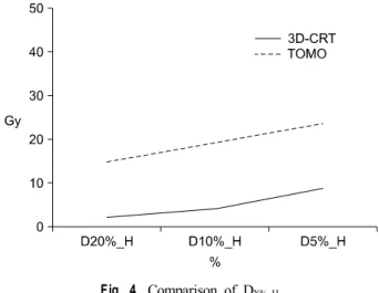
DX%_H의 선량 비교는 좌측 유방암을 대상으로 3D-CRT는 D20%_H: 2.1, D10%_H: 4, D5%_H: 8.8의 값을 TOMO는 D20%_H: 14.8, D10%_H: 19.3, D5%_H: 23.4로 나타났다(Table 8, Fig. 4).
고안 및 결론
3차원 CT영상을 이용하여 방사선치료계획을 어떻게 하느 냐에 따라 선량분포는 달라질 수 있다. 유방암 환자의 방사 선치료계획시 방사선이 조사되는 폐와 심장에는 저선량이
들어가고 표적에는 다량의 선량을 조사하는 것이 중요하다.2) 본 연구에서 유방암의 3D-CRT와 Tomotherapy의 방사선치
Fig. 4. Comparison of DX%_H.
료계획에 따른 선량-체적 히스토그램을 비교하였다.
두 방법 모두 VX_PTV의 값이 표적에 원하는 선량분포를 보 여준다. 3D-CRT 치료계획에서 prescribed dose를 초과하는 부분이 많은 부분을 차지하는 것을 조심해야 한다. Tomo- therapy는 Homogeneity Index, Conformity Index, 선량분포가 훌륭함을 보여주고 있고 V50_IB-NPTV의 값처럼 표적이 있는 유 방측도 보호하는 효과가 우수함을 알 수 있다. 폐와 심장에 조사되는 선량은 VX_IL, DX%_IL, VX_H, DX%_H의 값처럼 3D- CRT 보다 Tomotherapy가 많음을 보여준다.
유방암의 방사선치료계획시 주관적인 판단에 따라 다양한 결과로 나타나므로 적정한 치료계획을 결정해야 한다. Tomo- therapy는 표적에 대한 뛰어난 선량분포와 유방을 보호함을
보여주지만 폐와 심장에 많은 선량이 조사됨을 치료계획시 주의해야 하고 앞으로 해결해야 할 과제라 생각된다. 그리고 Tomotherapy 환자의 위치잡이, 치료계획기법 등 개선 방법들 이 제시되야 하며 본 연구를 통해 추후에 임상환자수의 증가 를 통하여 신빙성을 증가시키고 앞으로 Tomotherapy의 환자 치료에 기초 자료가 될 수 있을 거라고 사료된다.
참고문헌
1. Arthur DW, Vicini FA: Accelerated partial breast irradiation as a part breast conservation therapy. J Chin Oncol 2005;23:
1726-1735
2. Oliver M, Chen J, Wong E, et al.: A treatment planning study comparing whole breast radiation therapy against conformal, IMRT and tomotherapy for accelerated partial breast irra- diation. Radioother Oncol 2007;82:317-323
3. Langen KM, Buchholz DJ, Burch DR, et al.: Investigation of accelerated partial breast patient alignment and treatment with helical tomotherapy unit. Int J Radiat Oncol Biol Phys[In press]
4. Rusthoven KE, Carter DL, Howell K, et al.: Accelerated partial-breast intensity-modulated radiotherapy results in im- proved dose distribution when compared with three- dimensional treatment-planning techniques. Int J Radiat Oncol Biol Phys 2008;70:296-302
5. Weed DW, Edmundson GK, Vicini FA, et al.: Accelerated partial breast irradiation: a dosimetric comparison of three different techniques. Brachytherapy 2005;4:121-129
Abstract
Dosimetric Comparison of Three Dimensional Conformal Radiation Radiotherapy and Helical Tomotherapy Partial Breast Cancer
Dae Woong Kim, Jong Won Kim, Yun Kyeong Choi, Jung Soo Kim, Jae Woong Hwang, Kyeong Sik Jeong, Gye Suk Choi
Proton Therapy Center, National Cancer Center, Goyang, Korea
Purpose: The goal of radiation treatment is to deliver a prescribed radiation dose to the target volume accurately while minimizing dose to normal tissues. In this paper, we comparing the dose distribution between three dimensional conformal radiation radiotherapy (3D-CRT) and helical tomotherapy (TOMO) plan for partial breast cancer.
Materials and Methods: Twenty patients were included in the study, and plans for two techniques were developed for each patient (left breast:10 patients, right breast:10 patients). For each patient 3D-CRT planning was using pinnacle planning system, inverse plan was made using Tomotherapy Hi-Art system and using the same targets and optimization goals. We comparing the Homogeneity index (HI), Conformity index (CI) and sparing of the organs at risk for dose-volume histogram.
Results: Whereas the HI, CI of TOMO was significantly better than the other, 3D-CRT was observed to have significantly poorer HI, CI. The percentage ipsilateral non-PTV breast volume that was delivered 50% of the prescribed dose was 3D-CRT (mean: 40.4%), TOMO (mean: 18.3%). The average ipsilateral lung volume percentage receiving 20% of the PD was 3D-CRT (mean: 4.8%), TOMO (mean: 14.2), concerning the average heart volume receiving 20% and 10% of the PD during treatment of left breast cancer 3D-CRT (mean: 1.6%, 3.0%), TOMO (mean: 9.7%, 26.3%)
Conclusion: In summary, 3D-CRT and TOMO techniques were found to have acceptable PTV coverage in our study. However, in TOMO, high conformity to the PTV and effective breast tissue sparing was achieved at the expense of considerable dose exposure to the lung and heart.
Key words: breast cancer, 3D-CRT, helical tomotherapy