서 론
난자의 핵 이식을 위해서는 난자의 활성화가 필요하 며, 핵 이식 배를 이식할 수 있는 단계의 배로 발달시킬 수 있어야 한다.
성숙이 완료된 난자는 외부의 자극없이는 감수분열을
재개할 수 없어 난자의 활성화가 필요하다. 포유류 난자 의 활성화에 대한 연구는 Pincus와 Enzmann이 처음으로 보고된 이래 극체형성2, 3, 활성화물질4, 5, 배양조건6, 7, 8, 9, 발생단계10, 11, 12, 13
등이 활성화에 미치는 연구가 이루어 져 왔다. 소의 난자는 Ca2+Ionophore, ionomycin, ethanol, cyclohexamide 및 6-methylaminopurine 등을 이용한 여러
핵 이식을 위한 돼지난자의 활성화 및 핵 이식 배와 공배양에 따른 체외발생에 관한 연구
김상근*, 이만휘1 충남대 수의대 (게재승인 : 2002년 1월 18일)
Studies on the Porcine Oocytes Activation Regimed for Nuclear Transfer and Development Following Co-culture of Nuclear
Transferred Embryos
Sang-keun Kim
*, Man-hwi Rhee
1College of Veterinary Medicine, Chungnam National University
1
Dept. of Neurophysiology, Washington Uinversity
(Accepted : January 18, 2002)ABSTRACT : This study was carried out to investigate the optimal activation condition for
parthenogenetic development. In order to activate oocytes at 24 hrs post onset of maturation, the oocytes were cultured 3∼13 μM Ca2+ for 5 min., 5-8 ㎍/ml cytoclacin for 6 hrs, 0.5∼2.0 mM 6-dimethylaminopurine(DMAP) for 3 hrs alone or combination. The activated oocytes were cultured in TCM-199 media at 5% CO2, 95% air, 38℃.The cleavage rate after 48 hrs culture of oocytes treated with 3-13 μM Ca2+, 5-8 ㎍/ml cytoclacin and 0.
5∼2.0 mM DMAP for 5 min., 6 hrs and 3 hrs were 9.6%∼20.0%, 0.0%∼7.3% and 9.4%∼21.8%, 0.
0%∼7.3% and 9.1%∼21.8% and 0.0%∼7.3%, respectively. When oocyte were treated with 10 μM Ca2+, 10 ㎍/ml cytoclacin and 2.0 mM DMAP the blastocyst formation rate was significantly higher than other group. The cleavage rate after 48 hrs culture of oocytes treated with Ca2+ + cytoclacin, Ca2+ + DMAP, cytoclacin + DMAP were 75.9%∼93.5% and 9.7%∼19.0%, respectively. When oocytes were treated with Ca2+ followed by DMAP, the blastocyst formation rate was significantly higher than other group(p<0.05). When necleus transferred embryos co-cultured with bovine serum albumin(BSA), epithemal growth factor(EGF) and calf serum(CS), the developmental rate to blastocyst were higher than control group.
Key words : parthenogenesis, activation, Ca
2+, cytoclacin, DMAP*Corresponding author : Dr. Sangkeun Kim, Dept. of Vet. Med., Chungnam Natl. Univ. Daejeon 305-764, Korea [email protected]
29
가지 요인에 의해 활성화된다고 보고되었다14, 15.
Ca2+ 제재를 이용하여 난자를 활성화시키면 histone H1 kinase가 불활성화되어 감수분열이 재개되지만 일정 시간이 경과하면 kinase가 재활성화된다고 하였으며16, Ca2+ 농도의 증가는 감수분열의 재개를 유도하지만 전 핵형성을 억제한다는 보고16도 있다. 특히, Ca2+수준 증 가제 및 단백질합성억제제와 인산화억제제를 병행처리하 면 감수분열 재개와 난자활성화를 증가시킬 수 있다고 보 고되었다17. 그러나, 체외수정율과 배 발달율이 낮은 돼지 난자를 이용한 핵 이식 난자에 대해 여러 agent들이 활성 화에 미치는 영향과 핵 이식을 완료한 난자는 배양액에 albumin, 성장인자 및 혈청 등을 첨가하여 공배양시 배 발 달에 미치는 영향에 관한 보문은 접한 바 없었다.
이에 본 연구는 Ca2+, cytoclacin, 6-dimethylaminopurine (DMAP) 등의 처리가 난자의 활성화에 미치는 영향과 핵이식 배의 공배양에 따른 발달율을 구명하고자 실시 하였다.
재료 및 방법
난포란의 회수
도살 돼지의 난소를 적출하여, 100 IU/ml의 penicillin G(Sigma, U.S.A.)와, 100 ㎍/ml의 streptomycin sulfate (Sigma, U.S.A.)를 첨가한 38℃의 생리식염수에 침지하 여 실험실로 옮긴 다음 난포로부터 배양액이 들어있는 18 G 주사기로 난포액을 흡입하여 실체현미경(20∼40
× ) 하에서 난포란을 회수하였다.
난포란의 체외성숙 배양
회수한 난포란은 10%(v/v)의 fetal calf serum(FCS, Sigma, U.S.A.)와 1 ㎍/ml의 FSH(Sigma, U.S.A.), 2 IU/ml 의 hCG(Sigma, U.S.A.), 1 ㎍/ml의 β-estradiol (Sigma, U.S.A.), 100 IU/ml의 penicillin G 및 100 ㎍/ml의 streptomycin sulfate가 첨가된 TCM-199(Whittaker, U.S.A.) 배양액으로 배양하였다. 난포란의 체외성숙은 배양액 50 ㎕의 drop을 mineral oil (Squibb Co., U.S.A.)로 피복하 여 배양 2-3시간 전에 CO2 배양기내(5% CO2, 95% air, 38oC)에서 5-6시간 평형시킨 후 drop내에 5개의 난포란 을 주입하여 32∼40시간 성숙배양하였다.
핵이식을 위한 미세조작
공핵란은 0.5 ㎍/ml의 colcemid(Gibco., U.S.A.) + 10%
FCS + TCM-199 배양액에서 10시간 체외배양을 통해 cell stage를 G1기로 조절하였으며, 수핵과 탈핵의 미세 조작은 수핵란과 공핵란으로부터 분리된 할구세포를 각
농도의 cytoclacin (Sigma, U.S.A.)과 DMAP 및 Ca2+
Ionophore + 10% FCS + TCM-199 배양액으로 전처리를 한 후 미세조작을 위하여 micromanipulator(Narishige Co., Japan) 부착 도립현미경에 장착하여 성숙된 MII 난자를 micropipett으로 고정시키고 핵을 제거하기 위하여 30 ㎛ 의 micropipett을 투명대내로 진입시켜 제 1 극체와 염색 체를 원형질막에 싸여진 채로 흡입하여 제거하고, 공핵 란으로부터 분리된 할구세포 하나를 탈핵에 사용한 micropipett에 흡입하고 이를 미세조작으로 탈핵된 수핵 란의 원형질 외측 위란강에 주입하였다.
활성화 처리
40시간동안 성숙배양후 난구세포를 제거하기 위하여 0.1% hyaluronidase(Sigma, U.S.A.)가 첨가된 10% FCS + TCM-199 배지에서 난구세포를 제거하였다. 난구세포가 제거된 난자들중에 제 1극체가 방출된 난자만을 선별하 여 시험에 이용하였다. 난자의 활성화 처리를 위해 3, 5, 10, 13 μM Ca2+ Ionophore에 5분, 5, 8, 10, 13 ㎍/ml의 cytoclacin에 6시간, 0.5, 1.0, 1.5, 2.0 mM의 DMAP에 3시 간동안 단독 또는 병행처리 하였다.
배양
활성화 처리가 완료된 난자들을 배양액으로 세척한 후 10% FCS + TCM-199 배양액에서 배양을 하였으며 배양조건은 5% CO2, 95% air, 38℃였다.
전핵관찰
전핵형성 여부를 관찰하기 위하여 활성화 처리된 난 자를 처리 6시간후에 고정 염색하여 200 배율의 현미경 하에서 전핵형성 여부를 관찰하였다. 고정은 acetic acid : etanol(3 : 1)(Sigma, U.S.A.)의 고정액내에 24시간이상 침지시킨 후 1% aceto orcein(Sigma, U.S.A.) 액으로 염색 을 실시하였다.
공배양
핵 이식을 완료한 난자는 5% bovine serum albumin (BSA, Sigma, U.S.A.), 5 mM epithermal growth factor (EGF, Sigma, U.S.A.), calf serum(CS, Sigma, U.S.A.)를 첨 가한 TCM-199 배양액내에서 공배양하면서 배 발달을 관찰하였다.
통계분석
시험결과는 SAS package를 이용하여 분산분석을 하였 으며, 처리간의 유의성은 Duncan's multiple range test를
이용하여 검정하였다.
결 과
난자의 활성화 단용처리 Ca2+의 처리
핵 이식을 위한 난자의 활성화 물질로서 Ca2+이온을 5분간 처리후 배양했을 때 단위발생율은 Table 1과 같 다. 난자의 활성화제로서 3, 5, 10, 13 μM의 Ca2+ 이온 을 처리후 배양했을 때 2 cell 및 배반포로의 단위발생율 은 각각 9.6%∼20.0% 및 0.0%∼7.3%로서 10 μM Ca2+
농도에서 가장 높은 발생율을 나타냈다.
Table 1. Developmental rate of oocytes activated with a
various Ca2+ concentrationConcent. of Ca2+
(μM )
No. of oocytes treated
No. of oocytes developed to 2 cell Blastocyst
3 50 7(14.0) 1(2.0)
5 52 6(11.5) 2(3.8)
10 55 11(20.0) 4(7.3)
13 52 5( 9.6) 0(0.0)
* Oocytes activated a various Ca2+concentration for 5 min.
Cytoclacin의 처리
핵 이식을 위한 난자의 활성화 물질로서 cytoclacin를 처 리후 배양했을 때 단위발생율은 Table 2와 같다. 난자의 활 성화제로서 5, 8, 10, 13 ㎍/ml cytoclacin를 6시간 처리후 배양했을 때 2 cell 및 배반포로의 단위발생율은 각각 9.
4%∼21.8% 및 0.0%∼7.3%로서 10 ㎍/ml 농도에서 가장 높은 발생율을 나타냈다.
Table 2. Developmental rate of oocytes activated with a
various cytoclacin concentrationConcent. of cytoclacin(㎍/ml)
No. of oocytes treated
No. of oocytes developed to 2 cell Blastocyst
5 52 7(13.5) 0(0.0)
8 50 5(10.0) 1(2.0)
10 55 12(21.8) 4(7.3)
13 53 5( 9.4) 0(0.0)
*Oocytes activated a various cytoclacin concentration for 6 hrs
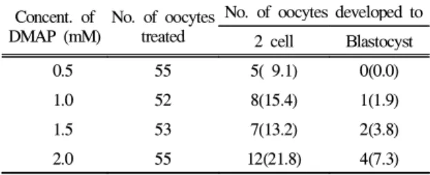
DMAP의 처리
핵 이식을 위한 난자의 활성화 물질로서 DMAP를 처
리한 후 배양했을 때 단위발생율은 Table 3과 같다.
Table 3. Developmental rate of oocytes activated with a
various DMAP concentrationConcent. of DMAP (mM)
No. of oocytes treated
No. of oocytes developed to 2 cell Blastocyst
0.5 55 5( 9.1) 0(0.0)
1.0 52 8(15.4) 1(1.9)
1.5 53 7(13.2) 2(3.8)
2.0 55 12(21.8) 4(7.3)
* Oocytes activated a various DMAP concentration for 3 hrs
난자의 활성화제로서 0.5, 1.0, 1.5, 2.0 mM의 DMAP를 3시간 처리후 배양했을 때 2 cell 및 배반포로의 단위발 생율은 각각 9.1%∼21.8% 및 0.0%∼7.3%로서 2.0 mM 농도에서 가장 높은 발생율을 나타냈다.
난자의 활성화 병용처리
핵 이식용 난자의 활성화를 위해 Ca2+ + cytoclacin, Ca2+ + DMAP, cytoclacin + DMAP를 각각 병용 처리후 배양했을 때 단위발생율은 Table 4와 같다. 난자의 활 성화를 위해 Ca2++ cytoclacin, Ca2++ DMAP, cytoclain + DMAP를 각각 처리후 배양했을 때 2 cell 및 배반포로의 단위발생율은 각각 75.9%∼93.5% 및 9.7%∼19.0%로서 단일처리군에 비해 높게 나타났다.
Table 4. Developmental rate of oocytes activated with a
various activation agentsActivation agents No. of oocytes treated
No. of oocytes developed to 2 cell Blastocyst Ca2+ + cytoclacin 60 54(90.0) 8(13.3)
Ca2+ + DMAP 58 44(75.9) 11(19.0)
cytoclacin +
DMAP 62 58(93.5) 6( 9.7)
*Oocytes activated with a various agents(10 μM Ca2+, 10 ㎍ /ml cytoclacin, 2.0 mM DMAP) for 3 hrs
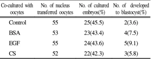
핵이식 배의 공배양
활성화 처리를 한 난자에 핵을 이식한 핵이식 배와 BSA, EGF 및 CS와 공배양했을 때 배반포로의 체외발생 율은 Table 5와 같다. 핵 이식 배와 BSA, EGF 및 CS와 공배양했을 때 배반포로의 체외발생율은 각각 7.5%, 9.1% 및 5.8%로서 대조군의 3.6%에 비해 높은 체외발생
율을 나타냈다.
Table 5. Developmental rates of nucleus transferred
oocytes by co-culture of BSA, EGF and CS Co-cultured withoocytes
No. of nucleus transferred oocytes
No. of cultured embryos(%)
No. of developed to blastocyst(%)
Control 55 25(45.5) 2(3.6)
BSA 53 23(43.4) 4(7.5)
EGF 55 24(43.6) 5(9.1)
CS 52 22(42.3) 3(5.8)
* nucleus transferred oocytes cocultured with BSA, EGF and CS
** BSA : Bovine serum albumin, EGF ; epithermal growth factor, CS ; calf serum
고 찰
난자의 핵 이식을 위해서는 난자의 활성화와 이식할 수 있는 단계의 배로 발달시킬 수 있어야 하며, 아울러 성숙이 완료된 난자는 외부의 자극 없이는 감수분열을 재개할 수 없어 난자의 활성화가 필요하다. 포유류 난자 의 활성화에 대한 연구는 Pincus와 Enzmann6이 처음으 로 보고된 이래 많은 연구가 이루어져 왔다4, 5, 18, 19, 20
. 그러나 돼지 난자의 활성화와 이식할 수 있는 단계로 발달시킨 핵 이식에 관한 연구보고는 거의 찾아 볼 수 없었다.
난자의 활성화제로서 Ca2+ 이온을 핵 이식난자에 처 리했을 때 단위발생율은 각각 10 μM 농도에서 가장 높은 발생율을 나타냈는데, Ca2+ Ionophhore의 처리로 Ca2+이온이 증가되면 성숙촉진인자는 불활화되어 난자 의 감수분열을 재개를 유도하고 아울러 염색체 응축을 유도하여 난자의 세포주기를 제 2차 성숙분열을 일으키 게 하지만 난자의 발육이 저조해지게 된다17. 그러나, Ca2+ 농도의 증가는 감수분열의 재개를 유도하지만 전 핵형성을 억제한다는 보고16와 일치되는 것으로 사료된 다. Ca2+처리시 핵 이식 배의 단위발생은 난자가 Ca2+수 준의 증가제 및 단백질 합성과 인산화 억제제를 병행 처리되었을 때에 한해서만 높게 나타났다. Cytoclacin 처 리군 역시 대조군에 비해 높은 발생율을 나타냈는데 cytoclacin은 단백질합성 및 인산화억제제로서 성숙촉진 인자를 불활성화시키므로 난자의 분할이 Ca2+ 이온에 비해 높게 나타났다는 보고15와 일치하는 것으로 사료되 었다. DMAP 역시 cytoclacin과 마찬가지로 단백질합성 및 인산화억제제로서 성숙촉진인자를 불활성화시키므 로 난자의 분할이 Ca2+처리군에 비해 다소 높게 나타냈 는데 이러한 결과는 시험동물은 다르지만 대체로 다른
연구자와의 결과14, 22와 일치하는 것으로 사료되었다.
핵 이식시 난자의 활성화제를 Ca2+ + cytoclacin, Ca2+
+ DMAP, cytoclacin + DMAP를 각각 병용 처리했을 때 배반포로의 단위발생율은 단일처리군에 비해 높게 나타 냈다. 이러한 결과는 Ca2+제제처리후 단백질합성 및 인 산산화 억제제인 cytoclacin과 DMAP에 의해 활성화됨으 로서 난자의 분할율을 높게 나타낸 것으로 사료된다16,
21. 역시, Ca2+과 DMAP 처리도 다른 처리군에 비해 난자 의 발생율이 유의하게 높았다. Ca2+제제와 다른 활성제 처리는 단백질합성 및 인산산화 억제제인 cytoclacin과 DMAP에 의해 활성화됨으로서 난자의 분할율을 높게 나타냈는데 이러한 결과는 소를 대상으로 한 다른 연구 자의 결과3, 16, 21와 본 시험의 공시동물인 돼지 난자에서 도 일치하는 것으로 사료되었다.
핵 이식 배와 BSA, EGF 및 CS와 공배양했을 때 배반 포로의 체외발생율은 각각 3.6%∼9.1%%로서 무첨가 대 조군의 3.6%에 비해 높은 체외발생율을 나타냈는데, 이 는 핵 이식 배와 cytoplast와 공배양했을 때 대조군에 비 해 높은 배발달율을 나타냈다는 다른 연구자의 보고와 유사한 결과이었다9.
결 론
본 연구는 Ca2+, cytoclacin, DMAP 등의 처리가 난자의 활성화에 미치는 영향과 핵이식 배와 BSA, EGF, CS와 의 공배양에 따른 체외발생율을 구명하고자 실시하였 다. 활성화 처리된 난자는 TCM-199 배양액에서 배양하 였으며, 배양조건은 5% CO2, 95% air, 38℃이었다.
1. Ca2+이온 3, 5, 10, 13 μM을 처리했을 때 배반포로의 단위발생율은 각각 9.6%∼20.0% 및 0.0%∼7.3%로서 10 μM 농도에서 가장 높은 발생율을 나타냈다.
2. Cytoclacin 5, 8, 10, 13 ㎍/ml을 처리했을 때 배반포로 의 단위발생율은 각각 9.4%∼21.8% 및 0.0%∼7.3%
로서 10 ㎍/ml 농도에서 가장 높게 나타났다.
3. DMAP 0.5, 1.0, 1.5, 2.0 mM을 처리했을 때 배반포로의 단위발생율은 각각 9.1%∼21.8% 및 0.0%∼7.3%로서 2.0 mM 농도에서 가장 높은 발생율을 나타냈다.
4. Ca2+ + cytoclacin, Ca2++ DMAP, cytoclacin + DMAP를 병용처리했을 때 배반포로의 단위발생율은 각각 75.9%∼93.5% 및 9.7%∼19.0%로서 단일처리군에 비 해 높게 나타났다.
5. 핵 이식 배와 BSA, EGF, CS 등이 첨가된 TCM-199 배 양액과 공배양했을 때 배발달율은 각각 7.5%, 9.1%, 5.8%로서 무첨가 대조군에 비해 약간 높게 나타났다.
참고문헌
1. Pincus G, Enzmann EV. The comaprative behaviour of mammalian eggs in vivo and in vitro. I. The activation of ovarian eggs. J of Experi Med, 62:665∼675, 1935.
2. Powell JW, Barnes FL. The kinetics of oocyte activation and polar body formation in bovine embryo clones.
Mol Reprod Dev, 33:53∼58, 1992.
3. Presicce GA, Yang X. Nuclear dynamics of oocyte activation and polar body formation in bovine embryo clones. Mol Reprod Dev, 37:61∼68, 1994.
4. Nagai T. Parthenogenic activation of cattle follicular oocytes with ethanol. Gamete Res, 16:243∼249, 1987.
5. Prochazka R, Durnford R, Fiser PS, et al. Parthenogenetic development of activated mature bovine oocytes.
Theriogenology, 39:1025∼1032, 1983.
6. Rho GJ, Wu B, Kawarsky S, et al. Activation regimens to prepare bovine oocyte. Mol Reprod Dev, 50:485∼
492, 1998.
7. Stice SL, Keefer CL, Matthews L. Bovine nuclear transfer embryos : Oocyte activation prior to blastomere fusion. Mol Reprod Dev, 38:61∼68, 1994.
8. Yang X, Jiang S, Foote RH, et al. Bovine oocyte development following different oocyte maturation and sperm capacitation procedures. Mol Reprod Dev, 34:9 4∼100, 1993.
9. Yang X, Jiang S, Farrell P, et al. Nuclear transfer in cattle : Effect of nuclear donor cells, cytoplast age co-culture and embryo transfer. Mol Reprod Dev, 35:2 9∼36, 1993.
10. Stice SL, Keefer CL. Improved developmental rates for bovine nucleus transfer embryos using cold shock activated oocytes. Biol. Reprod., 42(1):166, 1992(Abst) 11. Takano H, Koyama K, Kozai C, et al. Effect of aging of recipient oocytes on the development of bovine nuclear transfer embryos in vivo. Theriogenology, 39:
909∼917, 1993.
12. Ware CB, Barnes FL, Maiki-Laurila M, et al. Age dependance of bovine oocyte activation. Gamete Res, 22:265∼275, 1989.
13. Wu B, Lgnotz G, Currie WB, et al. Dynamics of maturation promoting factor and its constituent proteins during maturation of bovine oocyte. Biol Rerpod, 56:253∼259, 1997.
14. Samake S, Smith LC. Synchronization of cell division in eight-cell bovine embryos produced : effects of 6- methylaminopurine. J Reprod Fertil, 110:2 1∼27, 1997.
15. Shi Z, Jiang S, Yang X. Synergistic effect of A23187 and cycloheximide allows effective activation of freshly matured bovine oocytes. Theriogenology, 38:309, 1993 (Abst).
16. Collas P, Fissore R, Robl JM, et al. Electrically induced Ca2+ elevation, activation and parthenogenetic development of bovine oocytes. Mol Rerpod Dev, 34:212∼223, 1993.
17. Susko-Parrish JL, Leibfried-Ruttledge ML, Notthy DL,
et al. Inhibition of protein kinase after an induced Ca
2+transient causes transition of bovine oocytes to embryonic cycle without meiotic completion. Dev Biology, 166:
729∼739, 1994.
18. Campbell KHS, Ritchie WA, Wilmut I. Nuclear cytoplasmic interactions during the first cell cycle of nuclear transfer reconstructed bovine embryos : Implications for deoxyribonucleic acid replication and development. Biol Reprod, 49:933∼942, 1993.
19. First NL, Leibfried-Rutledge ML, Northey DL, et al.
Use of matured oocytes 24 hrs of age in bovine nuclear transfer. Theriogenology, 37:211, 1992(Abst).
20. Lavoir MC, Rumph ND, de la Fuente R, et al. The influence of cytoplasmic age on the development of embryos made by nuclear transfer. Theriogenology, 45:286, 1996 (Abst).
21. Liu L, Ju JC, Yang X. Parthenogenetic development and protein patterns of newly matured bovine oocytes after chemical activation. Mol Reprod Dev, 49:298∼
307, 1998.
22. Saeki K, Nagao Y, Kishi M, et al. Developmental capacity of bovine oocytes following inhibition of meiotic resumption by cycloheximide or 6-dimethylaminopurine.