Evaluation of Ciclopirox as a Virulence-modifying Agent Against Multidrug Resistant Pseudomonas aeruginosa Clinical Isolates from Egypt
Azza S. Zakaria*, Eva A. Edward, and Nelly M. Mohamed
Microbiology and Immunology Department, Faculty of Pharmacy, Alexandria University, Alexandria 21526, Egypt
Received: August 8, 2019 / Revised: August 18, 2019 / Accepted: August 20, 2019
Introduction
In the current battle between humankind and patho- gens, it seems that bugs are still having the upperhand.
The continuous rise in reported resistance to different antimicrobial agents and the shortage in the develop- ment of new antibiotics over the past decades are two facts weighing the results of this battle to the favor of the microorganisms [1]. Since the development of a novel antimicrobial agent may cost millions of dollars and may take a considerable number of years to happen, an alternative approach is becoming nowadays more
and more appealing. This approach intends to repurpose older drugs to combat infections caused by certain prob- lematic pathogens [2] or to hinder the bacterial virulence instead of bacterial viability [1].
One of the old drugs that has come back to the clinical stage is ciclopirox. Ciclopirox is an off-patent antifungal agent that has been developed about forty years ago and which possesses several advantages such as a wide safety margin, a profound efficacy and the ability to affect novel targets including resistant Gram-negative pathogens [2, 3]. In Gram-negative bacteria, ciclopirox was reported to affect galactose metabolism, as well as lipopolysaccharide (LPS) biosynthesis; two pathways extremely significant for bacterial growth and virulence [2].
Out of different members of Gram-negative bacteria, Targeting the pathogen viability using drugs is associated with development of drug resistance due to selective pressure. Hence, there is an increased interest in developing agents that target bacterial viru- lence. In this study, the inhibitory effect of ciclopirox, an antifungal agent with iron chelation potential, on the microbial virulence factors was evaluated in 26 clinical MDR Pseudomonas aeruginosa isolates col- lected from Alexandria Main University Hospital, a tertiary hospital in Egypt. Treatment with 9 µg/ml ciclopirox inhibited the hemolytic activity in 70% isolates, reduced pyocyanin production, decreased prote- ase secretion in 46% isolates, lowered twitching and swarming motility, and decreased biofilm formation by 1.5- to 4.5-fold. The quantitative real-time PCR analysis revealed that treatment with ciclopirox downregu- lated the expression levels of alkaline protease (aprA) and pyocyanin (phzA1). Ciclopirox is used to treat hematological malignancies and the systemic administration of ciclopirox is reported to have adequate oral absorption with a satisfactory drug safety profile. It is important to calculate the appropriate clinical dose and therapeutic index to reposition ciclopirox from a topical antifungal agent to a promising viru- lence-modifying agent agent against P. aeruginosa, a problematic Gram-negative pathogen.
Keywords: Ciclopirox, virulence factors, Pseudomonas aeruginosa, biofilm, gene expression
*Corresponding author
Tel: +203 4868482, Fax: +203 4871668 E-mail: [email protected]
© 2019, The Korean Society for Microbiology and Biotechnology
P. aeruginosa has been regarded as a cardinal opportu- nistic pathogen [4] responsible for about 10−15% of noso- comial infections all over the world [5]. P. aeruginosa can result in ventilator associated pneumonia, urinary tract infections, multi-organ system failure, burn wounds infection and sepsis in the ICU [6]. The high morbidity and mortality rates associated with P. aeruginosa infec- tions are due to the organism’s ability to accommodate to environmental changes, to develop resistance to various antimicrobial agents and to express multiple virulence factors [5].
P. aeruginosa is characterized by numerous cell-asso- ciated virulence factors, such as flagella, pili, biofilm and LPS. Aside from enabling motility, the flagellum indi- rectly participates in membrane permeabilization [4], while the type IV pili facilitate the bacterial adherence and are in charge of the twitching motility needed for the initial attachment and biofilm development [7].
Besides escaping the host immune system, biofilms pos- sess an antibiotic resistant nature that facilitates the bacterial endurance in chronic infections [4]. The fla- gellum and type IV pili, together, mediate the swarming motility of P. aeruginosa on semisolid surfaces [8]. The LPS of P. aeruginosa is invloved in the activation of the host immune responses [4]. This organism expresses, as well, various extracellular virulence factors including proteases, hemolysins, cytotoxin, pyocyanin, sidero- phores, exotoxin A and exoenzymes [5]. Proteases pro- duced by P. aeruginosa play a part in tissue destruction associated with eye and lung infections [4]. A blue redox- active exoproduct, pyocyanin, is responsible for inducing neutrophil apoptosis and impairing neutrophil-mediated host defenses in vivo [7]. The siderophores are used to scavenge iron required for the organism’s respiration and biofilm formation [9]. In addition, P. aeruginosa syn- thesizes a secretory apparatus (Type III) which enables it to inject the toxins from the cytoplasm into the target cell [4], thus, promoting the apoptosis of eukaryotic cells [10].
The present study was designed to evaluate a new role for ciclopirox; a virulence-modifying agent, and to evalu- ate its efficacy in inhibiting the virulence of selected P.
aeruginosa clinical isolates collected from Alexandria Main University Hospital (AMUH), a tertiary teaching hospital in Alexandria, Egypt.
Materials and Methods
Bacterial strains and culture conditions
Twenty-six P. aeruginosa strains isolated from pus, urine, bronchial lavage, pleural fluid and sputum were obtained from AMUH. These were numbered from PA1- PA26. All bacteria were stored as frozen stocks in 40%
glycerol at -80℃. A fresh culture was obtained by subcul- turing isolates on LB medium (tryptone 1%, yeast extract 0.5%, NaCl 1% and Bacto agar 1.5%) for 24 h at 37℃ prior to each experiment.
Identification of bacteria
The bacteria were cultured on a selective medium; cet- rimide agar base (Himedia, India), then were incubated at 37℃ for 24 h. The obtained colonies were subcultured and identified by standard microbiological methods [11].
Antibiotic sensitivity test
Antibiotic sensitivity was carried out by the disc diffu- sion method according to the Clinical Laboratory Standard Institute (CLSI) [12]. Different antibiotic discs were used; cefepime, ceftazidime, ceftriaxone ciprofloxa- cin, colistin, gentamycin, lomefloxacin, moxifloxacin, norfloxacin, ofloxacin, piperacillin and tobramycin (BD BBL, Sensi-Disc). Sensitivity test was analyzed for the identified strains by using Müller Hinton agar (Himedia) and incubated at 37℃ for 24 h.
Reagents and antimicrobial agent
All reagents and chemicals used were of a pure phar- maceutical grade. Ciclopirox olamine was purchased as Batrafen solution (10 mg/ml) (Global Napi Pharma- ceuticals, Egypt) and was diluted in sterile water to the required concentration immediately prior to each experi- ment.
Determination of Minimum Inhibitory Concentration (MIC) of ciclopirox
Ciclopirox MICs were determined using microbroth dilution protocols performed in accordance with CLSI [13]. A volume of 100 µl of each bacterial culture (grown in double strength nutrient broth (Oxoid, USA) and adjusted to 0.5 × 10⁶ CFU/ml using the 0.5 McFarland standard) was added to 96-well polystyrene microtiter plate. Aliquots of 100 µl of serial dilutions of ciclopirox
(2048 µg/ml) prepared in sterile water (the tested con- centrations ranged from 1024 to 2 µg/ml) were added to each well and incubated at 37℃ for 24 h. MIC was measured in triplicate and determined as reported before [14].
Phenotypic detection of different virulence factors Crystal violet biofilm assay. Quantitative determina- tion of biofilm formation before and after treatment with ciclopirox was tested by crystal violet microtiter plate assay [15, 16]. Briefly, a 96-well polystyrene plates were inoculated with a 100 µl culture of each of the isolates in double strength tryptic soy broth (Oxoid) to reach a final concentration of 0.5 × 10⁶ CFU/ml. A volume of 100 µl of 18 µg/ml ciclopirox was then added to each well [2]. Con- trol wells were also prepared without ciclopirox addition.
The plates were incubated aerobically for 48 h without shaking at 37℃. Next, wells were gently decanted and rinsed three times with sterile PBS (pH 7.2). After air drying, the wells were fixed with 100 µl of 99% methanol per well for 20 min. Biofilms were detected by staining the wells with 200 µl of 0.1% crystal violet (Sigma) for 15 min at room temperature and then washed thoroughly with distilled water to remove any residual dye. Then, 200 µl of glacial acetic acid (33%) were added and left for 30 min to elute crystal violet from the biofilms and the absorbance of the solubilized dye was measured at 520 nm using a microtiter plate reader. Each data point was averaged from three replicate wells and the follow- ing classification was used for the determination of bio- film formation: no biofilm production (ODs = ODnc), weak biofilm production (ODnc < ODs ≤ 2ODnc), moder- ate biofilm production (2ODnc < ODs ≤ 4ODnc) and strong biofilm production (4ODnc < ODs) [15].
Hemolytic activity on blood agar. P. aeruginosa isolates, before and after treatment with 9 µg/ml ciclopirox, were streaked onto blood agar medium (Nutrient agar supple- mented with 5% sheep blood, Oxoid). The formation of a hemolytic zone was indicative of hemolytic activity of tested isolates. Average hemolytic zones around the developed colonies of the control and treated isolates were then recorded after 24 h incubation at 37℃.
Protease assay. Protease activity was determined using skimmed milk agar plates containing 10% w/v of non-fat
dry milk and 1% w/v of Bacto-agar [17]. Aliquots of 40 µl from overnight culture of P. aeruginosa isolates, before and after treatment with 9 µg/ml of ciclopirox, were added into previously cut wells in the milk agar plates and incubated at 37℃ for 24 h. Protein digestion was shown as a clear zone around the wells. The average zone diameter of two independent readings was then recorded.
Qualitative and quantitative measurement of pyocyanin production
Pyocyanin pigment production was visualized by spreading P. aeruginosa isolates (without or with 9 µg/
ml ciclopirox) on P agar plates (peptone 20 g/l, magne- sium chloride 1.40 g/l, potassium sulfate 10 g/l, and Bacto agar 15 g/l (Oxoid). The final pH was adjusted to 7.2. This was followed by 24 h incubation at 37℃. Pyocy- anin producing isolates manifested deep blue colonies on P agar plates [18, 19].
The quantitative pyocyanin assay of P. aeruginosa was adopted from Essar et al. [20, 21]. After growth for 24 h in LB medium (without or with 9 µg/ml ciclopirox), culture supernatants were extracted with 1−2 ml chloro- form. The tubes were vortexed for a few seconds until the chloroform layer turned blue due to pyocyanin diffu- sion. The chloroform was acidified with a few drops of HCl, which resulted in a rapid change in color from blue to red, then analyzed spectrophotometrically [22]. All experiments were done in triplicates.
Bacterial motility
Swarming and twitching motilities of the tested P.
aeruginosa isolates, before and after treatment with ciclopirox, were assessed as described previously [23].
Overnight cultures grown in LB were directly trans- ferred to twitching plates (1.0 g tryptone, 0.5 g yeast extract, 0.5 g NaCl, 1.0 g Bacto agar in 100 ml distilled water) by using sterile toothpicks. For the swarming media plates (0.8 g nutrient broth N.2, 0.5 g glucose, 0.5 g Bacto agar in 100 ml distilled water), the bacterial culture was diluted 1:100 in fresh LB medium and an aliquot of 20 µl was spotted onto the surface of the plates.
After 16 h of growth at 37℃, swarming motilities were directly observed at the air-agar interface, while twitch- ing motility was measured at the agar-plastic interface
after the removal of the agar layer and staining with crystal violet [23]. Each experiment was performed using at least two independent cultures.
Molecular quantification of aprA and phzA1 genes by Real-time PCR
Quantitative real-time PCR was conducted for ten iso- lates selected according to the results of the phenotypic detection of their virulence factors using the Applied Biosystems 7500 Real-Time PCR System (Thermo Fisher Scientific Inc., USA) and used to assess the local- ized expression of aprA gene (encoding alkaline prote- ase) and phzA1 gene (necessary for pyocyanin biosynthesis) before and after treatment with 9 µg/ml ciclopirox. Gene specific primer pairs were synthesized in Macrogen, Korea, based on the previously published sequences [24]. The data were normalized against the house keeping gene Rpsl [25]. The primers of aprA, phzA1 and Rpsl gene for the RT-PCR amplification of cDNA are listed in Table 1.
RNA isolation and reverse transcription
Total RNA was extracted from each overnight subcul- ture of selected isolates using the TRIzol® MaxTM Bac- terial RNA Isolation Kit (Ambion by Life Technologies) in accordance with the manufacturer’s instructions.
RNA was quantified using Jenway Genova Nano, Keison products, UK.
Reverse transcription was carried out using the TOP- realTM One step RT qPCR Kit (Enzynomics). The real time PCR mixture consisted of 1 µl of TOPrealTM One step RT qPCR Enzyme Mix, 10 µl of 2X TOPrealTM One step RT qPCR Reaction Mix, 1 µl of each primer, 1 µl of total RNA, and sterile DNAse-free water made up to a total reaction volume of 20 µl.
After a preliminary holding step at 50℃ for 30 min, the cycling conditions used for the PCR reaction
included an initial denaturation step at 95℃ for 10 min, followed by 40 cycles of denaturation at 95℃ for 5 sec and annealing/extension at 60℃ for 30 sec. A negative (no template) control containing sterile DNAse-free water instead of the total RNA template was included in each RT- PCR run. Samples were run in triplicate; virtu- ally all individual results were within 0.5 Ct units of the averaged triplicate value.
Melting curves were then analyzed (to ensure the absence of primer dimers and other artifacts) in one cycle of 94℃, 53℃ and 94℃, one minute each. Amplifica- tion curves and cycle threshold (Ct) values were deter- mined by Stratagene MX3005P software. The Ct values for each gene amplification were converted into fold dif- ferences after normalization to the Ct of the Rpsl gene and according to the relative quantification method described previously [26].
Normalized expression of aprA and phzA1 in the ten selected isolates was calibrated against corresponding mRNA expression of the same isolate without ciclopirox treatment. Results were presented as the relative expres- sion of the mRNA compared to the untreated isolate.
Statistical analysis
Data of the RT-PCR data were expressed as means ± S.D. For multi-variable comparisons, one-way ANOVA was conducted, followed by Bonferroni testing using Prism 3 GRAPHPAD computer program. Differences were considered significant at p-value < 0.05. The data used in this experiment represents the mean of 3 biolog- ical replicates.
Results
Isolation and Identification
A total of 26 P. aeruginosa clinical isolates were col- lected from AMUH: 8 samples were obtained from pus, 8 Table 1. Primers of the selected genes chosen for transcript analysis using quantitative RT-PCR.
Gene Orientation Sequence (5’-3’) Product size (bp) Reference
aprA F GCTTCAGCCAGAACCAGAAGAT 78 24
R TCGACACATTGCCCTTCAAC
phzA1 F TAAAACGTAATCGCGAGTTCATG 74 24
R TTTTATTTGCGGAACGGCTATT
Rpsl F GCAAGCGCATGGTCGACAAGA 123 25
R CGCTGTGCTCTTGCAGGTTGTGA
from urine, 6 from sputum, 3 from bronchial lavage flu- ids and one isolate from the pleural fluid. Collected iso- lates were subjected to microscopic examination and their identity was confirmed by using preliminary and selective biochemical tests [11].
Ciclopirox MIC and antibiotic resistance profile of clinical isolates
The MICs of ciclopirox and the joined resistance pro- file data of the 26 clinical isolates are presented in Table 2. Overall, there was a clear correlation between the resistance of the strains to different antibiotics and the corresponding MICs of ciclopirox. The MIC values of ciclopirox against the tested isolates ranged from 8 µg/
ml to 64 µg/ml.
The clinical isolates exhibited different antibiotic resistance patterns. More than 73% of the isolates had ciclopirox MICs of < 16 µg/ml. All the isolates under test were resistant to ceftazidime, ceftriaxone, tobramycin, gentamycin and streptomycin. Sixty-two percent of the isolates were resistant to ciprofloxacin while only seven isolates (27%) shared ofloxacin resistance (Table 2).
However, most of the isolates tested (73%) were found to be susceptible to colistin.
Phenotypic detection of virulence factors
In the present study, 92% of the strains were biofilm formers. The majority of the isolates tested were moder- ate biofilm formers (20/26) (Fig. 1) and they were distrib- uted among different specimens. Only three isolates were found to be strong biofilm formers, one isolated
from sputum and two from urine samples. No significant correlation was found relating the collection sample site and biofilm formation (Fig. 1). Furthermore, no detected relationship between the biofilm intensity and the resis- tance pattern of the organisms was observed.
Changes in the virulence pattern of the studied P.
aeruginosa clinical isolates upon ciclopirox treatment are shown in Table 3. All of the twenty-six tested P.
aeruginosa isolates were found to possess hemolytic activity before treatment with ciclopirox olamine, while about 70% of the isolates lost their activity after treat- ment. When testing pyocyanin production, 24 isolates (92.3%) and 3 isolates (11.5%) were found to be pyocy- anin producers before and after ciclopirox treatment, respectively. As for protease production, a difference of 5 mm or more in proteolytic zone was considered an acceptable decrease in protease production [27]. Twelve isolates (46%) exhibited a prominent reduction in prote- ase secretion upon ciclopirox treatment while 5 isolates totally lost their protease production ability after treat- ment (Fig. 2). A total of 24 and 20 isolates were found to be positive for twitching and swarming motility, respec- tively. Furthermore, 6 and 10 isolates lost their twitch- ing and swarming motility, respectively, upon ciclopirox treatment (Table 3).
Ten isolates were selected for further tests since they displayed significant changes in virulence tests after treatment with 9 µg/ml of ciclopirox. The MIC of ciclopirox against these isolates ranged from 16−64 µg/
ml. The quantitative fold decrease in their biofilm forma- Table 2. Minimum inhibitory concentrations (MICs) of
ciclopirox against P. aeruginosa (N=26) and their joined resistance pattern.
MIC of ciclopirox (µg/ml) (No. of isolates)
Joined resistance pattern
8 (9) TOB, GM, CAZ, CRO, CIP 16 (10) TOB, GM, CAZ, CRO, FEP, PIP, CIP
32 (6) TOB, GM, CAZ, CRO, FEP, PIP, CIP, OFX, CL 64 (1) TOB, GM, CAZ, CRO, FEP, CIP, OFX, CL, NOR,
MFX, LOM
TOB; Tobramycin, GM; Gentamycin, CAZ; Ceftazidime, CRO; Ceftriaxone, FEP; Cefepime, PIP; Piperacillin, CIP; Ciprofloxacin, OFX; Ofloxacin, CL; Colistin, NOR; Norfloxacin, MFX; Moxifloxacin and LOM; Lome-
floxacin. Fig. 1. Distribution of isolates’ biofilm strength among dif-
ferent sample sites using crystal violet biofilm assay.
tion and pyocyanin production was demonstrated in Fig.
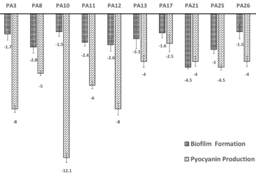
3. The selected strains were either moderate or strong biofilm formers. After ciclopirox overnight treatment, all of the isolates became non-biofilm formers with a decrease in biofilm formation ranging from 1.5- to 4.5- fold (Fig. 3). Among our selected strains, pyocyanin was significantly produced before treatment with ciclopirox.
That was manifested as a blue green pigment around the colonies grown on P agar plates and quantitatively upon extraction with chloroform. However, after ciclopirox treatment, there was a significant reduction in the amount of the pyocyanin produced ranging from 4- fold decrease in case of isolate PA26 to more than 12-fold decrease in case of PA10 (Fig. 3).
Table 3. Effect of ciclopirox olamine on different virulence factors of P. aeruginosa clinical isolates.
Isolate code
Swarming Twitching Hemolysis Pyocyanin production Protease production**
U T* U T U T U T U T
PA1 + - + - + - + - 11 10
PA2 + + + + + + + - 24 20
PA3 + - + - + - + - 20 11
PA4 + - + - + + + - 18 16
PA5 + + - - + + + - 22 16
PA6 + - + - + + + + 20 19
PA7 - - + - + - - - 14 11
PA8 - - + + + - + - 21 8
PA9 + + + + + + + - 22 20
PA10 - - + - + - + - 20 10
PA11 + - + + + - + - 19 0
PA12 + + + + + - + - 19 0
PA13 + - + + + - + - 20 11
PA14 + + + + + + + - 23 14
PA15 + + + + + - + + 21 15
PA16 - - + + + - + - 18 11
PA17 + - + - + - + - 19 12
PA18 + + + + + + + + 18 13
PA19 + + - - + + + - 18 18
PA20 + + + + + - - - 18 11
PA21 - - + + + - + - 20 12
PA22 + - + + + - + - 21 12
PA23 + - + + + - + - 18 0
PA24 - - + + + - + - 20 0
PA25 + - + + + - + - 18 9
PA26 + - + + + - + - 21 0
*U (Untreated sample), T (treated sample).
**Zone diameter in mm.
Fig. 2. Loss of protease activity in isolates PA23 and PA24 where (u) represents supernatant of untreated correspond- ing isolate and (t) represents supernatant of corresponding isolate treated with 9 μg/ml of ciclopirox olamine.
Effect of ciclopirox olamine on aprA and phzA1 gene expression
The effect of overnight treatment with 9 µg/ml ciclopirox on the expression of the virulence genes aprA (alkaline protease gene) (Fig. 4A) and phzA1 (pyocyanin biosynthesis gene) (Fig. 4B) among ten selected isolates
of P. aeruginosa grown at 37℃ was measured by quanti- tative RT-PCR (Fig. 4).
The levels of aprA and phzA1 gene expression were normalized to the levels of Rpsl gene expression and then compared with the levels of expression in corre- sponding untreated isolates. The transcripts of target Fig. 3. Fold change in biofilm formation and pyocyanin production after overnight treatment with 9 μg/ml ciclopirox among ten P. aeruginosa selected isolates. The error bars represent SDs.
Fig. 4. Relative gene expression of aprA (A) and phzA1 (B) determined by quantitative reverse transcriptase PCR for P. aeruginosa cultures. Total RNAs were prepared from ten selected isolates after overnight treatment with 9 μg/ml ciclopirox and calibrated against the corresponding untreated isolate. The levels of transcripts were normalized to the level of rpsl expression then compared to the level of expression by untreated cultures. No effect of the treatment corresponds to relative expression of 1. Results are expressed as the means and standard deviations of three independent determinations. The error bars represent SDs. The p-values indicate significance where *p < 0.05, **p < 0.01, ***p < 0.001 and ****p < 0.0001.
genes were calculated using the Pfaffle method or ΔΔCt method [26].
The results depicted in Fig. 4A clearly show that a sta- tistically significant but variable downregulation in the expression of aprA gene occurred upon treating the sam- ples with subinhibitory concentration of ciclopirox with fold change ranging from 0.45- to 0.03-fold. The p-value was < 0.05 among isolates PA25 and PA26, while iso- lates PA8, PA13 and PA21 showed a highly significant reduction in the expression of the target gene with a p- value of <0.001 compared to no effect of the treatment which corresponds to relative expression of 1.
The effect of subinhibitory concentration of ciclopirox on the ten selected pseudomonas cultures displayed dif- ferent results as respect to the relative expression of phzA1 gene as shown in Fig. 4B. Isolates PA10 and PA26 showed an unexpected significant upregulation in the gene under test compared to the expression of the isolates without treatment with fold changes in expres- sion equals to 3.73 and 1.93, respectively. In contrast, the significance of ciclopirox treatment of the other iso- lates ranged from highly downregulation values in both PA8 and PA21 to acceptable values, though significant, in case of PA25 and PA1. However, no significant effect was observed on the expression of isolate PA12.
Discussion
P. aeruginosa is one of the clinically significant oppor- tunistic pathogens causing infections that are life- threatening, and in most cases, difficult or challenging to eradicate [28]. The problem is aggravated by the fact that most of the isolated P. aeruginosa strains are reported to be multidrug resistant (MDR) ones [29]. In South Asia, the Middle East and the Mediterranean, modern medicine is already under threat from these MDR P. aeruginosa isolates [30].
In the present study, 26 clinical isolates of P. aerugi- nosa collected from hospitalized patients admitted to AMUH were found to be resistant to at least 3 classes of antibiotics and, thus, were recognized as MDR isolates [31]. A study conducted in the ICU of a parallel tertiary hospital, Alexandria University Students' Hospital, reported a percentage of 19% of MDR P. aeruginosa [32].
The tested isolates in the present study remained sus- ceptible to colistin, an antibiotic described as salvage
therapy for P. aeruginosa infections [33, 34]. However, because of its significant nephrotoxicity and neurotoxic- ity, colistin has a limited clinical use [35]. The lack of appearance of new classes of antibiotics for the treat- ment of Gram-negative infections for more than 40 years [36] has outlined the importance on focusing on agents that disarm bacteria, thereby decreasing or completely nullifying their pathogenicity.
Ciclopirox, an old antifungal agent, has been evaluated in this study for its ability to inhibit the virulence factors expressed by P. aeruginosa isolates under investigation.
Ciclopirox was previously reported to inhibit the growth of some Gram-negative bacteria [37, 38], but its efficacy had not been widely tested against antibiotic resistant bacteria. In the current study, the MIC50 of ciclopirox was found to be 16 µg/ml for MDR P. aeruginosa. An MIC range of 10 to >30 µg/ml was reported by Carlson- Banning et al. for clinical P. aeruginosa isolates [2]. A number of observations suggested that ciclopirox olamine might act as a chelator of iron ions [39−41], this could explain the inhibition of the cell growth observed in this work.
A concentration of 9 µg/ml of ciclopirox was used in this study to test its effect on virulence factors without affecting the bacterial growth. Hemolytic phospholi- pases secreted by Pseudomonas species are reported to directly cause lyse of human and sheep erythrocytes [42]. A percentage of 70% of tested isolates lost their hemolytic activity after ciclopirox treatment (Table 3).
However, among Candida albicans isolates, subinhibi- tory concentration of ciclopirox treatment caused a strong upregualtion of secreted phospholipases com- pared to the untreated ones [43].
The blue-green pyocyanin siderophore produced by P.
aeruginosa cultures has been shown in vitro to induce apoptosis in neutrophils, as well as to inhibit the phago- cytosis of apoptotic bodies by macrophages [44]. A per- centage of 93% of the tested isolates produced pyocyanin.
This percentage decreased to 12% upon ciclopirox treat- ment (Table 3) with a quantitative reduction ranging from 4-fold to more than12-fold (Fig. 3).
The proteases secreted by P. aeruginosa have estab- lished roles in sepsis and ocular infections where they can degrade immunoglobulins and fibrin and disrupt epithelial tight junctions [45]. A total of 12 isolates (46%) exhibited a prominent reduction in protease secretion
and 5 isolates totally lost their protease production abil- ity after treatment (Fig. 2). A similar finding has been reported when ciclopirox was tested against C. albicans where a decrease in protease secretion was reported [43]. Moreover, in a pancreatic cell model, ciclopirox was found to decrease caspases, a family of cysteine prote- ases, by forcing the cells to elevate the cleavage level of procaspase 3 [46].
During an infection, P. aeruginosa can adhere to host epithelial cells through the binding of its flagellum which is essential for the swimming and swarming motility of the organism [47]. The four pili localized at a cell pole of the organism are involved in twitching and swarming motility, as well as in the formation of bio- films [45]. Treatment with 9 µg/ml of ciclopirox resulted in decrease of twitching and swarming motility in 37.5%
and 85% of the isolates, respectively (Table 3). Similar results were previously proven among Salmonella typh- imurum strains where ciclopirox was found to decrease the bacterial motility by 91.4% compared to a control motile strain [48].
Twitching motility is an important factor in the devel- opment of biofilm, where highly organized structured communities of bacteria attached to one another and to a surface decrease the penetration of antibiotics and host defense molecules [49]. A percentage of 92% of the iso- lates under test were biofilm formers with different degrees of strength (Fig. 1). When ten strong to moder- ate biofilm formers were tested quantitatively, the fold decrease in biofilm formation ranged from 1.5 to 4.5-fold after ciclopirox treatment (Fig. 3). Similarly, a study investigating the effects of limiting iron levels in P. aeru- ginosa isolates found that biofilm development was sig- nificantly decreased by iron removal [50]. Disruption of biofilm formation is certainly a desirable goal that might prevent the repelling of antibiotics by biofilm structures of P. aeruginosa.
The effect of overnight treatment with ciclopirox on the expression of virulence genes aprA and phzA1 among ten selected isolates of P. aeruginosa was mea- sured by quantitative RT-PCR (Fig. 4). The levels of expression of the selected housekeeping gene; rpsl, a 30S ribosomal protein, did not change when cells were treated with subinhibitory concentrations of ciclopirox.
This indicated that essential functions of the bacterial cells were not directly affected by treatment with this
drug. However, the fact that genes encoding alkaline protease production and pyocyanin biosynthesis (aprA) and (phzA1), respectively, were downregulated in the presence of ciclopirox (Fig. 4) showed that this drug has multiple direct or indirect effects on the cells. It was interesting that although most of the strains treated with ciclopirox experienced downregulation with vari- able significance regarding phzA1 gene, the gene was markedly upregulated in PA10 and PA 26 (Fig. 4B). This can be explained by the fact that pyocyanin production in P. aeruginosa is a complex process involving two almost identical operons termed phz1 and phz2, which drive the production of phenazine-1-carboxylic acid which is further converted to pyocyanin [51].
The downregulatory effect of ciclopirox on genes encoding alkaline protease and pyocyanin production in P. aeruginosa has not been reported yet in the literature.
Thus, it needs further studies as it seems to be an encouraging pathway that might account for the antipseudomonal activity of ciclopirox in vivo.
Due to the current interest in ciclopirox for treatment of hematological malignancies such as multiple myeloma [52], systemic administration of this drug has been investigated in clinical trials with a resultant satis- factory drug safety profile and an adequate absorption following oral administration [53]. Calculation of the appropriate clinical dose and assessment of therapeutic index will be certainly required as a further step to per- mit the repositioning of ciclopirox from its role as merely a topical antifungal agent to a promising virulence-mod- ifying agent against one of the most problematic Gram- negative pathogens, P. aeruginosa.
Conflict of Interest
The authors have no financial conflicts of interest to declare.
References
1. Heras B, Scanlon MJ, Martin JL. 2014. Targeting virulence not via- bility in the search for future antibacterials. Br. J. Clin. Pharmacol.
79: 208-215.
2. Carlson-Banning KM, Chou A, Liu Z, Hamill RJ, Song Y, Zechied- rich L. 2013. Toward repurposing ciclopirox as an antibiotic against drug-resistant Acinetobacter baumannii, Escherichia coli, and Klebsiella pneumoniae. PLoS One 8: e69646.
3. Sonthalia S, Agrawal M. 2018. Topical ciclopirox - recalling a for-
gotten ally in the fight against cutaneous mycoses. EC Microbiology 14: 515-534.
4. Alnour TMS, Ahmed-Abakur EH. 2017. Multidrug resistant Pseu- domonas (P) aeruginosa: medical impact, pathogenicity, resis- tance mechanisms and epidemiology. JSM Microbiology 5: 1046.
5. Strateva T, Mitov I. 2011. Contribution of an arsenal of virulence factors to pathogenesis of Pseudomonas aeruginosa infections.
Ann. Microbiol. 61: 717-732.
6. Gad GF, El-Domany RA, Zaki S, Ashour HM. 2007. Characteriza- tion of Pseudomonas aeruginosa isolated from clinical and envi- ronmental samples in Minia, Egypt: prevalence, antibiogram and resistance mechanisms. J. Antimicrob. Chemother. 60: 1010-1017.
7. Aboushleib HM, Omar HM, Abozahra R, Elsheredy A, Baraka K.
2015. Correlation of quorum sensing and virulence factors in Pseudomonas aeruginosa isolates in Egypt. J. Infect. Dev. Ctries. 9:
1091-1099.
8. Overhage J, Bains M, Brazas MD, Hancock RE. 2008. Swarming of Pseudomonas aeruginosa is a complex adaptation leading to increased production of virulence factors and antibiotic resis- tance. J. Bacteriol. 190: 2671-2679.
9. van't Wout EF, van Schadewijk A, van Boxtel R, Dalton LE, Clarke HJ, Tommassen J, et al. 2015. Virulence factors of Pseudomonas aeruginosa induce both the unfolded protein and integrated stress responses in airway epithelial cells. PLoS Pathog. 11:
e1004946.
10. Pereira SG, Rosa AC, Ferreira AS, Moreira LM, Proenca DN, Morais PV, et al. 2014. Virulence factors and infection ability of Pseudo- monas aeruginosa isolates from a hydropathic facility and respi- ratory infections. J. Appl. Microbiol. 116: 1359-1368.
11. Kiska DL, Gilligan PH. 2003. Pseudomonas, pp. 719-728. In Mur- ray PR, Baron EJ, Jorgensen JH, Pfaller MA, Yolken RH (eds.), Man- ual of Clinical Microbiology, 8th Ed. American Society of Microbiology, Washington, D.C.
12. Clinical and Laboratory Standards Institute. 2015. Performance Standards for Antimicrobial Susceptibility Testing; Twenty-Fifth Informational Supplement. CLSI document M100-S25. Clinical and Laboratory Standards Institute, 950 West Valley Road, Suite 2500, Wayne, Pennsylvania 19087 USA.
13. Clinical and Laboratory Standards Institute. 2006. Performance Standards for Antimicrobial Susceptibility Testing; Sixteenth Informational Supplement. CLSI document M100-S16. Clinical and Laboratory Standards Institute, 940 West Valley Road, Suite 1400, Wayne, Pennsylvania 19087-1898 USA.
14. Ekanayaka SA, McClellan SA, Barrett RP, Kharotia S, Hazlett LD.
2016. Glycyrrhizin reduces HMGB1 and bacterial load in Pseudo- monas aeruginosa Keratitis. Invest. Ophthalmol. Vis. Sci. 57: 5799- 5809.
15. O'Toole GA, Pratt LA, Watnick PI, Newman DK, Weaver VB, Kolter R. 1999. Genetic approaches to study of biofilms. Methods Enzy- mol. 310: 91-109.
16. Merritt JH, Kadouri DE, O’Toole GA. 2011. Growing and analyzing static biofilms. Curr. Protoc. Microbiol. 22: 1-8.
17. Quiblier C, Zinkernagel AS, Schuepbach RA, Berger-Bachi B, Senn
MM. 2011. Contribution of SecDF to Staphylococcus aureus resis- tance and expression of virulence factors. BMC Microbiol. 11: 72.
18. King EO, Ward YM, Raney DE. 1954. Two simple media for the demonstration of pyocyanin and fluorescein. J. Lab. Clin. Med. 44:
301-307.
19. United States Pharmacopia. 2008. Microbial Limit Tests. pp. 2412- 2745. US Pharmacopeial Convention Inc. 31st Ed. Rockville: 19.
20. Essar DW, Eberly L, Hadero A, Crawford IP. 1990. Identification and characterization of genes for a second anthranilate synthase in Pseudomonas aeruginosa: interchangeability of the two anthranilate synthases and evolutionary implications. J. Bacteriol.
172: 884-900.
21. Essar DW, Eberly L, Han CY, Crawford IP. 1990. DNA sequences and characterization of four early genes of the tryptophan path- way in Pseudomonas aeruginosa. J. Bacteriol. 172: 853-866.
22. Rust L, Messing CR, Iglewski BH. 1994. Elastase assays. Methods Enzymol. 235: 554-562.
23. Rampioni G, Schuster M, Greenberg EP, Zennaro E, Leoni L. 2009.
Contribution of the RsaL global regulator to Pseudomonas aeru- ginosa virulence and biofilm formation. FEMS Microbiol. Lett. 301:
210-217.
24. Lenz AP, Williamson KS, Pitts B, Stewart PS, Franklin MJ. 2008.
Localized gene expression in Pseudomonas aeruginosa biofilms.
Appl. Environ. Microbiol. 74: 4463-4471.
25. Jazayeri JA, Nguyen K, Kotsanas D, Schneiders F, Tan C, Jazayeri M, et al. 2016. Comparison of virulence factors in Pseudomonas aeruginosa strains isolated from cystic fibrosis patients. J. Med.
Microb. Diagn. 5: 242.
26. Pfaffl MW. 2001. A new mathematical model for relative quantifi- cation in real-time RT-PCR. Nucleic Acids Res. 29: e45.
27. Fernandez L, Breidenstein EB, Song D, Hancock RE. 2012. Role of intracellular proteases in the antibiotic resistance, motility, and biofilm formation of Pseudomonas aeruginosa. Antimicrob.
Agents Chemother. 56: 1128-1132.
28. Driscoll JA, Brody SL, Kollef MH. 2007. The epidemiology, patho- genesis and treatment of Pseudomonas aeruginosa infections.
Drugs. 67: 351-368.
29. Maatallah M, Cheriaa J, Backhrouf A, Iversen A, Grundmann H, Do T, et al. 2011. Population structure of Pseudomonas aeruginosa from five Mediterranean countries: evidence for frequent recom- bination and epidemic occurrence of CC235. PLoS One 6: e25617.
30. Woodford N, Wareham DW, Guerra B, Teale C. 2014. Carbapene- mase-producing Enterobacteriaceae and non-Enterobacteriaceae from animals and the environment: an emerging public health risk of our own making? J. Antimicrob. Chemother. 69: 287-291.
31. Falagas ME, Koletsi PK, Bliziotis IA. 2006. The diversity of defini- tions of multidrug-resistant (MDR) and pandrug-resistant (PDR) Acinetobacter baumannii and Pseudomonas aeruginosa. J. Med.
Microbiol. 55: 1619-1629.
32. Abaza A. 2010. Multidrug resistant Pseudomonas aeruginosa in a health care setting in Alexandria. Bulletin of High Institute of Public Health 40: 333-347.
33. Mustafa MH, Chalhoub H, Denis O, Deplano A, Vergison A,
Rodriguez-Villalobos H, et al. 2016. Antimicrobial susceptibility of Pseudomonas aeruginosa isolated from cystic fibrosis patients in Northern Europe. Antimicrob. Agents Chemother. 60: 6735-6741.
34. Hachem RY, Chemaly RF, Ahmar CA, Jiang Y, Boktour MR, Rjaili GA, et al. 2007. Colistin is effective in treatment of infections caused by multidrug-resistant Pseudomonas aeruginosa in can- cer patients. Antimicrob. Agents Chemother. 51: 1905-1911.
35. Falagas ME, Kasiakou SK. 2005. Colistin: the revival of polymyxins for the management of multidrug-resistant Gram-negative bac- terial infections. Clin. Infect. Dis. 40: 1333-1341.
36. Brown D. 2015. Antibiotic resistance breakers: can repurposed drugs fill the antibiotic discovery void? Nat. Rev. Drug Discov. 14:
821-832.
37. Jue SG, Dawson GW, Brogden RN. 1985. Ciclopirox olamine 1%
cream. A preliminary review of its antimicrobial activity and ther- apeutic use. Drugs 29: 330-341.
38. Subissi A, Monti D, Togni G, Mailland F. 2010. Ciclopirox: recent nonclinical and clinical data relevant to its use as a topical anti- mycotic agent. Drugs 70: 2133-2152.
39. Abrams BB, Hanel H, Hoehler T. 1991. Ciclopirox olamine: a hydroxypyridone antifungal agent. Clin. Dermatol. 9: 471-477.
40. Korting HC, Grundmann-Kollmann M. 1997. The hydroxypyri- dones: a class of antimycotics of its own. Mycoses 40: 243-247.
41. Kruse R, Hengstenberg W, Hanel H, Raether W. 1991. Studies for the elucidation of the mode of action of the antimycotic hydroxypyridone compound, rilopirox. Pharmacology 43: 247- 255.
42. Ostroff RM, Vasil AI, Vasil ML. 1990. Molecular comparison of a nonhemolytic and a hemolytic phospholipase C from Pseudomo- nas aeruginosa. J. Bacteriol. 172: 5915-5923.
43. Niewerth M, Kunze D, Seibold M, Schaller M, Korting HC, Hube B.
2003. Ciclopirox olamine treatment affects the expression pat- tern of Candida albicans genes encoding virulence factors, iron metabolism proteins, and drug resistance factors. Antimicrob.
Agents Chemother. 47: 1805-1817.
44. Fuse K, Fujimura S, Kikuchi T, Gomi K, Iida Y, Nukiwa T, Watanabe
A. 2013. Reduction of virulence factor pyocyanin production in multidrug-resistant Pseudomonas aeruginosa. J. Infect. Chemo- ther. 19: 82-88.
45. Kipnis E, Sawa T, Wiener-Kronish J. 2006. Targeting mechanisms of Pseudomonas aeruginosa pathogenesis. Med. Mal. Infect. 36:
78-91.
46. Mihailidou C, Papakotoulas P, Papavassiliou AG, Karamouzis MV.
2018. Superior efficacy of the antifungal agent ciclopirox olamine over gemcitabine in pancreatic cancer models. Oncotar- get 9: 10360-10374.
47. Miao EA, Andersen-Nissen E, Warren SE, Aderem A. 2007. TLR5 and Ipaf: dual sensors of bacterial flagellin in the innate immune system. Semin. Immunopathol. 29: 275-288.
48. Malapaka VR, Barrese AA, Tripp BC. 2007. High-throughput screening for antimicrobial compounds using a 96-well format bacterial motility absorbance assay. J. Biomol. Screen. 12: 849- 854.
49. Bjarnsholt T, Tolker-Nielsen T, Hoiby N, Givskov M. 2010. Interfer- ence of Pseudomonas aeruginosa signalling and biofilm forma- tion for infection control. Expert. Rev. Mol. Med. 12: e11.
50. O'May CY, Sanderson K, Roddam LF, Kirov SM, Reid DW. 2009.
Iron-binding compounds impair Pseudomonas aeruginosa bio- film formation, especially under anaerobic conditions. J. Med.
Microbiol. 58: 765-773.
51. Higgins S, Heeb S, Rampioni G, Fletcher MP, Williams P, Camara M. 2018. Differential regulation of the phenazine biosynthetic operons by quorum sensing in Pseudomonas aeruginosa PAO1- N. Front Cell Infect. Microbiol. 8: 252.
52. Weir SJ, Patton L, Castle K, Rajewski L, Kasper J, Schimmer AD.
2011. The repositioning of the anti-fungal agent ciclopirox olamine as a novel therapeutic agent for the treatment of hae- matologic malignancy. J. Clin. Pharm. Ther. 36: 128-134.
53. Minden MD, Hogge DE, Weir SJ, Kasper J, Webster DA, Patton L, et al. 2014. Oral ciclopirox olamine displays biological activity in a phase I study in patients with advanced hematologic malignan- cies. Am. J. Hematol. 89: 363-368.