Received: 2011. 5.30. Accepted: 2011. 6.27.
Corresponding author: Young-Han Park, MD, PhD
Department of Obstetrics and Gynecology, Hallym University Sacred Heart Hospital, 896 Pyeongchon-dong, Dongan-gu, Anyang 431-061, Korea
Tel: +82-31-380-3820 Fax: +82-31-383-3820 E-mail: [email protected]
Th is is an Open Access article distributed under the terms of the Creative Commons Attribution Non-Commercial License (http://creativecommons.org/licenses/
by-nc/3.0/) which permits unrestricted non-commercial use, distribution, and reproduction in any medium, provided the original work is properly cited.
Copyright © 2011. Korean Society of Obstetrics and Gynecology
Human papillomavirus (HPV) is known as the causal agent of cer- vical cancer. The Papanicolaou smear is the most popular screen- ing tool for cervical cancer. Some low grade squamous intraepithe- lial lesions (LSILs) are the metaphase of the course going to high grade squamous intraepithelial lesion (HSIL) and probably will be presented as HSIL or cancer in the future.
Although screening methods have been improved in cytology, both conventional and liquid paps show relatively little difference in accuracy [1]. The sensitivity of cytology is 70-80% [2,3]. It is
pISSN 2233-5188 · eISSN 2233-5196
HIGH RISK TYPE HUMAN PAPILLOMAVIRUS INDICATES INCREASING HIGH GRADE INTRAEPITHELIAL LESION INCIDENCE IN CYTOLOGICAL LOW GRADE SQUAMOUS INTRAEPITHELIAL LESION PATIENTS
Young-Han Park, MD, Chae Choon Rhim, MD, Seong Ju Kim, MD, Jung Bae Kang, MD, Pong Rhim Jang, MD, Hong Bae Kim, MD, Keun Young Lee, MD, Chan Eun Park, MD, Yong Jo, MD, Eui Seon Rho, MD, Yong Woo Lee, MD, Seong Cheon Yang, MD, Soo Ran Choi, MD
Department of Obstetrics and Gynecology, Hallym University College of Medicine, Chuncheon, Korea
Objective
Human papillomavirus (HPV) is known as the causal agent of cervical cancer. The Papanicolaou smear is the most popular screening tool for cervical cancer. Some cytological low grade squamous intraepithelial lesions (LSILs) are in the developmental stage of high grade intraepithelial lesions (HSILs) and cervical cancer metaphases. Our purpose is to disclose the signifi cance of high risk type (HRT) HPV in LSIL.
Methods
Documentation of 200 cases of cone biopsy at Hallym University Sacred Heart Hospital from February 2006 to February 2008 was reviewed retrospectively. HPV typing with DNA microarray results were found in 20 of the LSIL patients. Chi-square and student- t tests were used in the statistical analysis.
Results
In the cytological LSIL patients, HRT-HPV positive patients were 16/20 (80%) and one low risk type (LRT) HPV positive patient was 1/20 (5%). In the cytological LSIL patients, postoperative pathological diagnoses were cervical intraepithelial neoplasia 1 (CIN 1) 16/26 (61.5%), CIN2 2/26 (7.7%), CIN3 5/26 (19.2%), carcinoma in situ 1/26 (3.8%). Among the 16 HRT-HPV positive patients, there were six that were above HSIL 6/16 (37.5%) in pathologic diagnoses. In the HRT-HPV negative 10 cytologic LSIL patients, there were two that were above HSIL 2/10 (20%) in pathologic diagnoses. HRT-HPV prevalences were not signifi cantly different between LSIL and HSIL.
Conclusion
In HRT-HPV positive LSIL, the risk of HSIL or cancer of the cervix seems to be increasing. The HPV test is helpful in LSIL patient strategies and should be done in LSIL patients in order to discriminate between the nature and prognosis of cytological LSIL.
Keywords: Human papillomavirus; Low grade squamous intraepithelial lesion
known that LSIL persists in 32% and progresses to carcinoma in situ (CIS) in 11% [4]. The HPV test is helpful in making decisions in cervical cytology. It is known that LSILs are related to low risk HPV types 6, 11, 40, 42, 43, 44, 54, 61, 70, 72, 81 and HSILs and cervical cancers are related to high risk HPV types 16, 18, 31, 33, 35, 39, 45, 51, 52, 56, 58, 59, 68, 73 and 82. But some cervical intraepithelial neoplasia 2 (CIN 2) and 3 lesions develop without a preexisting CIN 1 [5,6]. And HPVs are positive in CIN 1 about 85% [7,8]. Some CIN 1s and almost all CIN 2 and 3 lesions are associated with monoclonal “high-risk” HPV types [5,9].
For clinical convenience, cytological LSIL should be proven as pathological LSIL and cytological HSIL should be proven as patho- logical HSIL. But we could not agree to utilize such a rule in the clinic. The results of LSILs and HSILs in cytology are not always pathologic LSILs and HSILs. In cytological HSILs, the next step is simple. Cone biopsies always follow the cytologic HSILs. In cyto- logical LSIL, it is necessary to make a decision between the cone biopsy or the follow-up.
With the viewpoint that guidance is necessary in LSIL, HPV test- ing is one of the best choices for gynecologists before colposcopic consultation. A repeat pap follow up and immediate colposcopy are recommended as alternatives, if available [10].
We retrospectively reviewed the clinical characteristics of 200 cone biopsies and compared the results of cytological LSILs according to the HPV test fi ndings.
Materials and Methods
Two hundred cases of cone biopsy results at Hallym University Sacred Heart Hospital from February 2006 to February 2008 were
reviewed retrospectively. The mean age was 42.3±9.24 (range, 22-80) years.
During the review of pathologic reports, including punch and cone biopsies, severe reports were selected if two categories were presented in biopsies. There were 21 cervicitis (10.5%), 59 LSILs (29.5%), 99 HSILs (49.5%), and 21 cancers (10.5%). There were 26 pathological LSIL patients preoperatively. HPV typing with DNA microarrays (Biomedlab, Seoul, Korea) were performed in 20 patients preoperatively in LSIL patients. Available high risk type HPVs were 16, 18, 31, 33, 35, 39, 45, 51, 52, 56, 58, 59, 66, 68, and 69. Low risk type HPVs were 6, 11, 34, 40, 42, 43, 44 in our microarray kits. The HRT HPV positive results of DNA microarrays were 16 patients out of 20 patients with cytological LSIL.
Age
Patient numbers
P=0.012
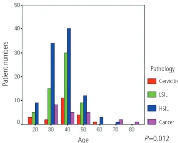
Fig. 1. The age and pathologic distribution of cone biopsies. HSILs are most frequent in the fi fth decade. LSIL, low grade squamous intraepithe- lial lesion; HSIL, high grade squamous intraepithelial lesion.
Cervicitis LSIL HSIL Cancer Pathology
Table 1. The age and pathologic distribution of cone biopsies (P=0.012)
Age Pathologic category in cone
Cervicitis LSIL HSIL Cancer Tatal
20 3 5 9 0 17
30 2 15 34 8 59
40 11 30 40 5 86
50 4 9 12 5 30
60 1 0 3 0 4
70 0 0 1 2 3
80 0 0 0 1 1
Total 21 59 99 21 200
The 40s and 50s are common ages for cone biopsy with cervical intraepithelial lesions. Cervical cancers were prevalent in the 30s-50s.
LSIL, low grade squamous intraepithelial lesion; HSIL, high grade squamous intraepithelial lesion; cancer, cervical cancer.
Chi-square and student-t tests were used in analysis. Results with
P-value less than 0.05 were regarded as statistically signifi cant. Results
1. Age and pathologic distribution (Table 1, Fig. 1) The distribution of pathologic fi ndings is shown according to age (Table 1, Fig. 1). The most frequent patient age for cone biopsies was in their 40s. The most frequent age for cancer patients was in their 30s. HSILs were most frequent in the 40s age group. LSILs were most frequent in 40s.
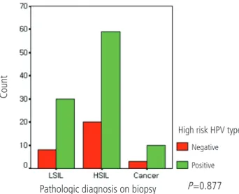
2. Cone biopsy results and high risk type (HRT) HPV positivity (Table 2, Fig. 2)
Pathologic diagnoses and HRT-HPV positivities were 78.9% (n=30) for LSIL, 74.7% (n=59) for HSIL (CIN 2, 3, CIS) and 76.9% (n=10) for cancer patients (P=0.877).
Pathologic diagnosis on biopsy
High risk HPV type Negative Positive
Count
P=0.877
Fig. 2. High risk HPV type positivity in all pathologic diagnosis of cone biopsies. High risk type HPVs are similar in prevalence among LSIL (78.9%), HSIL (74.7%) and cancer (76.9%) groups (P =0.877). LSIL, low grade squamous intraepithelial lesion, HSIL, high grade squamous intraepithelial lesion; HPV, human papillomavirus.
Pao smear
HPV Negative High risk HPV Low risk HPV
Patient numbers
Fig. 3. High risk HPV positivity according to the cytologic diagnoses of patients. LSIL, low grade squamous intraepithelial lesion; HSIL, high grade squamous intraepithelial lesion; ASCUS, atypical squamous cells of un- determined signifi cance; ASC-H, atypical squamous cells cannot exclude HSIL; HPV, human papillomavirus.
Table 2. High risk HPV type positivity in every pathologic diagnosis of cone biopsies ( P=0.877)
High risk HPV
Negative Positive Total
LSIL 8 (21.1) 30 (78.9) 38 (100)
HSIL 20 (25.3) 59 (74.7) 79 (100)
Cancer 3 (23.1) 10 (76.9) 13 (100)
Total 31 99 130
High risk type HPVs are similar in prevalence among LSIL (78.9%), HSIL (74.7%) and cancer (76.9%) groups ( P=0.877).
HPV, human papillomavirus; LSIL, low grade squamous intraepithelial le- sion; HSIL, high grade squamous intraepithelial lesion.
Table 3. High risk HPV positivity according to the cytologic diagnoses of patients
Negative High risk HPV Low risk HPV Total
Cervicitis 3 10 0 13
LSIL 3 16 (80%) 1 20
HSIL 7 31 (81.6%) 0 38
Cancer 2 2 0 4
ASCUS 2 19 0 21
ASC‐H 4 4 0 8
21 82 1 104
The prevalence of high risk HPVs are similar in HSIL (80%) and LSIL (81.6%).
HPV, human papillomavirus; LSIL, low grade squamous intraepithelial lesion; HSIL, high grade squamous intraepithelial lesion; ASCUS, atypical squamous
cells of undetermined signifi cance; ASC-H, atypical squamous cells cannot exclude HSIL.
3. HPV typing in every cytologic category (Table 3, Fig. 3) In the LSIL patients, HRT-HPV positive patients were 16/20 (80%) and one LRT-HPV positive patient was 1/20 (5%). This means that 75% of LSIL patients have HRT-HPV in their lesions. A further in- dication is that 75% of LSIL may develop into HSIL or cancer. Low risk HPV was only found in LSIL.
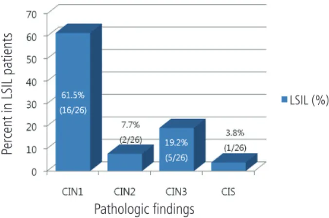
4. Pathologic results in cytological LSILs (Fig. 4)
Among the LSIL patients CIN 1 were 16/26 (61.5%), CIN 2 were 2/26 (7.7%), CIN3 were 5/26 (19.2%), CIS was 1/26 (3.8%), severe than
CIN2 were 8/26 (30.8%) and chronic cervicitis were 2 patients (7.7%). The concordance rate between cytology and pathology in LSIL was 61.5%.
5. HRT-HPV in cytologic LSIL is probably related to HSIL or Cancer (Fig. 5)
There were 26 cytological LSIL patients, and HPV test results were found in 20 of them. One low risk HPV positive patient 5% (1/20) was pathologically CIN 1. Sixteen (16/20) 80% of the cytologic LSIL patients were positive in HRT-HPVs (Fig. 5A).
In HRT- HPV positive cytologic LSIL 16 patients, 6/16 (37.5%) were more severe than HSIL upon pathologic diagnoses. The HSIL inci- dence was higher than the HRT-HPV negative group 2/10 (20%) and overall 8/26 (30.8%), but the rate of incidence has no statisti- cal signifi cance in our study (P=0.42) (Fig. 5B).
Discussion
In the management of cervical intraepithelial lesions, HPV tests are the preferred method of clinical aid. In cytological LSIL patients, we can do colposcopy directed punch biopsy and an HPV test in order to confi rm diagnosis and clinical guidance.
In Anyang city, cone biopsies are commonly performed under the diagnosis of HSIL from the ages of 30-40 years. In ALTS, 83% of the women of the LSIL group were positive for high-risk type HPVs [11]. HRT-HPV prevalence was similar in pathological LSILs (80%) and HSILs (81.6%) patients in our study (Table 3). The prevalence Fig. 4. Histologic diagnoses in cytologic LSIL patients. The cytologic LSILs
were pathological LSIL in biopsies (61.5%). The cytologic LSILs were his- tologic HSIL in 8/26 (30.7%). Vertical axis represents ratio (%) of every categories in cytologic LSILs. LSIL, low grade squamous intraepithelial lesion; HSIL, high grade squamous intraepithelial lesion; CIN, cervical in- traepithelial lesion; CIS, carcinoma in situ.
Pathologic fi ndings
Percent in LSIL patients
LSIL (%)
Fig. 5. High risk type HPVs in cytological LSILs are probably related to increasing histological HSIL. (A) 80% of high risk HPV positive cytologic LSILs were histologic HSIL. 5% of low risk HPV positive cytologic LSILs were histologic HSIL. The vertical axis represents ratio of each categories in LSIL pa- tients. (B) High risk HPV positive cytologic LSIL patients were higher than other groups-over all cytologic LSIL and high risk HPV negative cytologic LSIL patients. The vertical axis represents ration of every categories in LSIL patients. LSIL, low grade squamous intraepithelial lesion; HPV, human papillomavirus.
A B
High risk HPV Low risk HPV
LSIL (%)
HPV incidence (%)
HPV incidence in LSIL (%)
90 80 70 60 50 40 30 20 10
0 Over all High risk
HPV (+)
Low risk HPV (-) 40
30
20
10
0
of HRT HPVs were similar between ALTS group study and our study. This result confi rms that LSIL is a very specifi c indicator of the presence of HRT-HPV. LSIL is a disease induced by HPVs.
The life of LSIL is not as serious. 90% of adolescent LSIL will spontaneously regress (< 21 years of age) [12,13]. The HSIL rate is lower in post-menopause cytological LSIL women and they can be managed in the same way as ASC-US management in post- menopause [14,15]. 90% of high risk HPV positive cytological LSIL patients spontaneously regress within 24 months [16]. 70%
of LSIL with high risk HPV infected patients were spontaneously cleared of their infections [17]. Therefore, surgical treatment of LSIL is usually not preferred.
However, it is necessary for pathological LSIL patients whose his- tory was preceded by cytological HSIL or atypical glandular cells to undergo diagnostic excision biopsies or 6 month interval follow ups of colposcopy and cytology [10]. HRT HPV negative LSILs and low risk type HPV positive LSILs were found to be pathologically benign, as we expected. And a trend appears to exist in that high risk HPV positive cytological LSIL patients have increased rates of pathologically HSIL and cancer. It is not such a small portion as to be ignored and reaches approximately 30% [17]. Although our study has limited statistical infl uence, further research in HRT HPV positive LSIL patients would seem to be required.
Cytological LSIL women with high risk type HPV should be man- aged cautiously, with cytology and colposcopy, and by at least a 6 month interval follow-up. Histologic diagnosis by punch biopsy or excision is also recommended in HRT HPV positive LSILs. A large scale study in LSIL patients would seem to be mandated as being necessary in relation to high risk type HPVs.
References
1. Sulik SM, Kroeger K, Schultz JK, Brown JL, Becker LA, Grant WD. Are fluid-based cytologies superior to the conven- tional Papanicolaou test? A systematic review. J Fam Pract 2001;50:1040-6.
2. Taylor S, Kuhn L, Dupree W, Denny L, De Souza M, Wright TC Jr.
Direct comparison of liquid-based and conventional cytology in a South African screening trial. Int J Cancer 2006;118:957- 62.
3. Ronco G, Segnan N, Giorgi-Rossi P, Zappa M, Casadei GP, Carozzi F, et al. Human papillomavirus testing and liquid- based cytology: results at recruitment from the new technolo- gies for cervical cancer randomized controlled trial. J Natl
Cancer Inst 2006;98:765-74.
4. Mitchell MF, Tortolero-Luna G, Wright T, Sarkar A, Richards- Kortum R, Hong WK, et al. Cervical human papillomavirus infection and intraepithelial neoplasia: a review. J Natl Cancer Inst Monogr 1996:17-25.
5. ter Haar-van Eck SA, Rischen-Vos J, Chadha-Ajwani S, Huike- shoven FJ. The incidence of cervical intraepithelial neoplasia among women with renal transplant in relation to cyclospo- rine. Br J Obstet Gynaecol 1995;102:58-61.
6. ter Harmsel B, Smedts F, Kuijpers J, van Muyden R, Oosterhuis W, Quint W. Relationship between human papillomavirus type 16 in the cervix and intraepithelial neoplasia. Obstet Gynecol 1999;93:46-50.
7. Solomon D, Schiffman M, Tarone R; ALTS Study group.
Comparison of three management strategies for patients with atypical squamous cells of undetermined significance:
baseline results from a randomized trial. J Natl Cancer Inst 2001;93:293-9.
8. Cox JT, Schiffman M, Solomon D; ASCUS-LSIL Triage Study (ALTS) Group. Prospective follow-up suggests similar risk of subsequent cervical intraepithelial neoplasia grade 2 or 3 among women with cervical intraepithelial neoplasia grade 1 or negative colposcopy and directed biopsy. Am J Obstet Gy- necol 2003;188:1406-12.
9. Park TW, Fujiwara H, Wright TC. Molecular biology of cervical cancer and its precursors. Cancer 1995;76:1902-13.
10. Wright TC Jr, Massad LS, Dunton CJ, Spitzer M, Wilkinson EJ, Solomon D. 2006 consensus guidelines for the management of women with cervical intraepithelial neoplasia or adenocar- cinoma in situ. Am J Obstet Gynecol 2007;197:340-5.
11. Human papillomavirus testing for triage of women with cyto- logic evidence of low-grade squamous intraepithelial lesions:
baseline data from a randomized trial. The Atypical Squamous Cells of Undetermined Signifi cance/Low-Grade Squamous In- traepithelial Lesions Triage Study (ALTS) Group. J Natl Cancer Inst 2000;92:397-402.
12. Moscicki AB, Schiffman M, Kjaer S, Villa LL. Chapter 5: Updat- ing the natural history of HPV and anogenital cancer. Vaccine 2006;24 Suppl 3:S3/42-51.
13. Moscicki AB, Hills N, Shiboski S, Powell K, Jay N, Hanson E, et al. Risks for incident human papillomavirus infection and low- grade squamous intraepithelial lesion development in young females. JAMA 2001;285:2995-3002.
14. Sherman ME, Schiffman M, Cox JT; Atypical Squamous Cells
of Undetermined Signifi cance/Low-Grade Squamous Intraepi-
thelial Lesion Triage Study Group. Effects of age and human papilloma viral load on colposcopy triage: data from the randomized Atypical Squamous Cells of Undetermined Sig- nificance/Low-Grade Squamous Intraepithelial Lesion Triage Study (ALTS). J Natl Cancer Inst 2002;94:102-7.
15. Evans MF, Adamson CS, Papillo JL, St John TL, Leiman G, Coo- per K. Distribution of human papillomavirus types in ThinPrep Papanicolaou tests classifi ed according to the Bethesda 2001 terminology and correlations with patient age and biopsy out-
comes. Cancer 2006;106:1054-64.
16. Schlecht NF, Platt RW, Duarte-Franco E, Costa MC, Sobrinho JP, Prado JC, et al. Human papillomavirus infection and time to progression and regression of cervical intraepithelial neo- plasia. J Natl Cancer Inst 2003;95:1336-43.
17. Nobbenhuis MA, Helmerhorst TJ, van den Brule AJ, Rozendaal L, Voorhorst FJ, Bezemer PD, et al. Cytological regression and clearance of high-risk human papillomavirus in women with an abnormal cervical smear. Lancet 2001;358:1782-3.
세포검사상 저등급상피내 병변에서 고위험군 인유두종바이러스를 이용한 고등급상피내 종양의 예측
한림대학교 의과대학 산부인과학교실
박영한, 임채춘, 김성주, 강정배, 장봉림, 김홍배, 이근영, 박찬은, 조 용, 노의선, 이용우, 양성천, 최수란
목적
인유두종바이러스는 자궁경부암의 원인이다. Papanicolaou 검사는 자궁경부암의 선별검사이다. 일부 세포검사상 저등급상피내병변은 고 등급상피내병변이나 자궁경부암으로의 중간단계라고 할 수 있다. 본 연구는 저등급상피내 병변에서의 고위험군 인유두종바이러스형의 양 성이 갖는 의미를 규명하기 위한 연구이다.
연구방법
한림대학교성심병원을 2006년 2월부터 2008년 2월까지 방문하여 원추절제술을 받은 200명의 환자의 의무기록을 후향적으로 검토하였 다. DNA microarray를 이용한 인유두종바이러스 검사결과를 20명의 저등급상피내병변 환자에서 확인할 수 있었다. X2-검사와 student-t 검사를 사용하였다.
결과
세포검사상 저등급상피내병변(low grade squamous intraepithelial lesion, LSIL) 환자에서, 인유두종바이러스 고위험형 양성인 환자는 16/20명(80%)이며, 인유두종바이러스 저위험형 양성인 환자는 1/20명(5%)이었다. 세포검사상 LSIL 환자에서 원추생검술 후 조직학적 진 단에 따른 분류는 cervical intraepithelial neoplasia 1 (CIN 1) 16/26명(61.5%), CIN2 2/26명(7.7%), CIN3 5/26명(19.2%), CIS 1/26명(3.8%) 이었고, CIN2 이상의 병변은 8/26명(30.8%)였다. 인유두종 고위험형 양성인 16명의 환자는 고등급상피내병변(high grade intraepithelial lesion, HSIL) 이상이 6/16명(37.5%)이었다. 인유두종바이러스 고위험형 음성인 세포검사상 LSIL인 10명의 환자에서 조직검사상 HSIL 이 상은 2/10명(20%)이었다. 인유두종바이러스 고위험형의 유병률은 LSIL과 HSIL에서 비슷하였다.
결론
LSIL과 HSIL에서 인유두종바이러스 고위험형의 유병률은 LSIL과 HSIL에서 비슷하였다. 고위험형 바이러스 양성인 LSIL에서 HSIL이나 경 부암의 빈도는 증가하는 것처럼 보인다. LSIL환자에서 인유두종바이러스 검사는 세포검사상 LSIL의 성격과 예후를 구별하는 데 매우 도움 이 되는 검사로 보이므로 반드시 시행되어야 할 것이다.
중심단어: 인유두종바이러스, 저등급상피내병변