Kimura Disease Involving a Caruncle Woo Jin Kim1, Myoung Ja Chung2, In Cheon You1 1
4
0
0
전체 글
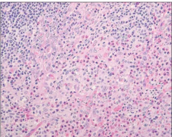
(2) Korean J Ophthalmol Vol.27, No.2, 2013. Fig. 1. A slit lamp biomicroscopic finding on the patient’s initial visit revealed a conjunctival nodule in the left medial canthal area.. Fig. 2. Histological findings of mass lesion (hematoxylin-eosin stain, ×100). Chronic inflammation with multiple follicular lymphoid hyperplasia with nodal germinal center.. vascular channels, especially the capillaries (Fig. 3). Vascular channels were lined by normal-appearing flat, spindleshaped endothelial cells (Fig. 4). Immunohistochemical staining for CD20 and CD3 showed T-cells surrounding well-formed lymphoid follicles with germinal centers containing B-cells (Fig. 5A and 5B). On the basis of these histopathologic findings, we established a diagnosis of Kimura disease. At the last followup 6 months after surgery, the patient had no evidence of a recurrence.. Discussion The caruncle is a modified cutaneous tissue containing fine hair, sebaceous glands, sweat glands, and goblet cells [7]. Although this large array of tissue elements can give 138. Fig. 3. Numerous infiltrated eosinophils with fibrous septa (hematoxylin-eosin stain, ×400).. Fig. 4. Multiple capillary proliferation with normal flat, spindleshaped vascular endothelium (hematoxylin-eosin stain, ×400).. rise to a wide variety of lesions, caruncular tumors are uncommon [7]. This is the first report describing Kimura disease localized to a caruncle. Kimura disease typically presents as firm, painless, pruritic, single-to-multiple subcutaneous nodules [4]. Kimura disease has a predilection for the skin of the pre-auricular, parotid, and submandibular regions. Kimura disease of the orbit and ocular adnexa is uncommon, but well-documented by Buggage et al. [5]. The orbit, especially in the superior orbital space, may be a preferred site for Kimura disease. The disease was first described in Chinese literature as an eosinophilic hyperplastic lymphograuloma and was later characterized by Kimura et al. in 1948 [1]. Hundreds of cases under a variety of names have been reported in Asia, including China, Japan, and Indonesia..
(3) WJ Kim, et al. Kimura Disease in Caruncle. A. B. Fig. 5. Immunohistochemical staining for CD20 and CD3 in Kimura disease (×200). The germinal center B-lymphocytes of follicles is positive for CD20 (A) and CD3 stained in the surrounding mantle zone of T-lymphocytes (B).. Kimura disease occurs mainly in Asian males, presenting as large (>2 cm) subcutaneous papules or nodules, elevated blood eosinophilia (up to 54%), hypergammaglobulinemia (IgE), regional lymphadenopathy, and salivary gland involvement [8,9]. Systemic symptoms (fever, night sweats, and weight loss) are uncommon. The patient described herein was female and had a normal peripheral blood eosinophil count and serum IgE level. The etiology and pathogenesis of Kimura disease are unknown. Allergic reactions, infections, and autoimmune reactions with an aberrant immune response have been suggested [10]. Because of the non-specific presentation of Kimura disease, strict histopathologic examination is crucial to confirming the diagnosis. Histopathologically, Kimura disease is characterized by an inflammatory infiltrate with prominent collections of eosinophils and scattered lymphoid follicles, increased fibrocollagenous tissue, and vascular proliferation, with vessels lined by normal endothelial cells [11]. Our case also showed the typical pathologic findings which occur in Kimura disease. The differential diagnosis of Kimura disease is broad and includes angiolymphoid hyperplasia with eosinophilia (ALHE), Hodgkin lymphoma, Langerhans cell histiocytosis, lymphadenopathy of a drug reaction, and parasitic lymphadenitis. However, distinguishing Kimura disease from neoplasms is not difficult when Reed-Sternberg cells, atypical lymphocytes, and Langerhans cells are identified. A positive history of drug use, and seropositivity for parasitic infections can be helpful for differentiating diagnoses. Kimura disease and ALHE were often confused in a number of early reports [5,11]. Over the years, the clinical features of Kimura disease overlap significantly with those of ALHE. Clinically, both present as relatively asymptomatic soft tissue swellings in the head and neck. However,. there are characteristic and distinctive clinicopathological features between the two diseases. In brief, Kimura disease occurs predominantly in Asian males. Patients mostly demonstrate a peripheral eosinophilia and elevated serum IgE levels. The solitary lesions are usually in the deep subcutaneous tissues, and are frequently associated with regional lymphadenopathy and salivary gland involvement. By contrast, ALHE occurs in all racial groups with a slight female predilection. Patients present with small, superficial dermal papulonodules, which are frequently erythematous, accompanied by bleeding, pruritis, and tumor growth. Regional lymphadenopathy, serum eosinophilia, and elevated IgE levels are rare [11]. Kimura disease and ALHE can be differentiated on a histopathological basis, especially with their vascular endothelial morphology. Kimura disease is a chronic allergic or autoimmune inflammatory disorder, whereas ALHE is a neoplasm of blood vessels [10]. Histopathologically, the most important diagnostic feature of ALHE is the appearance of endothelial cells, which have a vesicular nucleus, abundant cytoplasm, and an ovalto-cuboidal shape [5]. These endothelial cells can project into vascular spaces or form deposits in vessels [5,12,13]. Stromal fibrosis is uncommon in ALHE [8]. In our patient, the histopathologic examination of the surgical specimen revealed flat, spindle-shaped endothelial cells which had normal-appearing nuclei and cytoplasm; findings which are consistent with Kimura disease. Interestingly, Kimura disease is associated with renal disease, which may develop before or concurrently with disease progression [14]. Googe et al. [8] demonstrated IgE in the renal glomeruli of a patient with Kimura disease who manifested nephritic syndrome. Our patient had microalbuminuria, but declined additional work-up. The optimal treatment for Kimura disease has not been 139.
(4) Korean J Ophthalmol Vol.27, No.2, 2013. determined. Excision is the most widely used therapy and oral or intralesional steroids, radiation, and chemotherapy can also be used. Recurrence after excision is common because of incomplete excision [3]. Considered an inflammatory process, the disease has an excellent prognosis. We treated the patient with complete excision and recurrence has not been detected through the 6-month follow-up visit. There is a tendency for caruncular lesions to have inconsistencies between clinical and histopathologic diagnosis, which can be as high as 50% [15]. Our patient is a further unusual example of the confounding nature of the caruncular lesions. Despite its rarity, Kimura disease should be included in the differential diagnosis of caruncular lesions. Caruncular lesions are rare and diverse, making clinical diagnosis difficult. With a strict histopathologic examination of excised tissue, a correct diagnosis can be achieved.. Conflict of Interest No potential conflict of interest relevant to this article was reported.. References 1. Kimura T, Yoshimura S, Ishikawa E. On the unusual granulation combined with hyperplastic changes of lymphatic tissue. Trans Soc Pathol Jpn 1948;37:179-80. 2. Kung IT, Chan JK. Kimura’s disease or Kimm’s disease? Am J Surg Pathol 1988;12:804-5. 3. Kung IT, Gibson JB, Bannatyne PM. Kimura’s disease: a clinico-pathological study of 21 cases and its distinction from angiolymphoid hyperplasia with eosinophilia. Pathology 1984;16:39-44.. 140. 4. Kuo TT, Shih LY, Chan HL. Kimura’s disease: involvement of regional lymph nodes and distinction from angiolymphoid hyperplasia with eosinophilia. Am J Surg Pathol 1988;12:843-54. 5. Buggage RR, Spraul CW, Wojno TH, Grossniklaus HE. Kimura disease of the orbit and ocular adnexa. Surv Ophthalmol 1999;44:79-91. 6. Lee SJ, Song JH, Kim SD. Kimura’s disease involving the ipsilateral face and extraocular muscles. Korean J Ophthalmol 2009;23:219-23. 7. Kiratli H, Kocabeyoglu S, Saglam A, Soylemezoglu F. Langerhans cell histiocytosis of the caruncle. Clin Experiment Ophthalmol 2007;35:661-3. 8. Googe PB, Harris NL, Mihm MC Jr. Kimura’s disease and angiolymphoid hyperplasia with eosinophilia: two distinct histopathological entities. J Cutan Pathol 1987;14:263-71. 9. Urabe A, Tsuneyoshi M, Enjoji M. Epithelioid hemangioma versus Kimura’s disease: a comparative clinicopathologic study. Am J Surg Pathol 1987;11:758-66. 10. Abuel-Haija M, Hurford MT. Kimura disease. Arch Pathol Lab Med 2007;131:650-1. 11. Chen H, Thompson LD, Aguilera NS, Abbondanzo SL. Kimura disease: a clinicopathologic study of 21 cases. Am J Surg Pathol 2004;28:505-13. 12. Rosai J. Angiolymphoid hyperplasia with eosinophilia of the skin: its nosological position in the spectrum of histiocytoid hemangioma. Am J Dermatopathol 1982;4:175-84. 13. Rosai J, Gold J, Landy R. The histiocytoid hemangiomas: a unifying concept embracing several previously described entities of skin, soft tissue, large vessels, bone, and heart. Hum Pathol 1979;10:707-30. 14. Rajpoot DK, Pahl M, Clark J. Nephrotic syndrome associated with Kimura disease. Pediatr Nephrol 2000;14:486-8. 15. Ostergaard J, Prause JU, Heegaard S. Caruncular lesions in Denmark 1978-2002: a histopathological study with correlation to clinical referral diagnosis. Acta Ophthalmol Scand 2006;84:130-6..
(5)
수치

관련 문서
–The AABB suggests adhering to a restrictive strategy in hospitalized patients with preexisting cardiovascular disease and considering transfusion for patients with symptoms or
KDIGO Clinical Practice Guideline for the Diagnosis, Evaluation, Prevention, and Treatment of Chronic Kidney Disease–Mineral and Bone Disorder (CKD–MBD). Kidney Int 2009:
• Given similar degrees of hyperglycemia only a third of patients with diabetes develop clinically important renal disease genetic determinantQ. • Relatively increased
After first field tests, we expect electric passenger drones or eVTOL aircraft (short for electric vertical take-off and landing) to start providing commercial mobility
Chronic kidney disease (partial update): Early identification and management of chronic kidney disease in adults in primary and secondary care (Clinical
1) Global Initiative for Chronic Obstructive Lung Disease (GOLD). Global strategy for the diagnosis, management and prevention of COPD. Yates, B.S., Jørgen Vestbo, M.D.,
1 John Owen, Justification by Faith Alone, in The Works of John Owen, ed. John Bolt, trans. Scott Clark, "Do This and Live: Christ's Active Obedience as the
Severe disease activity and cy- tomegalovirus colitis are predictive of a nonresponse to infliximab in patients with ulcerative colitis. Guidelines for the management of
