© 2018 The Korean Ophthalmological Society
This is an Open Access article distributed under the terms of the Creative Commons Attribution Non-Commercial License (http://creativecommons.org/licenses /by-nc/3.0/) which permits unrestricted non-commercial use, distribution, and reproduction in any medium, provided the original work is properly cited.
Original Article
Lacrimation is a very common ophthalmic symptom, which is often caused by the closure of the lacrimal outflow
system. Epiphora may result from various causes, such as trauma from repeated rivus lacrimaris enlargement proce- dures, burns, radiotherapy, conjunctivochalasis, lacrimal duct inflammation, xeroma, and drugs [1]. Silicone tube in- tubation has been widely used with dacryocystorhinostomy procedures since Keith [2] reported on it in 1968. It has been successful in patients of all ages with varied presenta- tions, such as congenital nasolacrimal duct obstruction in
Received: April 24, 2018 Accepted: July 8, 2018
Corresponding Author: Hee Bae Ahn, MD, PhD. Department of Ophthal- mology, Dong-A University College of Medicine, #32 Daesingongwon-ro, Seo-gu, Busan 49201, Korea. Tel: 82-51-240-5222 , Fax: 82-51-254-1987, E-mail: [email protected]
Combination Surgery of Silicone Tube Intubation and Conjunctival Resection in Patients with Epiphora
Seon Tae Kim, Long Yu Jin, Hee Bae Ahn
Department of Ophthalmology, Dong-A University College of Medicine, Busan, Korea
Purpose: To compare the success rates of performing only silicone tube intubation versus carrying out both conjunctival resection and silicone tube intubation.
Methods: The subjects of this study involved 62 patients (96 eyes) between October 2015 and May 2017 who were diagnosed as having punctal stricture or nasolacrimal duct stenosis. Out of 96 eyes, 47 underwent only silicone tube intubation, and 49 underwent both silicone tube intubation and conjunctival resection. Three parameters were measured at 1, 3, and 6 months after the surgery: the area of the tear meniscus using RT- Vue-100 anterior segment optical coherence tomography, the height of the tear meniscus using a slit lamp microscope, and the subjective satisfaction of patients as a result of improved sympotms like epiphora. The surgery was considered successful when the patients’ experienced the resolution of symptoms and reduction of the area and height of the tear meniscus.
Results: The area of the tear meniscus, height of the tear meniscus, and subjective satisfaction of patients was superior in the group that underwent both silicone tube intubation and conjunctival resection compared sili- cone tube intubation only. Based on these results, the success rate of the surgery was 68.9% in the group that underwent only silicone tube intubation and 78.7% in the group that underwent both silicone tube intubation and conjunctival resection.
Conclusions: The resection of relaxed plica semilunares seems to increase the success rate of silicone tube intubation through the reduction of the area and height of the tear meniscus. Therefore, after determining the degree of conjunctivochalasis, if it was found to be severe, a combination with conjunctival resection was ex- pected to increase the success rate of the surgery.
Key Words: Conjunctival resection; Plica semilunaris; Silicone tube intubation; Success rate; Tear meniscus
infants and acquired nasolacrimal duct obstruction in adults [3].
There have been many studies to increase the success rate of silicone tube intubation. Jung et al. [4] compared the success rates of silicone tube intubation only and the com- bination with punctoplasties. Park et al. [5] found that two-stranded silicone tubes increased the success rate of surgery. Lee et al. [6] reported that the success rate varied according to the thickness of the silicone tube. In addition, the use of mitomycin C for silicone tube intubation resulted in a high success rate [7-11]. According to previous studies, the success rate of silicone tube intubation is estimated at 75% to 95% [12-14].
Oh et al. [15] reported that scleral fixation of conjunctivo- chalasis resolved the symptoms in patients with epiphora.
Based to this result, we focused on conjunctivochalasis to improve the success rate of silicone tube intubation. We ex- pected conjunctivochalasis around the lacrimal caruncle to specifically block the lower punctum and affect tear flow.
Therefore, we aimed to compare the success rates of carrying out only silicone tube intubation vs. carrying out both con- junctival resection and silicone tube intubation. Afterwards, we identified the clinical effects from both procedures.
Materials and Methods
The subjects of this study consisted of 62 patients (96 eyes) who visited Dong-A University Hospital for lacrima-
tion between October 2015 and May 2017 and were diag- nosed with nasolacrimal duct stenosis. All the patients were examined with a lacrimal syringing test and was only in- cluded if the syringing passed through a nasolacrimal duct.
As a result of the syringing test, patients with regurgitation due to complete nasolacrimal duct obstruction were exclud- ed. Their medical records were prospectively analyzed.
This study was conducted with the approval of the institu- tional review board of Dong-A Univiersity Hospital (DAUHIRB-17-065). Informed consent was obtained from each patient. A total of 96 eyes were allocated in a random- ized controlled fashion into two groups. Out of the 96 eyes, 47 underwent only silicone tube intubation, and 49 under- went both silicone tube intubation and conjunctival resec- tion (Fig. 1). Patients who underwent silicone tube intuba- tion and dacryocystorhinostomy for a diagnosis of lacrimal canaliculus and nasolacrimal duct stenosis as well as those with a history of cornea or conjunctival disease that may cause epiphora were excluded from this study. Silicone
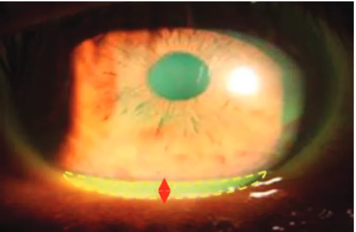
Fig. 1. The arrow showing the conjunctivochalasis. Bulged plica semilunaris is blocking lower punctum.
Fig. 2. The process of conjunctival resection. (A) Preoperative state. (B) The conjunctivochalasis of plical semilunaris was marked by pen. (C) Marked area was resected and followed by electrocau- terization. (D) Postoperative state after 6 months.
A
C
B
D
Fig. 3. The area of the tear meniscus. (A) The arrow shows the longitudinal plane of tear meniscus. (B) The area was measured bymanually drawing a line along the boundary of the tear meniscu- susing RTVue-100 anterior segment optical coherence tomography.
A B
tube intubation was endoscopically performed. Lidocaine and epinephrine were mixed under the inferior nasal con- cha, and a wet cotton swab was placed for 5 minutes to al- low for the nasal mucosa to contract. The infratrochlear and infraorbital nerves were then treated with 2% lidocaine as a local anesthetic. The lacrimal punctum was enlarged, and a lacrimal probe was used to clear the obstructions.
The silicone tube was inserted into the nasal cavity through the superior and inferior nasolacrimal canaliculus while accurately identifying the Hasner valve in the inferior me- atus using an endoscopy. The silicone tube taken out of the nose was knotted with a black silk thread, and the knot was fixed to the mucous membrane of the inferior meatus. Af- ter silicone tube intubation, the plica semilunaris was in- cised with a 2-mm × 4-mm rectangle and resected, fol- lowed by electrocauterization (Fig. 2A-2D). Although we did not completely remove the bulged plica semilunaris, the resection of the plica semilunaris was only performed if there was a mechanically complete obstruction of the lacri- mal punctum due to an anteriorly bulged plica semilunaris.
The obstruction was judged by one experienced operator
using a slit lamp examination.
After the surgery, 0.5% moxifloxacin and 0.5% lotepred- nol etabonate eye lotion was administered to the eyes three times a day. The silicone tubes were maintained for at least 6 months and were removed when the lacrimation resolved and perfusion functioned well in the saline perfusion test.
Three parameters were measured at 1, 3, and 6 months af- ter the surgery: the area of the tear meniscus using RT- Vue-100 anterior segment optical coherence tomography (Optovue, Fremont, CA, USA), the height of the tear me- niscus using a slit lamp microscope, and the subjective sat- isfaction of patients according to the presence or absence of symptoms. The results were analyzed and compared be- tween the patient group that underwent silicone tube intu- bation alone and the patient group that underwent both sili- cone tube intubation and conjunctival resection. The surgery was considered successful when the patients’ expe- rienced a resolution of symptoms and a reduction of the area and height of the tear meniscus. RTVue-100 anterior segment optical coherence tomography was performed 3 seconds after natural flickering after turning off the light.
The area of the tear meniscus was measured by manually drawing a line along the boundary of the tear meniscus us- ing the embedded program (Fig. 3A, 3B). The height of the tear meniscus was measured using the caliper of the slit lamp microscope (Fig. 4). The patients’ subjective percep- tion of symptoms and results of postoperative syringing tests was classified as ‘good,’ ‘normal,’ and ‘fail.’ The re- sults were defined as ‘good’ if the symptoms were elimi- nated after surgery and passed the syringing test; ‘normal’
if the symptoms somewhat remained, but improved and passed the syringing test; and ‘fail’ if the symptoms were not improved and did not pass the syringing test. Clinical success was defined as ‘good’ or ‘normal.’
Statistical analysis was carried out using SPSS ver. 12.0 (SPSS Inc., Chicago, IL, USA). The indices in both groups Fig. 4. The height of the tear meniscus was measured using the
caliper of the slit lamp microscope. The yellow dot line and arrow- head show the area and heights of tear meniscus.
Table 1. Baseline characteristics of the patients
Silicone tube intubation only Combined conjunctival resection p-value
Patients (eye) 29 (47) 33 (49)
Sex (male : female) 12 : 17 14 : 19 0.591
Mean age (yr) 59.8 ± 7.18 64.3 ± 9.92 0.051
Initial area of tear meniscus (mm²) 0.052 ± 0.046 0.059 ± 0.058 0.349
Initial height of tear meniscus (mm) 0.71± 0.75 0.76 ± 0.69 0.243
Values are presented as number or mean ± standard deviation.
before and at 1, 3, and 6 months after the surgery were ana- lyzed and compared through the Student t-test and chi- square test. A p-value <0.05 was considered to indicate sta- tistical significance.
Results
There was no significant difference in gender or age be- tween the 29 patients (47 eyes) who underwent silicone tube intubation and the 33 patients (49 eyes) who under- went both conjunctival resection and silicone tube intuba- tion. The initial area of the tear meniscus before surgery was 0.052 ± 0.046 mm² in the group that underwent only silicone tube intubation and 0.059 ± 0.058 mm² in the group that underwent both intubation and conjunctival resection.
The initial height of the tear meniscus before surgery in the groups was 0.71 ± 0.75 and 0.76 ± 0.69 mm, respectively.
There was no statistically significant difference in the area and height of the tear meniscus between the two groups before surgery (Table 1).
The area of the tear meniscus decreased at 1, 3, and 6 months after the surgery in both groups. There was no dif- ference between the two groups at one month after the sur- gery. However, at 3 and 6 months after the surgery, the group that underwent both silicone tube intubation and conjuncti- val resection exhibited a statistically significant decrease in
the area of the tear meniscus compared to the group that un- derwent only silicone tube intubation (Table 2 and Fig. 5A, 5B). The height of the tear meniscus also decreased in both groups after the surgery. There was no difference in the height of the tear meniscus between the two groups at 1 and 3 months after the surgery. However, at 6 months, the group that also underwent conjunctival resection exhibited a statis- tically significant decrease in this regard (Table 3 and Fig.
6A, 6B). There was no significant difference in the subjective satisfaction of patients (success of surgery which was rated as ‘good’ or ‘normal’) between the two groups at one month after the surgery. However, the satisfaction was higher in the group that underwent both silicone tube intubation and con- junctival resection at 3 and 6 months after the surgery (Table 4). Based on these results, the success rate of the surgery was 68.9% in the group that underwent only silicone tube intuba- tion and 78.7% in the group that underwent both silicone tube intubation and conjunctival resection. Postoperatively, wound dehiscence occurred in one patient at the area of the resected conjunctiva that healed naturally. In one patient, a conjunctival granuloma formed, which was resected.
Discussion
Although the success rate of silicone tube intubation has been reported to be lower than that of a dacryocystorhinos- tomy, silicone tube intubation protects the normal anatomi- cal structure of the nasolacrimal duct. It has been widely used due to its technical simplicity, short surgery time, low pain, low bleeding risk, and fast postoperative recovery [2].
The silicone tube serves as a stent that reduces lacrimal outflow resistance and enlarges the diameter of the soft tis- sue of the nasolacrimal duct to increase lacrimal outflow.
The mechanical obstruction of the lower punctum because of the relaxed conjunctiva from conjunctivochalasis, par- ticularly the plica semilunaris, may reduce the effective- Fig. 5. The RTVue-100 anterior segment optical coherence images
show that the area of tear meniscus was de creased after surgery.
(A) Preoperative state and (B) postoperative state.
A B
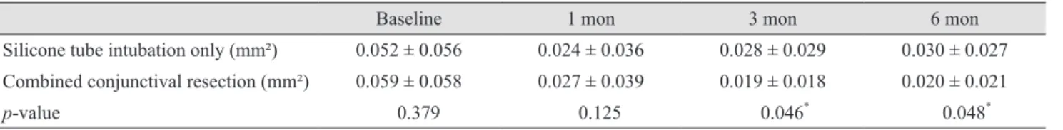
Table 2. Comparison of the area of the tear meniscus between two groups
Baseline 1 mon 3 mon 6 mon
Silicone tube intubation only (mm²) 0.052 ± 0.056 0.024 ± 0.036 0.028 ± 0.029 0.030 ± 0.027 Combined conjunctival resection (mm²) 0.059 ± 0.058 0.027 ± 0.039 0.019 ± 0.018 0.020 ± 0.021
p-value 0.379 0.125 0.046* 0.048*
Values are presented as mean ± standard deviation.
*Statistical significance is p < 0.05 for the Student t-test.
ness of silicone tube intubation and may interrupt lacrimal outflow. The plica semilunaris is a small wrinkle located in the bulbar conjunctiva of the inner canthus. It is retained as the nictitating membrane to protect the eyes in both birds and fishes and is referred to as the third eyelid. However, it is atrophied in primates, and consequently, the conjunctiva is not directly attached to the eyeball, thereby enabling greater rotation of the eyeball. It also forms a lacrimal lake during movement of the eyeball to prevent lacrimal out- flow [16]. In this study, patients who underwent silicone tube intubation had a history conjunctivochalasis and an enlarged or elongated plica semilunaris. In this study, we identified that the plica semilunaris was more elongated than the bulbar conjunctiva, but the cause has yet to be elu- cidated. Therefore, further research is needed to clarify the cause, including histopathologic findings. Previous studies on conjunctivochalasis have reported the following causes:
elastic fiber degeneration associated with aging [17], chron-
ic inflammation [18-20], lymphangiectasia according to a disorder of lymphatic vessel leakage, and a reduction of goblet cell count and collagen fiber density [21-24]. A com- parison of the relaxed tissues in the plica semilunaris and bulbar conjunctiva in other areas may reveal the reason be- hind this finding.
Many previous studies have reported that anterior seg- ment optical coherence tomography has high accuracy and good reproducibility for tear meniscus measurements [25,26]. Based on these reports, we used anterior segment optical coherence tomography to measure the area of the tear meniscus. There was no significant difference in the area or height of the tear meniscus between the two groups immediately after the surgery. However, 3 months after the surgery, the group that underwent both silicone tube intu- bation and conjunctival resection exhibited a significant de- crease in the tear compared to the group that underwent only silicone tube intubation. Immediately after the surgery, there may not have been a significant difference in the area and height of the tear meniscus between the two groups due to the inflammation and edema of the resected conjunctiva.
However, over time, the enlarged and elongated conjunctiva was removed and the wound healed, thereby enabling smoother operation of the lacrimal outflow system through the lower punctum. Consequently, more effective tear per- fusion occurred in the group that underwent both silicone tube intubation and conjunctival resection. The patients’
symptoms resolved as the tear perfusion began to function Fig. 6. The height of the tear meniscus, as shown through the yel-
low dot line, using the caliper of the slit lamp microscope was de- creased after surgery. (A) Preoperative state and (B) postoperative state.
A B
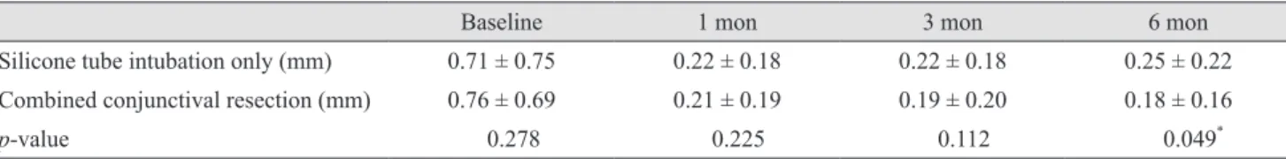
Table 3. Comparison of the height of the tear meniscus between the two groups
Baseline 1 mon 3 mon 6 mon
Silicone tube intubation only (mm) 0.71 ± 0.75 0.22 ± 0.18 0.22 ± 0.18 0.25 ± 0.22 Combined conjunctival resection (mm) 0.76 ± 0.69 0.21 ± 0.19 0.19 ± 0.20 0.18 ± 0.16
p-value 0.278 0.225 0.112 0.049*
Values are presented as mean ± standard deviation.
*Statistical significance is p < 0.05 for the Student t-test.
Table 4. The number of patients with subjective satisfaction between the two groups after surgery
1 mon 3 mon 6 mon
Success Fail Success Fail Success Fail
Silicone tube intubation only 24 (82.7) 5 (17.3) 20 (68.9) 9 (31.1) 20 (68.9) 9 (31.1) Combined conjunctival resection 25 (75.7) 8 (24.3) 25 (75.7) 8 (24.3) 26 (78.7) 7 (21.3)
p-value 0.062 0.049 0.042*
Values are presented as number (%).
*Statistical significance is p < 0.05 for the chi-square test.
more smoothly. Therefore, the symptoms seemed to be cor- related with the area and height of the tear meniscus.
Limitations of this study were that the degree of the re- laxed conjunctiva was not quantified and that the conjuncti- va was resected under the subjective judgment of the surgeon.
Kim et al. [27] reported that the degree of conjunctivochala- sis could be objectively assessed using an anterior segment optical coherence tomography. This limitation may be im- proved by objectively quantifying the condition of the re- laxed conjunctiva through anterior segment optical coher- ence tomography before silicone tube intubation. Moreover, although we included a relatively large number of samples (96 eyes), long-term follow-up for over 6 months was not performed. Therefore, re-evaluations through longer fol- low-ups may be necessary.
The low success rate of surgery reported herein may have resulted from the high standards of what was consid- ered a successful surgery, i.e., one that satisfies all three factors (improvement in the area and height of the tear me- niscus and symptoms). However, in terms of the complica- tions of conjunctival resection, only two of the 96 eyes ex- hibited wound dehiscence (n = 1) and a conjunctival granuloma (n = 1). Therefore, the surgery is considered a safe procedure with low complication rates.
In conclusion, relaxed plica semilunaris resection seems to increase the success rate of silicone tube intubation through the reduction of the area and height of the tear me- niscus, similar to other factors in other studies for increas- ing the success rate of silicone tube intubation. Therefore, if silicone tube intubation is planned, the extent of conjunc- tiva relaxation should be determined. If the bulged plica semilunaris is mechanically blocking the punctum, a com- bination with conjunctival resection is expected to increase the success rate of the surgery.
Conflict of Interest
No potential conflict of interest relevant to this article was reported.
Acknowledgements
This article was supported by the research fund of the Dong-A University.
References
1. Oum JS, Park JW, Choi YK, et al. Result of partial naso- lacrimal duct obstruction after silicone tube intubation. J Korean Ophthalmol Soc 2004;45:1777-82.
2. Keith CG. Intubation of the lacrimal passages. Am J Oph- thalmol 1968;65:70-4.
3. Beigi B, O'Keefe M. Results of Crawford tube intubation in children. Acta Ophthalmol (Copenh) 1993;71:405-7.
4. Jung JJ, Jang SY, Jang JW, In JH. Comparison results of silicone tube intubation according to syringing and dacryo- cystography. J Korean Ophthalmol Soc 2014;55:1584-8.
5. Park JJ, Shin DS, Hong SP, Lee KW. Effects of double sil- icone tube intubation for nasolacrimal duct obstruction in adults. J Korean Ophthalmol Soc 2005;46:1951-6.
6. Lee DH, Choi HY, Ahn JH. Comparison of results and com- plications between 0.64 mm and 0.94 mm silicone tube intu- bation in adults. J Korean Ophthalmol Soc 2016;57:1193-8.
7. Lee KS, Byun YJ. Dacryocystorhinostomy with intraopera- tive mitomycin C. J Korean Ophthalmol Soc 1998;39:1909-14.
8. Kim YT, Chung WS. The effect of mitomycin C in endo- nasal dacryocystorhinostomy. J Korean Ophthalmol Soc 2002;43:728-32.
9. Lee JM, Lee YJ, Kim JH. The effect of mitomycin C con- centration in endonasal dacryocystorhinostomy. J Korean Ophthalmol Soc 2004;45:1609-1614.
10. Song BY, Kim JD, Kim S. Silicone intubation and postop- erative mitomycin application for partial nasolacrimal duct obstruction in adults. J Korean Ophthalmol Soc 2005;46:16-21.
11. Kim DS, Lee YJ. Efficacy of silicone nasolacrimal intuba- tion with mitomycin C treatmentfor treatment of incomplete nasolacrimal duct obstruction. J Korean Ophthalmol Soc 2006;47:181-5.
12. Lim CS, Martin F, Beckenham T, Cumming RG. Nasolac- rimal duct obstruction in children: outcome of intubation. J AAPOS 2004;8:466-72.
13. Kaufman LM, Guay-Bhatia LA. Monocanalicular intubation with Monoka tubes for the treatment of congenital nasolacri- mal duct obstruction. Ophthalmology 1998;105:336-41.
14. Engel JM, Hichie-Schmidt C, Khammar A, et al. Monocana- licular silastic intubation for the initial correction of congen- ital nasolacrimal duct obstruction. J AAPOS 2007;11:183-6.
15. Oh SH, Park JY, Yim HB, Lee NY. Treatment of epiphora in patients with conjunctivochalasis using conjunctival fixa- tion to the sclera. J Korean Ophthalmol Soc 2012;53:1063-7.
16. Dartt DA. The conjunctiva: structure and function. In:
Duane TD, Tasman W, Jaeger EA. Duane's foundations of clinical ophthalmology. Philadelphia: Lippincott, Williams
& Wilkins; 2012. p. 11.
17. Watanabe A, Yokoi N, Kinoshita S, et al. Clinicopathologic study of conjunctivochalasis. Cornea 2004;23:294-8.
18. Francis IC, Chan DG, Kim P, et al. Case-controlled clinical and histopathological study of conjunctivochalasis. Br J Ophthalmol 2005;89:302-5.
19. Meller D, Li DQ, Tseng SC. Regulation of collagenase, strome- lysin, and gelatinase B in human conjunctival and conjuncti- vochalasis fibroblasts by interleukin-1beta and tumor necrosis factor-alpha. Invest Ophthalmol Vis Sci 2000;41:2922-9.
20. Erdogan-Poyraz C, Mocan MC, Bozkurt B, et al. Elevated tear interleukin-6 and interleukin-8 levels in patients with conjunctivochalasis. Cornea 2009;28:189-93.
21. Ellis S. Structure and function of the lymphatic system: an overview. Br J Community Nurs 2006;11:S4-6.
22. Skandalakis JE, Skandalakis LJ, Skandalakis PN. Anatomy
of the lymphatics. Surg Oncol Clin N Am 2007;16:1-16.
23. Kim JT, Kim JH, Kim JC. Visualization of subconjunctival lymphatics and its significance. J Korean Ophthalmol Soc 2008;49:1215-9.
24. Bae JB, Park WC. Histopathologic characteristics of con- junctivochalasis. J Korean Ophthalmol Soc 2013;54:1165-74.
25. Wang J, Aquavella J, Palakuru J, Chung S. Repeated mea- surements of dynamic tear distribution on the ocular sur- face after instillation of artificial tears. Invest Ophthalmol Vis Sci 2006;47:3325-9.
26. Zhou S, Li Y, Lu AT, et al. Reproducibility of tear meniscus measurement by Fourier-domain optical coherence to- mography: a pilot study. Ophthalmic Surg Lasers Imaging 2009;40:442-7.
27. Kim JS, Bae JB, Seo JW, Park WC. Changes in area of con- junctiva and tear meniscus measured using anterior segment optical coherence tomography after conjunctivochalasis surgery. J Korean Ophthalmol Soc 2015;56:509-14.