https://doi.org/10.20307/nps.2017.23.2.119
119
Neolignan Derivatives from the Flower of Magnolia biondii Pamp.
and their Effects on IL-2 expression in T-cells
Thi Tuyet Mai Nguyen1, Thi Thu Nguyen1, Hyun-Su Lee2,3, Chang-Duk Jun2, Byung Sun Min3, and Jeong Ah Kim1,*
1College of Pharmacy, Research Institute of Pharmaceutical Sciences, Kyungpook National University, Daegu 41566, Republic of Korea
2School of Life Sciences, Immune Synapse Research Center and Cell Dynamics Research Center, Gwangju Institute of Science and Technology, Gwangju 61005, Republic of Korea
3College of Pharmacy, Drug Research and Development Center, Catholic University of Daegu, Gyeongbuk 38430, Republic of Korea
Abstract − The isolation of the MeOH extract from the flower bud of Magnolia biondii Pamp. using various column chromatographies and HPLC led to eleven neoglignan derivatives (1 - 11). Their structures were mainly determined by 1D and 2D NMR spectral data analysis and physiological methods. The isolated compounds (1 - 11) were tested for anti-allergic effects using IL-2 inhibitory assay in Jurkat T cells.
Keywords − Magnolia biondii Pamp.; Magnoliaceae; Neolingnan; Anti-allergy; IL-2
Introduction
Magnoliae Flos is a flower bud of Magnolia biondii Pamp., commonly known as Sin-yi in Korea. It belongs to the family of Magnoliaceae and is widely distributed in Japan, China, and Korea. The flower buds of M. biondii traditionally were used for symptomatic management of allergic rhinitis, sinusitis, and headache in Asia countries1. Extensive studies have been reported that several secondary metabolites such as lignans, neolignans, sesqui- terpends, and terpenoids from this plant have various biological activities2-4. In particular, several types of neolignans have been studied to have antiperoxidative, antimicrobial, cytotoxic, and inflammatory activities5-7.
Mast cell-derived interleukin-2 (IL-2) is an essential cytokine for the T-cell immunity, including differentiation and effector functions. This cytokine is also implicated in the generation and maintenance of Treg cells which antagonize immune responses8. Recent study showed evidence that IL-2 of mast cell contributes to the suppression of chronic allergic dermatitis by a regulatory mechanism. The secretion of IL-2 by mast cell enhances the proportion of Treg cells at the site of inflammation
and decreases the fraction of activated T-cells at the inflammatory site9.
In our ongoing to find new anti-allergic agents from natural sources, the flower buds of M. biondii have been phytochemically investigated to isolate eleven neolignans (1 - 11). All the isolates (1 - 11) were evaluated for their anti-allergic activity by inhibition of IL-2 in Jurkat T cells.
Experimental
General experimental procedures – The optical rotations were measured using a JASCO DIP-1000 spectropolari- meter (JASCO, Tokyo, Japan). The NMR spectra were recorded using a Bruker Advance Digital 500 MHz NMR spectrometer using TMS as the internal standard. Silica gel 60 (Merck, 60 - 200µm) and reversed phase (RP)-C18
silica gel (Merck, 40 - 63µm) were used for column chromatography (CC). TLC was performed using Merck precoated silica gel F254 plates and RP-18 F254s plates.
HPLC was performed with a HPLC Waters system (Waters, Middleton, USA): 1525 Binary pump; Water 2998 photo- diode array detector; YMC Pak ODS column (20 × 250 mm, 5µm); tR in min. HPLC solvents were purchased from Burdick & Jackson (USA).
Plant materials – The flower buds of M. biondii were purchased from Kyungdong traditional market in Seoul (February 2015) and identified by Professor Byung Sun Min at Catholic university of Daegu. It has been deposited
*Author for correspondence
Jeong Ah Kim, College of Pharmacy, Research Institute of Pharma- ceutical Sciences, Kyungpook National University, Daegu 41566, Republic of Korea
Tel: +82-53-950-8574; E-mail: [email protected]
at pharmacognosy laboratory (voucher no. 17AMG40) in the college of pharmacy, Kyungpook national university, Korea.
Extraction and isolation – The dried flower buds of M. biondii (9.0 kg) were extracted three times refluxing with MeOH (3 h × 4 L) at 60oC. The filtered MeOH- soluble part was concentrated in vacuo to obtain brown residue (1.2 kg) which was suspended in water and partitioned with organic solvents, then afford extracts of chloroform (591.7 g), ethylacetate (EtOAc, 28.2 g), and aqueous extract (580.1 g), successfully. Chloroform-soluble extract was chromatographed on a silica gel vacuum liquid chromatography (VLC) and eluting with a gradient mixture of methylene chloride (MC):hexane (3:7 → 1:0) and EtOAc:MC (1:99 → 1:19) to afford nine fractions (MB-1 to MB-9). Fraction MB4 (78.8 g) was chromato- graphed on a silica gel VLC and eluting with gradient mixture of 5% to 20% acetone in hexanes to yield six fractions (MB4-1 to MB4-6). Fraction MB4-4 (15.4 g) was fractionated on a silica gel column with a stepwise gradient of 55% to 60% acetone in hexanes to afford six fractions (MB44-1 to MB44-6). Fraction MB44-3 (751.1 mg) was purified using preparative HPLC (6 mL/min, 60 min) with gradient mixture of 60% to 90% MeOH in water to afford compounds 10 (2.0 mg, tR= 38.4 min) and 8 (3.8 mg, tR= 45.1 min). Fraction MB4-5 (36.0 g) was chromatographed on silica gel column with gradient of 15% to 55% EtOAc in hexanes to afford seven fractions (MB45-1 to MB45-7). Fraction MB45-6 (2.3 g) was sub- jected to reversed phase gel column chromatography and eluting with gradient mixture of 45% to 55% acetone in water to afford six fractions (MB456-1 to MB456-6).
Fraction MB456-5 (20.6 mg) was purified using preparative HPLC (6 mL/min, 60 min) with gradient elution of 60%
to 90% MeOH in water to afford compounds 5 (1.0 mg, tR= 33.5 min), 6 (7.8 mg, tR= 35.1 min), and 7 (2.1 mg, tR= 36.8 min). Fraction MB5 (42.0 g) was chromatographed over silica gel column with a gradient mixture of 5% to 20% acetone in hexanes by using VLC to afford seven fractions (MB5-1 to MB5-7). Fraction MB5-4 (3.2 g) was further subjected to reverse phase gel column chromato- graphy with gradient elution of 40% to 50% MeOH in water to afford seven fractions (MB54-1 to MB54-7).
Fraction MB54-3 (1.3 g) was separated by RP-C18 with gradient elution of 50% to 70% MeOH in water to afford nine fractions (MB543-1 to MB543-9). Fraction MB543- 4 (10.0 mg) was purified by Waters HPLC (6 ml/min, 60 min) with gradient of 85% to 100% MeOH in water to obtain compound 3 (3.3 mg, tR= 21.3 min). Fraction MB543-5 (30.0 mg) was purified by using preparative
Waters HPLC (6 ml/min, 60 min) with an isocratic elution of 58% acetonitrile(ACN) in water to afford compound 4 (6.9 mg, tR= 49.8 min). Compounds 1 (5.7 mg; tR= 35.5 min) and 2 (2.7 mg, tR= 43.5 min) were isolated from fraction MB54-7 (100.1 mg) by using Waters HPLC (6 ml/min, 80 min) with gradient mixture of 65% to 70%
ACN in water. Fraction MB9 (176.5 g) was fractionated on a silica gel VLC and eluting with gradient mixture of 10% to 40% acetone in hexanes to gain six fractions (MB9-1 to MB9-6). Fraction MB9-5 (60.3 g) was chro- matographed on silica gel column and eluting with gradient mixture of 20% to 90% acetone in hexanes to yield nine fractions (MB95-1 to MB95-9). Fraction MB95-5 (8.3 g) was separated on silica gel column using a gradient elution of 1% to 5% MeOH in MC to afford nine fractions (MB955-1 to MB955-9). Fraction MB955-1 (178.6 mg) was isolated by using preparative HPLC (6 mL/min, 60 min) with gradient mixture of 40% to 70% MeOH in water to afford compound 11 (3.5 mg, tR= 49.3 min).
Fraction MB955-6 (2.4 g) was subjected to reversed phase column chromatography and eluting with a stepwise gradient of 40% to 50% MeOH in water to afford compound 9 (613.4 mg).
Fargesone A (1)− Colorless oil; −106.2 (c 0.1, MeOH); 1H and 13C NMR data, see Tables 1 and 3.
Fargesone B (2)− White powder; −19.5 (c 0.1, MeOH); 1H and 13C NMR data, see Tables 1 and 3.
Kadsurin A (3)− Colorless oil; −67.3 (c 0.1, MeOH); 1H and 13C NMR data, see Tables 1 and 3.
Denudatin B (4)− White wax; +55.2 (c 0.1, MeOH); 1H and 13C NMR data, see Tables 1 and 3.
Burchellin (5)− Viscous oil; 1H and 13C NMR data, see Tables 1 and 3.
cis-Burchellin (6) − Viscous oil; −39.0 (c 0.1, MeOH); 1H and 13C NMR data, see Tables 1 and 3.
1'-epi-Burchellin (7) − Viscous oil; 1H and 13C NMR data, see Tables 1 and 3.
Acuminatin (8)− Viscous oil; +10.6 (c 0.1, MeOH); 1H and 13C NMR data, see Tables 2 and 3.
3',4-O-Dimethylcedrusin (9) − Red brown wax;
+1.9 (c 0.1, MeOH); 1H and 13C NMR data, see Tables 2 and 3.
Denudadione A (10)− Viscous oil; 1H and 13C NMR data, see Tables 2 and 3.
Magliflonenone (11)− White powder; −70.5 (c 0.1, MeOH); 1H and 13C NMR data, see Tables 2 and 3.
Cell culture− Jurkat T cells (ATCC TIB-152, Manassas, VA) were grown in RPMI medium (Gibco-BRL, Gaither- sburg, MD) supplemented with 10% fetal bovine serum (FBS), penicillin G (100 units/ml), streptomycin (100µg/
α [ ]D20
α [ ]D20 α [ ]D20
α [ ]D20
α [ ]D20
α [ ]D20
α [ ]D20
α [ ]D20
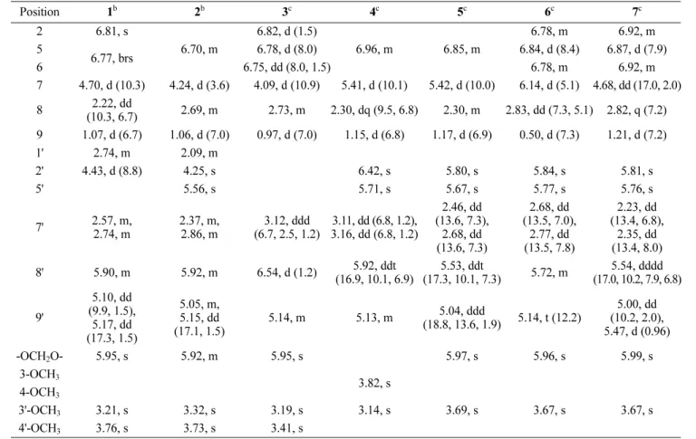
Table 1. 1H NMR spectroscopic data for compounds 1 – 7 (δ values)a
Position 1b 2b 3c 4c 5c 6c 7c
2 6.81, s
6.70, m
6.82, d (1.5)
6.96, m 6.85, m
6.78, m 6.92, m
5 6.77, brs 6.78, d (8.0) 6.84, d (8.4) 6.87, d (7.9)
6 6.75, dd (8.0, 1.5) 6.78, m 6.92, m
7 4.70, d (10.3) 4.24, d (3.6) 4.09, d (10.9) 5.41, d (10.1) 5.42, d (10.0) 6.14, d (5.1) 4.68, dd (17.0, 2.0)
8 2.22, dd
(10.3, 6.7) 2.69, m 2.73, m 2.30, dq (9.5, 6.8) 2.30, m 2.83, dd (7.3, 5.1) 2.82, q (7.2) 9 1.07, d (6.7) 1.06, d (7.0) 0.97, d (7.0) 1.15, d (6.8) 1.17, d (6.9) 0.50, d (7.3) 1.21, d (7.2)
1' 2.74, m 2.09, m
2' 4.43, d (8.8) 4.25, s 6.42, s 5.80, s 5.84, s 5.81, s
5' 5.56, s 5.71, s 5.67, s 5.77, s 5.76, s
7' 2.57, m,
2.74, m
2.37, m, 2.86, m
3.12, ddd (6.7, 2.5, 1.2)
3.11, dd (6.8, 1.2), 3.16, dd (6.8, 1.2)
2.46, dd (13.6, 7.3),
2.68, dd (13.6, 7.3)
2.68, dd (13.5, 7.0),
2.77, dd (13.5, 7.8)
2.23, dd (13.4, 6.8),
2.35, dd (13.4, 8.0)
8' 5.90, m 5.92, m 6.54, d (1.2) 5.92, ddt
(16.9, 10.1, 6.9)
5.53, ddt
(17.3, 10.1, 7.3) 5.72, m 5.54, dddd (17.0, 10.2, 7.9, 6.8) 9'
5.10, dd (9.9, 1.5), 5.17, dd (17.3, 1.5)
5.05, m, 5.15, dd (17.1, 1.5)
5.14, m 5.13, m 5.04, ddd
(18.8, 13.6, 1.9) 5.14, t (12.2)
5.00, dd (10.2, 2.0), 5.47, d (0.96)
-OCH2O- 5.95, s 5.92, m 5.95, s 5.97, s 5.96, s 5.99, s
3-OCH3
3.82, s 4-OCH3
3'-OCH3 3.21, s 3.32, s 3.19, s 3.14, s 3.69, s 3.67, s 3.67, s
4'-OCH3 3.76, s 3.73, s 3.41, s
a δ value recorded in ppm (J in Hz).
b 1H NMR measured at 500 MHz in CDCl3.
c 1H NMR measured at 500 MHz in CD3OD.
Table 2. 1H NMR spectroscopic data for compounds 8 – 11 (δ values)a
Position 8 9 10 11
2 6.98, d (2.0) 6.95, s 6.58, d (2.0) 6.39, s
5 6.84, d (8.2) 6.82, d (8.7) 6.79, d (8.2)
6 6.96, dd (8.2, 2.0) 6.95, s 6.66, dd (8.2, 2.1) 6.39, s
7 5.12, d (9.5) 5.55, d (7.4)
2.46, m 2.57, d (6.1)
8 3.46, m 3.60, dd (12.2, 5.8) 2.07, m
9 1.38, d (6.8) 3.90, d (4.6),
3.96, dd (10.9, 5.8) 1.07, d (6.1) 0.62, d (6.5)
2' 6.79, s 6.67, d (2.2)
3' 3.52, m 5.80, s
6' 6.77, s 6.67, d (2.2) 7.04, s 5.49, s
7' 6.36, dd (15.7, 1.6) 2.65, d (7.9) 3.12, dd (16.4, 8.6) 2.22, d (11.3),
2.39, ddd (11.3, 6.4, 1.4)
8' 6.11, dq (15.7, 6.6) 1.88, m 5.86, ddt (17.6, 10.9, 6.8) 5.05, m
9' 1.87, dd (6.6, 1.6) 3.68, d (6.3) 5.17, d (6.3), 5.20, s 1.76, dd (12.0, 11.9), 2.32, m 3-OCH3
3.89, s 3.86, s
3.86, s 3.85, s
4-OCH3 3.85, s 3.82, s
5-OCH3 3.85, s
3'-OCH3 3.89, s 3.88, s
5'-OCH3 3.64, s 3.68, s
aδ value recorded in ppm (J in Hz) and 1H NMR measured at 500 MHz in CDCl3.
ml), and L-glutamine (2 mM). The cells were cultured at 37oC in a humidified incubator containing 5% CO2 and 95% air.
Reverse transcription PCR and conventional PCR – Jurkat T cells (1× 10⁶) were incubated with indicated concentrations of compounds for 30 min at 37oC. Incubated cells were stimulated with PMA (200 nM) and A23187 (1 µM) for 6 h for PCR. Cells were harvested and total RNAs were isolated with TRIZOL reagent (JBI, Korea).
Reverse transcription of the RNA was performed using RT PreMix (enzynomics, Korea). For conventional PCR, the primers and PCR conditions for each gene were used as following: human IL-2, 5'-CAC GTC TTG CAC TTG TCA C-3' and 5'-CCT TCT TGG GCA TGT AAA ACT- 3'. Human GAPDH, 5'-CGG AGT CAA CGG ATT TGG TCG TAT-3' and 5'-AGC CTT CTC CAT GGT GGT GAA GAC-3'. The amplification profile was composed of denaturation at 94oC for 30 s, annealing at 60oC for 20 s,
and extension at 72oC for 40 s. The 30 cycles were preceded by denaturation at 72oC for 7 min. All experi- ments were performed at least three times unless otherwise indicated.
Cell viability assay− Jurkat T cells (3 × 10⁵) were seeded in a 24 well-plate and incubated with isolates (1 - 11) for 24 h. After incubation, cells (180µl) were added with MTT solution (20µl, 5 mg/ml). After 2 h of incubation on 37oC incubator, cells were centrifuged and supernatants were taken out. 150µl of DMSO were added and incubated for 15 min on RT. After incubation, absorbance was detected in 590 nm wavelength.
Result and Discussion
The MeOH extract of dried flower buds of M. biondii was partitioned into chloroform, EtOAc-, and water- soluble fractions. Chromatographic purification of chloro- Table 3. 13C NMR spectroscopic data for compounds 1 – 11 (δ values)a
Position 1b 2b 3c 4c 5c 6c 7c 8b 9b 10b 11b
1 134.4 134.2 135.5 131.3 133.1 133.2 136.4 132.8 133.9 134.0 139.1
2 106.9 106.7 108.8 111.2 107.8 107.3 106.0 109.7 109.5 110.2 104.8
3 148.0 147.9 149.5 151.3 149.8 149.4 149.7 149.3 149.3 148.6 153.4
4 147.6 147.5 149.1 150.8 149.6 148.8 148.6 146.8 149.1 149.6 137.0
5 108.2 108.1 108.1 112.8 109.1 109.2 109.3 110.0 111.1 111.7 153.4
6 120.5 120.1 122.2 121.2 120.2 120.4 120.3 119.4 118.8 119.5 104.8
7 87.2 86.1 86.8 93.3 92.8 89.3 94.9 93.8 87.9 49.0 45.4
8 51.4 48.5 50.6 51.5 37.1 44.2 46.4 45.8 53.9 45.5 37.9
9 8.9 11.2 9.4 6.9 8.3 12.4 18.8 17.8 64.1 13.8 14.6
1' 52.8 54.1 144.5 143.9 53.2 55.8 55.0 132.4 135.5 140.6 50.4
2' 80.0 81.1 140.2 133.6 111.1 112.3 112.1 109.4 112.6 194.5 180.0
3' 84.7 82.3 83.3 79.3 154.3 153.8 153.7 144.3 144.3 70.0 101.5
4' 172.4 173.4 103.4 177.4 185.7 185.5 185.4 149.3 146.7 202.4 183.1
5' 105.7 103.5 44.2 102.9 102.8 102.6 102.7 133.4 127.9 89.5 153.4
6' 196.4 196.9 196.1 189.2 185.3 185.1 185.3 113.5 116.1 147.3 108.9
7' 31.3 29.2 34.5 34.6 50.9 46.5 46.7 131.1 32.1 32.9 60.9
8' 135.4 136.4 136.4 136.6 132.6 132.2 132.9 123.7 34.7 133.9 81.9
9' 117.9 117.0 117.6 117.5 122.1 120.2 118.4 18.5 62.4 118.3 43.7
-OCH2O- 101.2 101.2 102.5 − 101.9 102.2 102.7 − − − −
3-OCH3 − − − 56.5 − − − 56.1 56.2 56.1 56.2
4-OCH3 − − − 56.5 − − − 56.1 56.2 56.1 60.9
5-OCH3 − − − − − − − − − − 56.2
3'-OCH3 51.5 53.4 47.7 51.6 55.8 56.2 55.8 56.8 56.1 − −
4'-OCH3 56.4 56.1 51.4 − − − − − − − −
5'-OCH3 − − − − − − − − − 54.1 55.3
aδ value recorded in ppm.
b 13C NMR measured at 125 MHz in CDCl3.
c 13C NMR measured at 125 MHz in CD3OD.
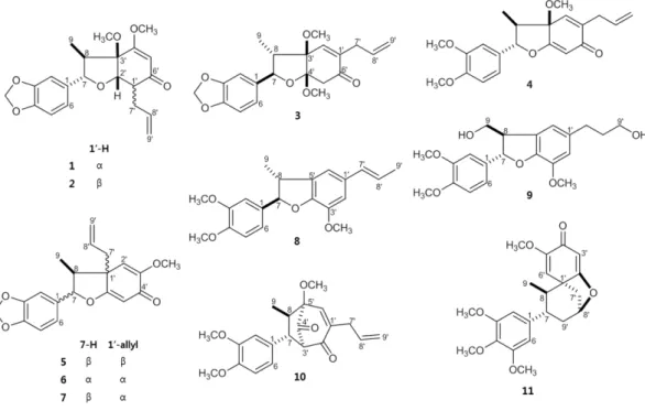
form-soluble fraction led to the isolation and determi- nation of eleven neolignan derivatives as fargesone A (1), fargesone B (2)10, (+)-kadsurin A (3)11, denudatin B (4)12, burchellin (5), cis-burchellin (6)13,14, 1'-epi-burchellin (7)15, (±)-acuminatin (8)16, 3',4-O-dimethylcedrusin (9)17, denudadione A (10)18, and maglifloenone (11)19 by com- paring their physicochemical and spectroscopic data with those reported in the literature (Fig. 1).
The cytotoxic effects of 1 - 11 were evaluated using an MTT assay and all isolated compounds were tested to inhibit of IL-2 production in Jurkat T-cells, which were stimulated with stimulated with PMA (200 nM) and A23187 (1µM). Among tested neolignans (1 - 11), denu-
datin B (4) showed strong inhibitory effect on production of IL-2 (Fig. 2A). The MTT assay indicated that none of the neolignans showed cytotoxicity in activated Jurkat T- cells at concentrations effective for the inhibition of IL-2 production (Fig. 2B).
Denudatin B is an epimer of 5'-OCH3 on kadsurenone which has been demonstrated to block platelet-activating factor (PAF) receptor12. PAF has been implicated as a mediator of inflammation and anaphylaxis20. Furthermore, denudatin B reported to have inhibitory effects on NO production through the decreased expression of the iNOS gene21 and free radical scavenging activity22. Denudatin B from M. biondii which were traditionally used for allergy Fig. 1. Chemical structures of neolignans 1 - 11 isolated from the flower of Magnolia biondii Pamp.
Fig. 2. Inhibition of IL-2 expression and cell viability by neolignans 1-11 isolated from M. biondii.
A) Jurkat T cells (1× 10⁶) were treated with 50µM concentrations of 1 - 11 for 30 min and stimulated with PMA (100 nM)/A23187 (1µM) for 6 h. After incubation, cells were harvested and total RNA was isolated from harvested cells. Human IL-2 mRNA levels were detected by conventional PCR. B) Jurkat T cells (3 × 10⁵) were seeded in a 24 well-plate and incubated with 50µM concentrations of 1 - 11 for 24 h. After incubation, cell viability was examined by MTT assay. Data was shown as percentages compared to cell viability treated with Mock control.
disease were effective on inhibition of IL-2. The secretion of IL-2 triggers off allergic reaction9. The allergy has generally and/or occasionally attended by consequential inflammatory reaction.
Taken together, these findings support denudatin B to be used as a useful natural candidate for inflammation and allergy related activities on the further mechanism. Thus, our study suggests that denudatin B isolated from M.
biondii may probably be used as an inhibitor of inflam- matory and allergic diseases.
Acknowledgments
This research was supported by the National Research Foundation of Korea (NRF), funded by the Ministry of Science, ICT, and Future Planning (NRF-2015M3A9 A5031271).
References
(1) Park, C. S.; Kim, T. B.; Lee, J. Y.; Park, J. Y.; Lee, Y. C.; Jeong, S.
S.; Lee, Y. D.; Cho, Y. S.; Moon, H. B. Korean J. Intern. Med. 2012, 27, 84-90.
(2) Song, Q.; Fischer, N. H. Rev. Soc. Quím. Mex. 1999, 43, 211-218.
(3) Marin, G. H.; Mansilla, E.; Ciocchini, S.; Quiroga, N.; Cardozo, N.;
Weiss, M.; Gustavo, H. M.; Trebucq, H. Annalen der Chemischen Forschung. 2013, 1, 50-55.
(4) Lee, S.; Chappell, J. Plant Physiol. 2008, 147, 1017-1033.
(5) Haraguchi, H.; Ishikawa, H.; Shirataki, N.; Fukuda, A. J. Pharm.
Pharmacol. 1997, 49, 209-212.
(6) Syu, W. J.; Shen, C. C.; Lu, J. J.; Lee, G. H.; Sun, C. M. Chem.
Biodivers. 2004, 1, 530-537.
(7) Kuo, W. L.; Chung, C. Y.; Hwang, T. L.; Chen, J. J. Phytochemistry
2013, 85, 153-160.
(8) Akber, U.; Na, B. R.; Ko, Y. S.; Lee, H. S.; Kim, H. R.; Kwon, M.
S.; Park, Z. Y.; Choi, E. J.; Han, W. C.; Lee, S. H.; Oh, H. M.; Jun, C. D.
Int. Immunopharmacol. 2015, 25, 130-140.
(9) Hershko, A. Y.; Suzuki, R.; Charles, N.; Alvarez-Errico, D.; Sargent, J. L.; Laurence, A.; Rivera, J. Immunity. 2011, 35, 562-571.
(10) Chen, C. C.; Huang, Y. L.; Chen, Y. P.; Hsu, H. Y.; Kuo, Y. H.
Chem. Pharm. Bull. 1988, 36, 1791-1795.
(11) Tyagi, O. D.; Jensen, S.; Boll, P. M.; Sharma, N. K.; Bisht, K. S.;
Parmar, V. S. Phytochemistry 1993, 32, 445-448.
(12) Ponpipom, M. M.; Bugianesi, R. L.; Brooker, D. R.; Yue, B. Z.;
Hwang, S. B.; Shen, T. Y. J. Med. Chem. 1987, 30, 136-142.
(13) Wenkert, E.; Gottlieb, H. E.; Gottlieb, O. R.; Pereira, M. O. S.;
Formiga, M. D. Phytochemistry 1976, 15, 1547-1551.
(14) Jensen, S.; Olsen, C. E.; Tyagi, O. D.; Boll, P. M.; Hussaini, F. A.;
Gupta, S.; Bisht, K. S.; Parmar, V. S. Phytochemistry 1994, 36, 789-792.
(15) Tyagi, O. D.; Wengel, J.; Prasad, A. K.; Boll, P. M.; Olsen, C. E.;
Pati, H. N.; Bisht, K. S.; Parmar, V. S. Acta Chem. Scand. 1994, 48, 1007- 1011.
(16) El-Feraly, F. S.; Cheatham, S. F.; Hufford, C. D.; Li, W. S.
Phytochemistry 1982, 21, 1133-1135.
(17) Pieters, L.; de Bruyne, T.; Claeys, M.; Vlietinck, A.; Calomme, M.;
vanden Berghe, D. J. Nat. Prod. 1993, 56, 899-906.
(18) Kuroyanagi, M.; Yoshida, K.; Yamamoto, A.; Miwa, M. Chem.
Pharm. Bull. 2000, 48, 832-837.
(19) Du, J.; Wang, M. L.; Chen, R. Y.; Yu, D. Q. J. Asian Nat. Prod.
Res. 2001, 3, 313-319.
(20) Kasperska-Zajac, A.; Brzoza, Z.; Rogala, B. Recent Pat. Inflamm.
Allergy Drug Discov. 2008, 2, 72-76.
(21) Noshita, T.; Funayama, S.; Hirakawa, T.; Kidachi, Y.; Ryoyama, K.
Biosci. Biotechnol. Biochem. 2008, 72, 2775-2778.
(22) Reddy, S. D.; Siva, B.; Poornima, B.; Kumar, D. A.; Tiwari, A. K.;
Ramesh, U.; Babu, K. S. Pharmacogn. Mag. 2015, 11, 235-241.
Received January 6, 2017 Revised April 3, 2017 Accepted April 3, 2017