Anti-atherosclerotic effects of perilla oil in rabbits fed a high-cholesterol diet Yeseul Cha1
9
0
0
전체 글
(2) 172. Yeseul Cha et al.. natural products with minimal adverse-effects to control blood cholesterol level and to improve blood flow are required. Since the consumption of high-fat diets is the major cause of disturbances in blood flow, selection of fats and oils might be very important. It is well known that unsaturated fatty acids (UFA) properly regulate blood lipid profiles, and thereby improved coronary heart disease [14-17]. Fish oil ω-3 polyunsaturated fatty acids (PUFA) prevented vasoconstriction [18], and suppressed vascular inflammatory response by decreasing production of reactive oxygen species (ROS) [19,20]. Notably, αlinolenic acid (ALA), a well-known ω-3 PUFA rich in perilla oil improved insulin sensitivity and hyperlipidemia, and prevented coronary heart disease [21,22]. Especially, in recent studies, we demonstrated that perilla oil possessing a low linoleic acid/α-linolenic acid (ω-6/ω-3) ratio not only inhibited platelet aggregation and improved blood flow [23], but also delayed and attenuated brain hemorrhage in stroke-prone spontaneously hypertensive rats (SHR-SP), thereby extending their lifespan [24]. Since PUFA affects both platelets and endothelial cells that play a crucial role in the regulation of thrombosis and haemostasis [25], we investigated the effects of perilla oil on the high-cholesterol diet (HCD)-induced hypercholesterolemia and atherosclerosis.. Materials and Methods Materials. Perilla oil was obtained from Anydoctor Healthcare Co., Ltd. (Cheonan, Korea). Perilla oil was extracted under a cold-pressed method at 30-48oC, and analyzed with Varian 3800 gas chromatograph (Varian Inc., Walnut Creek, CA, USA) equipped with a Supelcowax 10 fused-silica capillary column (Supelco, Bellefonte, PA, USA). From the fatty acid analysis, it was found that perilla oil contains 72.12% PUFA, 19.1% monounsaturated fatty acids (MUFA), and 8.49% saturated fatty acids (SFA). Especially, among PUFA, 57.47% was ω-3 αlinolenic acid [ALA, 18:2(n-3)] [23,24]. Animals. Seven-month-old male New Zealand white rabbits (body weight 2.5 kg) were procured from Samtaco Co. (Osan, Korea), and subjected to the experiment after 2week acclimation to the laboratory environment. The animals were housed in each cage with free access to Lab Anim Res | September, 2016 | Vol. 32, No. 3. feed and water under constant environmental conditions (23±2oC temperature; 45-65% relative humidity; 12hour light-dark cycle; 150-300 lux brightness). All the animal experiments were conducted according to the Standard Operation Procedures, and approved by the Institutional Animal Care and Use Committee of Chungbuk National University, Korea. Induction of hypercholesterolemia and treatment. Acute dietary hypercholesterolemia was induced by feeding rabbits with a powdered HCD containing 0.5% cholesterol and 1% corn oil for 2 weeks, followed by 0.5% cholesterol for additional 10 weeks during treatment period [26-28]. After 2-week induction period, the animals were grouped (n=6/group) according to their blood cholesterol levels to adjust to similar mean values. To assess therapeutic efficacy of perilla oil against hypercholesterolemia and atherosclerosis, the rabbits were fed the HCD containing 0.1 or 0.3% perilla oil during the 10-week period. Blood biochemistry. Body weights were recorded every week from immediately before starting the experiment. After 16hour fasting at the end of 2-week hypercholesterolemia induction period and every 2 week during 10-week treatment period, blood sample was collected from auricular artery, and lipid profiles and parameters of hepatic and renal functions were measured in sera using a blood chemistry analyzer (Hitachi-747; Hitachi Korea, Seoul, Korea). The parameters include TC, LDL, HDL, triglycerides (TG), alanine transaminase (ALT), aspartate transaminase (AST), lactate dehydrogenase (LDH), alkaline phosphatase (ALP), total proteins (TP), glucose, blood urea nitrogen (BUN), and creatinine. Measurement of atheroma. The aortic arch (10 cm) was dissected from the aortic valve of the heart and washed with saline. The sample was dissected in 5 cm long from the orifice of carotid artery, and opened longitudinally. After removing fats and tissues adhering to the adventitia, the aorta was dehydrated with 100% propylene glycol for 10 min, and stained with 0.7% Sudan IV (in propylene glycol) for 10 min [26-28]. Then, it was hydrated with 85% propylene glycol for 5 min, washed with distilled water, and photographed and analyzed with Digital Image Analyzer (Image Inside; Focus, Seoul, Korea) for red atheromatous.
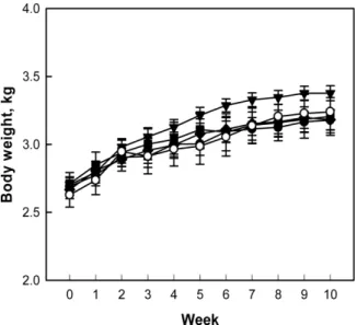
(3) Perilla oil improves atherosclerosis. 173. plaques. The extent of lipid accumulation (atherosclerosis index; AI, %) was calculated as the percent Sudanpositive area to the total area of the aortic wall. Microscopic examination. After weighing, liver and kidney tissues were frozen at −70oC, and cryosections (10 μm in thickness) were mounted on gelatin-coated slides. The sections were washed with phosphate-buffered saline (PBS, pH6.5) followed by propylene glycol for 2 min, and stained with 0.5% Oil red O for 1 hour. After washing with 85% propylene glycol, the slides were counterstained with Harris’ hematoxylin, washed again with distilled water, and observed under a light microscope. Measurement of lipid peroxidation. The liver and kidney tissues were dissected on an ice block. The tissues were homogenized in 19 volumes of 10 mM PBS (pH7.4) to make a 5% homogenate at 4oC. Into the tissue homogenate (250 μL), 250 μL sodium dodecyl sulphate (SDS, 8.1% solution) and 500 μL 20% acetic acid (adjusted to pH3.5) were added. After adding 250 μL 2-thiobarbituric acid (TBA, 0.75% solution), the mixture was boiled in a glass tube capped with aluminum foil for 30 min [29]. Samples were cooled on ice, centrifuged at 13,000 g for 10 min, and absorbance of the supernatant was read at 532 nm for the quantification of malondialdehyde (MDA). Statistical analysis. The results are presented as means±standard error. The significance of differences of all results was analyzed by one-way analysis of variance followed by the Dunnett’s multiple-range test correction, using SPSS version 12.0 (SPSS Inc., Chicago, IL, USA). Statistical significance was set a priority at P<0.05.. Results During 10-week treatment period, following 2-week hypercholesterolemia-induction period, there were no abnormal symptoms in control and treated rabbits. The rabbits exhibited a gradual increase in their body weights, although the body weight gain of rabbits fed 0.1% perilla oil was slightly higher than that of control animals without statistical significance (P>0.05) (Figure 1). After 2-week feeding of 0.5% cholesterol and 1% corn oil for hypercholesterolemia induction, blood TC was. Figure 1. Change in body weights of hypercholesterolemic rabbits fed a high-cholesterol diet (HCD) containing perilla oil or lovastatin for 10 weeks. ○, Normal diet; ●, HCD alone; ▼, HCD+0.1% perilla oil; ■, HCD+0.3% perilla oil; ◆, HCD+ 0.002% lovastatin.. dramatically elevated to 738-754 mg/dL from 59 mg/dL of normal level. LDL also increased to 237 mg/dL from 19 mg/dL in normal animals. In comparison, there were slight changes in blood HDL (from 30 to 38-50 mg/dL) and TG (from 47 to 29-47 mg/dL). During additional 10week feeding only 0.5% cholesterol, the TC, LDL, and TG levels doubled, but HDL decreased to 40% of initial concentration (Figure 2). Notably, addition of perilla oil attenuated the increases in TC, LDL, and TG in a concentration-dependent manner, without affecting HDL level. Such effects were also achieved with lovastatin. A long-term feeding of HCD caused extensive atheromatous plaques covering 74% of the aortic arch and abdominal artery (Figure 3). The degree of atheromatous plaques formation was markedly attenuated by 10-week treatment with perilla oil to 52% and 45% at 0.1% and 0.3% in diet, respectively. Lovastatin (0.002% in diet) also decreased the atheroma area to 31% of HCD group. In blood biochemical analysis, a long-term hypercholesterolemia significantly reduced TP level, in comparison with slight increases in ALT, AST, and LDH, indicative of hepatic dysfunction (protein synthesis) rather than acute hepatocytic injury (Table 1). ALP, glucose, BUN, and creatinine levels were not affected by hypercholesterolemia. Notably, perilla oil and lovastatin remarkably restored the blood levels of hepatic dysfunction to normal levels. The long-term feeding of HCD significantly increased Lab Anim Res | September, 2016 | Vol. 32, No. 3.
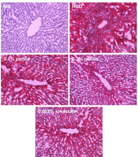
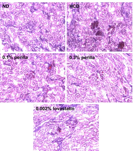
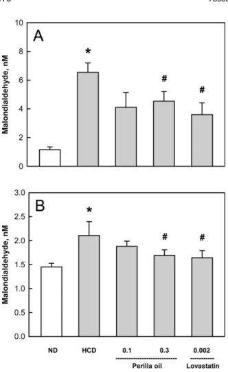
(4) 174. Yeseul Cha et al.. Figure 2. Effects of perilla oil and lovastatin on the blood total cholesterol (TC), low-density lipoproteins (LDL), high-density lipoproteins (HDL), and triglycerides (TG) of high-cholesterol diet (HCD)-fed rabbits (% of initial concentration). ○, Normal diet; ●, HCD alone; ▼, HCD+0.1% perilla oil; ■, HCD+0.3% perilla oil; ◆, HCD+0.002% lovastatin. *Significantly different from normal, P<0.05. #Significantly different from HCD alone, P<0.05.. the weights of the liver and spleen, which were not affected by treatment with perilla oil or lovastatin (Table 2). In microscopic findings, extensive accumulation of lipids were observed in the liver (Figure 4). It is of interest to note that the hepatic steatosis was markedly attenuated by feeding perilla oil in a concentrationdependent manner, in which the effect of 0.3% perilla oil was higher than that of 0.002% lovastatin. Lipid accumulation was also observed focally around the glomerular structures (Figure 5). The glomerular lipid accumulation was remarkably inhibited by perilla oil and lovastatin. Particularly, 0.3% perilla oil fully prevented the renal lipidosis. In the analysis of lipid peroxidation in the organs showing lipidosis, the MDA concentrations in the liver and kidneys was increased 4 folds and 1.5 folds, respectively, by the long-term HCD feeding (Figure 6). Interestingly, perilla oil (0.1-0.3%) displayed a marked Lab Anim Res | September, 2016 | Vol. 32, No. 3. anti-oxidative activity on the MDA formation, comparable with the effect of lovastatin (0.002%).. Discussion In the present study, perilla oil substantially inhibited hypercholesterolemia-mediated atheroma formation and lipid accumulation in the liver and kidneys. In addition, perilla oil attenuated the oxidative membrane injury (lipid peroxidation) in the organs showing lipidosis. Actually, statins such as lovastatin is preferentially prescribed to suppress hepatic cholesterol synthesis, remarkably improving blood lipid profiles [30,31]. However, it is well known that over-dosage and longterm administration of the statins cause severe hepatotoxicity, and that low doses could not effectively control the blood cholesterol from diets [12,27]. Notably, management of risk factors in preventive mode, rather than therapeutic.
(5) 175. Perilla oil improves atherosclerosis. Figure 3. Findings of atheroma of rabbits fed a high-cholesterol diet (HCD) containing perilla oil or lovastatin for 10 weeks. *Significantly different from normal diet (ND), P<0.05. #Significantly different from HCD alone, P<0.05. Table 1. Effects of perilla oil and lovastatin on the blood biochemistry of high-cholesterol diet (HCD)-fed rabbits Treatment ALT (U/L) AST (U/L) LDH (U/L) ALP (U/L) TP (g/d/L) Glucose (g/dL) BUN (mg/dL) Creatinine (mg/dL). Normal diet 42.3±11.0 46.7±14.0 132.2±13.2 50.8±8.3 06.0±0.1 122.0±6.3 21.8±0.7 01.5±0.1. HCD 56.7±5.1 78.0±4.7 172.8±39.4 57.4±4.3 03.7±0.3* 124.0±6.9 24.0±0.3 01.1±0.3. +0.1% Perilla oil 34.4±4.1 48.8±14.2 147.7±18.5 51.9±3.6 05.7±0.9# 112.9±4.4 24.0±1.4 01.2±0.2. +0.3% Perilla oil 30.3±3.9 49.0±7.8 153.7±17.1 53.4±6.5 05.0±0.4# 123.6±5.8 22.9±0.7 01.3±0.1. +0.002% Lovastatin 39.9±8.5 46.6±13.1 133.4±18.5 51.1±6.1 05.5±0.3# 130.3±4.9 21.6±0.5 01.4±0.1. ALT, alanine transaminase; AST, aspartate transaminase; ALP, alkaline phosphatase; LDH, lactate dehydrogenase; TP, total proteins; BUN, blood urea nitrogen. *Significantly different from normal diet (P<0.05). #Significantly different from HCD alone (P<0.05).. mode, of the cardiovascular and cerebrovascular diseases is extremely important, because the time to death after. outbreak of the diseases is very short [32]. Accordingly, dietary restriction and appropriate choice of fats or oils Lab Anim Res | September, 2016 | Vol. 32, No. 3.
(6) 176. Yeseul Cha et al.. Table 2. Effects of perilla oil and lovastatin on the organ weights of high-cholesterol diet (HCD)-fed rabbits Normal diet. HCD. +0.1% Perilla oil. +0.3% Perilla oil. +0.002% Lovastatin. 118.90±7.7900 6.39±0.32 6.94±0.37 2.23±0.23. 116.73±3.7600 7.41±0.28 7.56±0.62 1.73±0.16. 3.70±0.15 0.20±0.00 0.22±0.01 0.07±0.01. 3.69±0.11 024±0.01 0.24±0.02 0.05±0.00. Liver Kidneys Heart Spleen. 69.63±5.820 6.27±0.40 7.54±0.60 1.16±0.11. Absolute weight (g) 116.08±8.66*0 116.81±4.6500 6.53±0.31 6.80±0.33 7.23±0.45 8.00±0.28 02.05±0.23* 2.17±0.23. Liver Kidneys Heart Spleen. 2.19±0.19 0.20±0.01 0.24±0.01 0.03±0.00. Relative weight (%) 03.68±0.27* 3.50±0.12 0.21±0.01 0.21±0.01 0.23±0.01 0.24±0.01 00.07±0.01* 0.06±0.01. *Significantly different from normal diet (P<0.05).. have been recommended. While excessive consumption of fats and oils, especially containing high levels of saturated fatty acids (SFA), is known to be harmful for vascular diseases, UFA such as α-linolenic acid (ALA, ω-3) are believed to be beneficial. In fact, it has been reported that perilla oil containing a. high level ω-3 PUFA decreased blood TG, TC, and LDL in animals and humans [20,33]. Also in human studies, perilla oil not only improved blood HDL and recovered the function of arterial endothelial cells [34], but also reduced blood TG and the risk of cardiac infarction [35]. Also, in the present study, perilla oil containing 72.12%. Figure 4. Representative microscopic findings of the liver of rabbits fed a high-cholesterol diet (HCD) containing perilla oil or lovastatin for 10 weeks. ND, normal diet. Lab Anim Res | September, 2016 | Vol. 32, No. 3.
(7) Perilla oil improves atherosclerosis. 177. Figure 5. Representative microscopic findings of the kidney of rabbits fed a high-cholesterol diet (HCD) containing perilla oil or lovastatin for 10 weeks. ND, normal diet.. PUFA improved the blood lipid profiles and lipid accumulation in tissues, leading to atherosclerosis and hepatic and renal steatosis. Notably, in our gas chromatographic analysis of perilla oil, 57.47% was ALA out of 72.12% PUFA. Supportively, it was demonstrated that ALA inhibited platelet activation and arterial thrombus formation [36]. In our recent study, perilla oil markedly suppressed platelet aggregation as well as thrombus formation in the FeCl3-induced endothelial injury model, too [23]. Activated platelets attach to vascular endothelial walls injured during oxidative reaction mediated by OxLDL, aggregate there, and form thrombus and atherosclerosis. Therefore, perilla oil has attracted investigators’ attention, because a diet rich in PUFA may be helpful in preventing heart diseases [22,37,38] and blood coagulation [17]. More importantly, it was demonstrated that most of the plant oils with high ω-6/ω-3 fatty acid ratios including canola oil, safflower oil, olive oil, corn oil, and soybean. oil, increased hemorrhagic stroke in SHR-SP and shortened their life span, except only perilla oil with a low ω-6/ω3 fatty acid ratio [15,16,39,40]. In our previous study, perilla oil with 0.25 ω-6/ω-3 fatty acid ratio delayed and attenuated brain hemorrhagic stroke and renal lesions in SHR-SP, whereas canola oil with 2.63 ω-6/ω-3 ratio aggravated the lesions, advancing time to death [24]. It was reported that ω-3 PUFA has anti-oxidative and anti-inflammatory activities; it inhibited C-reactive protein in an atherosclerosis model [20] and increased mucosal blood flow by inhibiting leukotriene production in an inflammatory bowel disease model [41]. In the present study, the anti-oxidative activity of perilla oil was also confirmed: i.e., feeding perilla oil markedly suppressed lipid peroxidation in the fatty liver and kidneys following HCD-induced hypercholesterolemia. To date, statins and anti-coagulants have been used to improve hyperlipidemia and blood flow, and thereby to prevent atherosclerosis and cardiovascular diseases [42Lab Anim Res | September, 2016 | Vol. 32, No. 3.
(8) 178. Yeseul Cha et al.. Acknowledgments This work was supported by “Food Functionality Evaluation program” under the Ministry of Agriculture, Food and Rural Affairs and partly Korea Food Research Institute (G2015). Conflict of interests The authors declare that there is no financial conflict of interests to publish these results.. References. Figure 6. Effects of perilla oil (0.1 or 0.3%) and lovastatin (0.002%) on the lipid peroxidation of liver (A) and kidney (B) tissues of high-cholesterol diet (HCD)-fed rabbits. ND, normal diet.. 44]. In fact, it is well known that platelet activation and aggregation is an initial feature of thrombus formation on the injured arterial endothelium undergoing atherosclerosis [45]. However, the most important triggering factor of oxidative injury of arterial intimal layer, leading to platelet aggregation and clonal expansion of vascular smooth muscle cells, is OxLDL containing a high level of cholesterol. By comparison with harmful effects of various fats and plant oils containing high ω-6/ω-3 fatty acid ratios [39,40], it was confirmed that perilla oil, only an oil containing a low ω-6/ω-3 ratio, exerted beneficial effects on hemorrhagic stroke and atherosclerosis in our previous and present studies. Therefore, it is strongly recommended that perilla oil could be the first choice on modern food tables consuming high amount of fats and oils.. Lab Anim Res | September, 2016 | Vol. 32, No. 3. 1. Huh KB. The present status of nutrition-related diseases and its countermeasures. Kor J Nutr 1990; 23(3): 197-207. 2. Lee HK. Pattern of disease incidence and nutrition in Korea. Kor J Nutr 1996; 29(4): 381-383. 3. Lee YC. Hypercholesterolemia in Korea and nutritional factors. Korean Soc Lipidol Atherosc 1991; 1(1): 111-122. 4. Spady DK, Woollett LA, Dietschy JM. Regulation of plasma LDL-cholesterol levels by dietary cholesterol and fatty acids. Annu Rev Nutr 1993; 13: 355-381. 5. Natio HK. Atherogenesis: current topics on etiology and risk factors. Clin Chem 1995; 41(1): 132-133. 6. Bemis CE, Gorlin R, Kemp HG, Herman MV. Progression of coronary artery disease. A clinical arteriographic study. Circulation 1973; 47(3): 455-464. 7. Kannel WB, Castelli WP, Gordon T, McNamara PM. Serum cholesterol, lipoproteins, and the risk of coronary heart disease. The Framingham study. Ann Intern Med 1971; 74(1): 1-12. 8. Jialal I, Devaraj S. The role of oxidized low density lipoprotein in atherogenesis. J Nutr 1996; 126(4 suppl): 1053S-1057S. 9. Glass CK, Witztum JL. Atherosclerosis. the road ahead. Cell 2001; 104(4): 503-516. 10. Blum A, Simsolo C, Hasin Y. 3-Hydroxy-3-methylglutaryl coenzyme a (HMG-CoA) reductase inhibitors (statins), atherosclerosis and coronary syndromes. Atherosclerosis 2004; 175(1): 1-5. 11. Larsen ML, Illingworth DR. Drug treatment of dyslipoproteinemia. Med Clin North Am 1994; 78(1): 225-245. 12. Cho JH, Lee NJ, Chai HY, Kim TM, Park JH, Kang JK, Kim YB, Hwang SY. Effect of Hwalgidan SJ-101 on atherosclerosis in hypercholesterolemic rabbits. Lab Anim Res 2005; 21(2): 149157. 13. Hong SH, Chai HY, Kim TM, Lee NJ, Kim DK, Cho JH, Park JH, Kim YB, Kang JK, Hwang SY. Therapeutic Effects of Mulberry Root-Bark (Mori radicis Cortex) Ethanol Extract on Atherosclerosis in Hypercholesterolemic Rabbits. Lab Anim Res 2005; 21(3): 273-279. 14. Kim HK, Choi S, Choi H. Suppression of hepatic fatty acid synthase by feeding alpha-linolenic acid rich perilla oil lowers plasma triacylglycerol level in rats. J Nutr Biochem 2004; 15(8): 485-492. 15. Huang MZ, Watanabe S, Kobayashi T, Nagatsu A, Sakakibara J, Okuyama H. Unusual effects of some vegetable oils on the survival time of stroke-prone spontaneously hypertensive rats. Lipids 1997; 32(7): 745-751. 16. Okuyama H, Yamada K, Miyazawa D, Yasui Y, Ohara N. Dietary lipids impacts on healthy ageing. Lipids 2007; 42(9): 821-825. 17. Lanzmann-Petithory D. Alpha-linolenic acid and cardiovascular diseases. J Nutr Health Aging 2001; 5(3): 179-183. 18. Vanschoonbeek K, de Maat MP, Heemskerk JW. Fish oil consumption and reduction of arterial disease. J Nutr 2003;.
(9) Perilla oil improves atherosclerosis. 133(3): 657-660. 19. De Caterina R, Cybulsky MA, Clinton SK, Gimbrone MA Jr, Libby P. Omega-3 fatty acids and endothelial leukocyte adhesion molecules. Prostaglandins Leukot Essent Fatty Acids 1995; 52(23): 191-195. 20. Zhang L, Geng Y, Yin M, Mao L, Zhang S, Pan J. Low omega-6/ omega-3 polyunsaturated fatty acid ratios reduce hepatic Creactive protein expression in apolipoprotein E-null mice. Nutrition 2010; 26(7-8): 829-834. 21. Griffin MD, Sanders TA, Davies IG, Morgan LM, Millward DJ, Lewis F, Slaughter S, Cooper JA, Miller GJ, Griffin BA. Effects of altering the ratio of dietary n-6 to n-3 fatty acids on insulin sensitivity, lipoprotein size, and postprandial lipemia in men and postmenopausal women aged 45-70 y: the OPTILIP Study. Am J Clin Nutr 2006; 84(6): 1290-1298. 22. De Lorgeril M, Renaud S, Mamelle N, Salen P, Martin JL, Monjaud I, Guidollet J, Touboul P, Delaye J. Mediterranean alphalinolenic acid-rich diet in secondary prevention of coronary heart disease. Lancet 1994; 343(8911): 1454-1459. 23. Jang JY, Kim TS, Cai J, Kim J, Kim Y, Shin K, Kim KS, Lee SP, Kang MH, Choi EK, Rhee MH, Kim YB. Perilla oil improves blood flow through inhibition of platelet aggregation and thrombus formation. Lab Anim Res 2014; 30(1): 21-27. 24. Cai J, Jang JY, Kim J, Shin K, Kim KS, Park D, Kim TS, Lee SP, Ahn B, Choi EK, Lee J, Kim YB. Comparative effects of plant oils on the cerebral hemorrhage in stroke-prone spontaneously hypertensive rats. Nutr Neurosci 2016; 19(7): 318-326. 25. Heemskerk JW, Vossen RC, van Dam-Mieras MC. Polyunsaturated fatty acids and function of platelets and endothelial cells. Curr Opin Lipidol 1996; 7(1): 24-29. 26. Cheong SH, Kim MY, Sok DE, Hwang SY, Kim JH, Kim HR, Lee JH, Kim YB, Kim MR. Spirulina prevents atherosclerosis by reducing hypercholesterolemia in rabbits fed a high-cholesterol diet. J Nutr Sci Vitaminol (Tokyo) 2010; 56(1): 34-40. 27. Park D, Kyung J, Kim D, Hwang SY, Choi EK, Kim YB. Antihypercholesterolemic and anti-atherosclerotic effects of polarizedlight therapy in rabbits fed a high-cholesterol diet. Lab Anim Res 2012; 28(1): 39-46. 28. Jang JY, Kim J, Cai J, Kim Y, Shin K, Kim TS, Lee SP, Park SK, Choi EK, Kim YB. An ethanolic extract of Angelica gigas improves atherosclerosis by inhibiting vascular smooth muscle cell proliferation. Lab Anim Res 2014; 30(2): 84-89. 29. Kwon SC, Kim YB. Antioxidative and aldose reductaseinhibitory effects of a fermentation filtrate of Rubus coreanus. Lab Anim Res 2011; 27(4): 365-368. 30. McClelland GA, Stubbs RJ, Fix JA, Pogany SA, Zentner GM. Enhancement of 3-hydroxy-3-methylglutaryl-coenzyme A (HMGCoA) reductase inhibitor efficacy through administration of a controlled-porosity osmotic pump dosage form. Pharm Res 1991; 8(7): 873-876. 31. Yim JE, Choue RW, Kim YS. Effects of dietary counceling and HMG-CoA reductase inhibitor treatment on serum lipid levels in hyperlipidemic patients. Kor Nutr Soc 1998; 8(1): 61-76. 32. Blaha MJ, Bansal S, Rouf R, Golden SH, Blumenthal RS, Defilippis AP. A practical “ABCDE” approach to the metabolic. 179. syndrome. Mayo Clin Proc 2008; 83(3): 932-941. 33. Kurowska EM, Dresser GK, Deutsch L, Vachon D, Khalil W. Bioavailability of omega-3 essential fatty acids from perilla seed oil. Prostaglandins Leukot Essent Fatty Acids 2003; 68(3): 207212. 34. Wei M, Xiong P, Zhang L, Fei M, Chen A, Li F. Perilla oil and exercise decrease expressions of tumor necrosis factor-alpha, plasminogen activator inhibitor-1 and highly sensitive C-reactive protein in patients with hyperlipidemia. J Tradit Chin Med 2013; 33(2): 170-175. 35. Eussen SR, Geleijnse JM, Giltay EJ, Rompelberg CJ, Klungel OH, Kromhout D. Effects of n-3 fatty acids on major cardiovascular events in statin users and non-users with a history of myocardial infarction. Eur Heart J 2012; 33(13): 1582-1588. 36. Holy EW, Forestier M, Richter EK, Akhmedov A, Leiber F, Camici GG, Mocharla P, Luscher TF, Beer JH, Tanner FC. Dietary α-linolenic acid inhibits arterial thrombus formation, tissue factor expression, and platelet activation. Arterioscler Thromb Vasc Biol 2011; 31(8): 1772-1780. 37. Ascherio A, Rimm EB, Giovannucci EL, Spiegelman D, Stampfer M, Willett WC. Dietary fat and risk of coronary heart disease in men: cohort follow up study in the United States. BMJ 1996; 313(7049): 84-90. 38. Hu FB, Stampfer MJ, Manson JE, Rimm EB, Wolk A, Colditz GA, Hennekens CH, Willett WC. Dietary intake of alphalinolenic acid and risk of fatal ischemic heart disease among women. Am J Clin Nutr 1999; 69(5): 890-897. 39. Huang MZ, Naito Y, Watanabe S, Kobayashi T, Kanai H, Nagai H, Okuyama H. Effect of rapeseed and dietary oils on the mean survival time of stroke-prone spontaneously hypertensive rats. Biol Pharm Bull 1996; 19(4): 554-557. 40. Ratnayake S, Lewandowski P. Rapid bioassay-guided screening of toxic substances in vegetable oils that shorten the life of SHRSP rats. Lipids Health Dis 2010; 9: 13. 41. Shimizu T, Igarashi J, Ohtuka Y, Oguchi S, Kaneko K, Yamashiro Y. Effects of n-3 polyunsaturated fatty acids and vitamin E on colonic mucosal leukotriene generation, lipid peroxidation, and microcirculation in rats with experimental colitis. Digestion 2001; 63(1): 49-54. 42. Zhao G, Zang SY, Jiang ZH, Chen YY, Ji XH, Lu BF, Wu JH, Qin GW, Guo LH. Postischemic administration of liposome-encapsulated luteolin prevents against ischemia-reperfusion injury in a rat middle cerebral artery occlusion model. J Nutr Biochem 2011; 22(10): 929-936. 43. Pan CH, Tsai CH, Lin WH, Chen GY, Wu CH. Ethanolic Extract of Vitis thunbergii Exhibits Lipid Lowering Properties via Modulation of the AMPK-ACC Pathway in Hypercholesterolemic Rabbits. Evid Based Complement Alternat Med 2012; 2012: 436786. 44. Sylvester KW, Cheng JW, Mehra MR. Esomeprazole and aspirin fixed combination for the prevention of cardiovascular events. Vasc Health Risk Manag 2013; 9: 245-254. 45. Fintel DJ. Oral antiplatelet therapy for atherothrombotic disease: overview of current and emerging treatment options. Vasc Health Risk Manag 2012; 8: 77-89.. Lab Anim Res | September, 2016 | Vol. 32, No. 3.
(10)
수치


+3
관련 문서
Moreover, high density lipoprotein (HDL)-cholesterol contents on serum of rats fed a hyperlipidemic diet were increased 10% and low density lipoprotein (LDL)-cholesterol
Total lipid, triglyceride, cholesterol, HDL-cholesterol and LDL-cholesterol in serum of rats fed high fat-high cholesterol diet containing Salvia plebeia
Oral administration of Sophorae Fructus extract significantly decreased serum total cholesterol (TC), LDL-cholesterol, trig- lyceride (TG), GOT and GPT levels, and
Total lipid, triglyceride, cholesterol, HDL-cholesterol and LDL-cholesterol in serum of rats fed high fat-high cholesterol diet containing Salvia plebeia
The low density lipoprotein cholesterol (LDL), high density lipoprotein cholesterol (HDL), total cholesterol (TC), triglyceride (TG), systolic blood pressure (SBP), diastolic
HEFS, hydro-alcoholic extract of fenugreek seeds; MetS, metabolic syndrome; FPG, fasting plasma glucose; TG, triglycerides; TC, total cholesterol; LDL-C, low-density lipoprotein
Results: The serum levels of triglycerides, total cholesterol (TC), low-density lipoprotein cholesterol (LDL-C), high-density lipo- protein cholesterol (HDL-C),
Contents of total cholesterol, triglyceride, LDL-cholesterol and HDL-cholesterol in serum of rats fed a high fat diet containing ethanol extract from the leaf of Lythrum
