The Effects of Hydro-Alcoholic Extract of Fenugreek Seeds on the Lipid Profile and Oxidative Stress in Fructose-Fed Rats
Maryam Mohammad-Sadeghipour1,2, Mehdi Afsharinasab3,4, Maryam Mohamadi4, Mehdi Mahmoodi2, Soudeh Khanamani Falahati-pour5, Mohammad Reza Hajizadeh3,4,*
1Student Research Committee and 2Department of Clinical Biochemistry, Afzalipour School of Medicine, Kerman University of Medical Sciences, Kerman;
3Department of Clinical Biochemistry, Faculty of Medicine, Rafsanjan University of Medical Sciences, Rafsanjan; 4Molecular Medicine Research Center, Institute of Basic Medical Sciences Research and 5Pistachio Safety Research Center, Rafsanjan University of Medical Sciences, Rafsanjan, Iran
Background: Metabolic syndrome (MetS) is a complex clinical disorder that can lead to an increase in oxida- tive stress. Patients with this syndrome are at risk of diabetes and cardiovascular disease. The Trigonella foenum- graecum L. (fenugreek) plant has many therapeutic effects, including anti-diabetic and antioxidant. The present study aimed to investigate the effects of the hydro-alcoholic extract of fenugreek seeds (HEFS) on dyslipidemia and oxidative stress due to high-fructose diet-induced MetS.
Methods: In this experimental study, to induce MetS, animals received water containing 20% fructose for 8 weeks. After induction of MetS, 48 male Wistar rats (200–250 g) were randomized into six groups. HEFS was ad- ministered to animals at doses of 100 and 200 mg/kg orally for 4 weeks. Animal blood samples were collected to measure biochemical and antioxidant parameters of fasting plasma glucose (FPG), total cholesterol (TC), high- density lipoprotein cholesterol (HDL-C), low-density lipoprotein cholesterol (LDL-C), triglycerides (TG), malondi- aldehyde (MDA), glutathione peroxidase (GPX), catalase (CAT), and total antioxidant capacity (TAC).
Results: The findings showed that the serum levels of FPG, TC, LDL-C, TG, and MDA were significantly reduced in HEFS-exposed groups compared with the control group (P<0.05). Also, significant increases in HDL-C, GPX, CAT, and TAC levels (P<0.05) were observed.
Conclusion: Our results revealed that treatment with HEFS increases the levels of antioxidant enzymes, de- creases FPG level, and at the same time, modifies the lipid profile in MetS. Therefore, HEFS may help to alleviate the risk of some chronic complications of this disease.
Key words: Antioxidants, Dyslipidemia, Trigonella, Metabolic syndrome, Oxidative stress
Received September 7, 2019 Reviewed October 11, 2019 Accepted July 16, 2020
* Corresponding author Mohammad Reza Hajizadeh
https://orcid.org/0000-0002-1199-9601 Molecular Medicine Research Center, Rafsanjan University of Medical Sciences, Rafsanjan 7719617996, Iran Tel: +98-3431315023
Fax: +98-3431315003 E-mail: [email protected]
INTRODUCTION
Metabolic syndrome (MetS) is characterized by clustering of metabolic abnormalities of central obesity, hypertension, elevated fasting plasma glucose (FPG), dyslipidemia, high body mass index, insulin resistance, and oxidative stress.1 MetS has become a signifi- cant problem in public health, with a prevalence rate of 20% to 25% among adults worldwide.2 Patients with this syndrome are at
risk of various diseases, including diabetes, cardiovascular disease,3 non-alcoholic fatty liver disease,4 and cancer.5 Excessive consump- tion of fructose, a simple monosaccharide, is closely related to obe- sity. Fructose-fed rats have been reported as a rat model of MetS6 and were used as such in the present study.
One of the products of biological systems is reactive oxygen spe- cies (ROS), which are highly reactive and short-lived. One of the primary methods for removal of ROS is the antioxidant system.7,8
Copyright © 2020 Korean Society for the Study of Obesity
This is an Open Access article distributed under the terms of the Creative Commons Attribution Non-Commercial License (https://creativecommons.org/licenses/by-nc/4.0/) which permits unrestricted non-commercial use, distribution, and reproduction in any medium, provided the original work is properly cited.
2017-03-16 https://crossmark-cdn.crossref.org/widget/v2.0/logos/CROSSMARK_Color_square.svg
The Effects of Hydro-Alcoholic Extract of Fenugreek Seeds on the Lipid Profile and Oxidative Stress in Fructose-Fed Rats
Maryam Mohammad-Sadeghipour1,2, Mehdi Afsharinasab3,4, Maryam Mohamadi4, Mehdi Mahmoodi2, Soudeh Khanamani Falahati-pour5, Mohammad Reza Hajizadeh3,4,*
1Student Research Committee and 2Department of Clinical Biochemistry, Afzalipour School of Medicine, Kerman University of Medical Sciences, Kerman;
3Department of Clinical Biochemistry, Faculty of Medicine, Rafsanjan University of Medical Sciences, Rafsanjan; 4Molecular Medicine Research Center, Institute of Basic Medical Sciences Research and 5Pistachio Safety Research Center, Rafsanjan University of Medical Sciences, Rafsanjan, Iran
Background: Metabolic syndrome (MetS) is a complex clinical disorder that can lead to an increase in oxida- tive stress. Patients with this syndrome are at risk of diabetes and cardiovascular disease. The Trigonella foenum- graecum L. (fenugreek) plant has many therapeutic effects, including anti-diabetic and antioxidant. The present study aimed to investigate the effects of the hydro-alcoholic extract of fenugreek seeds (HEFS) on dyslipidemia and oxidative stress due to high-fructose diet-induced MetS.
Methods: In this experimental study, to induce MetS, animals received water containing 20% fructose for 8 weeks. After induction of MetS, 48 male Wistar rats (200–250 g) were randomized into six groups. HEFS was ad- ministered to animals at doses of 100 and 200 mg/kg orally for 4 weeks. Animal blood samples were collected to measure biochemical and antioxidant parameters of fasting plasma glucose (FPG), total cholesterol (TC), high- density lipoprotein cholesterol (HDL-C), low-density lipoprotein cholesterol (LDL-C), triglycerides (TG), malondi- aldehyde (MDA), glutathione peroxidase (GPX), catalase (CAT), and total antioxidant capacity (TAC).
Results: The findings showed that the serum levels of FPG, TC, LDL-C, TG, and MDA were significantly reduced in HEFS-exposed groups compared with the control group (P<0.05). Also, significant increases in HDL-C, GPX, CAT, and TAC levels (P<0.05) were observed.
Conclusion: Our results revealed that treatment with HEFS increases the levels of antioxidant enzymes, de- creases FPG level, and at the same time, modifies the lipid profile in MetS. Therefore, HEFS may help to alleviate the risk of some chronic complications of this disease.
Key words: Antioxidants, Dyslipidemia, Trigonella, Metabolic syndrome, Oxidative stress
Received September 7, 2019 Reviewed October 11, 2019 Accepted July 16, 2020
* Corresponding author Mohammad Reza Hajizadeh
https://orcid.org/0000-0002-1199-9601 Molecular Medicine Research Center, Rafsanjan University of Medical Sciences, Rafsanjan 7719617996, Iran Tel: +98-3431315023
Fax: +98-3431315003 E-mail: [email protected]
The most important antioxidant enzymes include catalase (CAT), glutathione peroxidase (GPX), superoxide dismutase (SOD), and ceruloplasmin.9,10 Imbalance between ROS (increased level) and the antioxidant system (reduced antioxidants) is the leading cause of oxidative stress.7,10 Malondialdehyde (MDA) resulting from oxi- dation of unsaturated fatty acids is an important marker of oxida- tive stress.11
There is a relationship between oxidative stress and MetS, which aggravates MetS factors such as insulin resistance, hypertension, hypertriglyceridemia, and reduction of high-density lipoprotein cholesterol (HDL-C).12,13 The risk of MetS increases with an in- crease in oxidative stress.14 Recent studies have shown that MetS patients exhibit a significant reduction in activity/expression of an- tioxidant systems, including SOD, GPX, CAT, and reduced gluta- thione (GSH).15 Currently, many chemical medications are sug- gested for treatment and control of MetS factors, including lipid- lowering agents (statins),16 insulin sensitizers (metformin, acarbose, and thiazolidinedione),17 and weight loss medications, but their various side effects limit their use. Finding a therapeutic approach with few side effects is a challenge for researchers that has prompt- ed consideration of herbal medicines as treatment.18
Trigonella foenum-graecum L. (fenugreek) is an annual plant be- longing to the family Leguminosae.19 Properties of fenugreek seeds include lowering total cholesterol (TC), triglycerides (TG), low- density lipoprotein cholesterol (LDL-C), and very-low-density li- poprotein cholesterol (VLDL-C)20 and being anti-diabetic,21 anti- tympanites, anti-diarrheal,20,22 diuretic,23 anti-inflammatory, and an- ti-cough.24 Beneficial compounds, including trigonelline, 4-hy- droxyisoleucine, diosgenin, orientin, luteolin flavonoids, steroid sa- ponins, vitamins, and minerals such as iron and calcium, have been found in fenugreek seeds,25 indicating its use as a valuable therapeu- tic food. Moreover, studies of fenugreek in type 2 diabetes showed that active ingredients such as 4-hydroxyisoleucine, diosgenin, and galactomannan had positive effects on this pathologic state.24
Various studies have shown the effect of fenugreek on oxidative stress.26-28 One study reported that administration of aqueous ex- tract of fenugreek plays a protective role against carbon tetrachlo- ride-induced liver injury in rats. In that study, the ameliorative ef- fects of the extract improved levels of serum transaminases, attenu- ated hepatic lipid peroxidation, and activated hepatic antioxidant
enzymes.29 Another study indicated that the combination of endur- ance swimming training and fenugreek seed extract could signifi- cantly decrease plasma glucose level and elevate the activity of car- diac antioxidant enzymes in diabetic rats.30 Fenugreek seeds im- prove irradiation-induced oxidative stress by increasing GSH and SOD levels.31 Zingerone and gingerol are the most abundant com- pounds in the hydro-alcoholic extract of fenugreek seeds (HEFS) that have been reported to show antioxidant activity.32
Considering the seriousness and prevalence of MetS, finding a new treatment strategy to deal with this problem in modern society is important. On the other hand, no study has been performed on the antioxidant effects of fenugreek on MetS. Therefore, the pres- ent study aimed to investigate the protective effects of fenugreek seeds on the lipid profile and oxidative stress in fructose-fed rats.
The extract was expected to alter the levels of antioxidants and bio- chemical parameters involved in MetS.
METHODS Chemicals
Fructose, thiobarbituric acid (TBA), 1,1,3,3-tetra methoxy pro- pane (TEP), reduced GSH, nicotinamide adenine dinucleotide phosphate (NADPH), glutathione reductase (GR), and other high-grade chemicals were purchased from Sigma Chemical Com- pany (St. Louis, MO, USA). Hydrogen peroxide (H2O2) was pur- chased from Merck (Darmstadt, Germany). FPG, TC, TG, LDL- C, and HDL-C were calculated using Pars Azmoon kits with an automatic analyzer (Biochemistry analyzer BT 4500; Farasamed Co., Biotechnica, Rome, Italy) for biochemical testing.
Animals
In this experimental study, adult male Wistar rats (weighing 200–250g) were obtained from the animal house of Rafsanjan University of Medical Sciences (RUMS). All the animals were kept on a 12/12 light-dark schedule with a humidity of 65%–70% and a temperature of 22°C± 2°C and were given free access to water and food. All procedures of this study were approved by the Institution- al Animal Care and Use Committee of Rafsanjan University of Medical Sciences with the IR code: IR.RUMS.REC.1395.11.
Preparation of HEFS
The fenugreek seed was taken from the Davaran region located in Rafsanjan, Iran, and was confirmed by the Botany Research Di- vision of the Valiasr University of Rafsanjan. The seeds were dried at room temperature and powdered in a laboratory blender (War- ing Products Division, Torrington, USA). The extraction process was performed according to our previous work,33 using a Soxhlet (BAKHSHI Laboratory Industrial Co., Tehran, Iran) with a 75%
(v/v) ethanol solution. The extract yield was calculated to be 6.67%.
Study design and induction of MetS
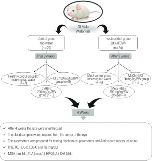
Fig. 1 depicts the study design used in the present work. Forty- eight male Wistar rats were randomly separated into control and
fructose diet groups. The control group received tap water, and the fructose diet group was fed 20% fructose drinking water (FDW) daily for 8 weeks.6 The FDW was prepared by diluting 20 g of fruc- tose in 100 mL of tap water at weight to volume; to induce MetS, D-fructose > 99% was used. The FDW was freshly prepared daily.
To prevent evaporation, the bottle was covered with aluminum foil.
After 8 weeks, the weight of the rats was measured using a digital scale (FEW, Tokyo, Japan), and blood samples were obtained after 12 hours fasting by penetrating the retro-orbital plexus with a capil- lary tube.
Subsequently, to confirm induction of MetS in rats, we analyzed indicators of MetS of body weight, FPG, TG, and TC changes in the case group after administration of 20% FDW for 8 weeks, as
Figure 1. Schematic of the study. C: healthy control group; C+HEFS 100: healthy control group that received 100 mg/kg/BW of HEFS; C+HEFS 200: healthy control group that received 200 mg/kg/BW of HEFS; MetS: metabolic syndrome group that did not receive HEFS; MetS+HEFS 100: metabolic syndrome group that received 100 mg/kg/
BW of HEFS; MetS+HEFS 200: metabolic syndrome group that received 200 mg/kg/BW of HEFS. FDW, fructose drinking water; HEFS, hydro-alcoholic extract of fenugreek seeds; FPG, fasting plasma glucose; TC, total cholesterol; HDL-C, high-density lipoprotein cholesterol; LDL-C, low-density lipoprotein cholesterol; TG, triglycerides; MDA, malondialdehyde; TCA, trichloroacetic acid; GPX, glutathione peroxidase; CAT, catalase.
48 Male Wistar rats Control group
tap water (n= 24)
Healthy control group (C) receiving tap water
(n= 8)
MetS control group receiving tap water
(n= 8) C+HEFS 100 mg/kg/BW
group (n= 8)
MetS+HEFS 100 mg/kg/BW group
(n= 8) C+HEFS
200 mg/kg/BW group (n= 8)
MetS+HEFS 200 mg/kg/BW
group (n= 8) Fructose diet group
20% (FDW) (n= 24)
4 Weeks
After 4 weeks the rats were anesthetized
The blood samples were prepared from the corner of the eye
The supernatant was prepared for testing biochemical parameters and Antioxidant assays including:
FPG, TC, HDL-C, LDL-C and TG (mg/dL) MDA (nmol/L), TCA (nmol/L), GPX (U/L), CAT (U/L)
After 8 weeks After 8 weeks
contrasted with the control group.34,35 The fructose-fed rats had sig- nificantly higher body weight, FPG, TG, and TC compared with the control rats. Therefore, the rats were enrolled in the study. Af- terward, the MetS rats were randomly divided into three groups (eight rats/group): a MetS group that received no extract (MetS), a MetS group that received 100 mg/kg/BW of HEFS (MetS+HEFS 100), and a MetS group that received 200 mg/kg/BW of HEFS (MetS+HEFS 200). The healthy rats in the control group were also divided into three groups: a healthy group that received no ex- tract (C), a healthy group that received 100 mg/kg/BW of HEFS (C+HEFS 100), and a healthy group that received 200 mg/kg/BW of HEFS (C+HEFS 200). All rats were fed a standard diet during the study. The doses of 100 and 200 mg/kg of HEFS were ob- tained by diluting the HEFS in physiological saline, and rats underwent intra-gastric gavage at a volume of 1 mL per day for 4 weeks.36 The doses of 100 and 200 mg/kg of HEFS were selected based on Gupta’s study.37 All experiments were performed between 10:00 AM and 1:00 PM.
Biochemical assays
Following ethical considerations, the rats were anesthetized, and blood samples were collected from the corner of the eye in glass as- say tubes without anticoagulant. The tubes were placed at room temperature to allow the blood to clot and then centrifuged at 3,000 RPM for 10 minutes. The transparent and non-hemolytic supernatant was prepared for testing biochemical parameters of FPG, TC, HDL-C, LDL-C, and TG with fitting kits (Pars Azmoun, Tehran, Iran), and the results are expressed as mg/dL.
Antioxidant assays
MDA was determined by thiobarbituric acid reactive substances (TBARS) using a colorimetric method.38 Briefly, 0.5 mL of plasma was added to 2 mL of TBA reagent containing 0.25 M/L HCl, 15 g trichloroacetic acid, and 375 mg TBA. The mixture was boiled in a water bath at 95°C for 30 minutes and, after fast cooling, was cen- trifuged at 8,000 g and 4°C 15 minutes. The MDA was determined based on the absorbance of TBA reactive substances at 532 nm.
The TBARS concentration was calculated using TEP as a standard and expressed as nmol/L of plasma.
The GPX activity of the serum samples was defined by the
coupled enzymatic reaction at 25°C according to the method of Paglia and Valentine.39 Briefly, the enzymatic reaction in a cuvette that contained NADPH, NaN3, GSH, and GR was initiated by adding H2O2, and the change in absorbance was monitored spec- trophotometrically at 340 nm. The result was expressed as U/L of plasma.
Serum CAT activity was determined according to the proce- dure of Aebi with H2O2 (30 mM) as the substrate.40 To 100 mL of 30 mmol/L H2O2 solution in 350 mL phosphate buffer (pH 7.0), 50 mL of serum sample was added, and the consumption of H2O2 was followed spectrophotometrically at 240 nm for 3 minutes at 25°C and expressed as U/L of plasma. Total antioxidant capacity (TAC) was determined by the ferric reducing antioxidant power assay (FRAP assay).41 Data were expressed as mmol/L in plasma.
Statistical analysis
Results are reported as mean± standard error of the mean where indicated. One-way analysis of variance followed by Tukey’s multi- ple comparisons test was used to analyze the data from the bio- chemical and oxidative stress assessments before and after treat- ment. Paired sample t-test was used to compare the variables before and after the intervention. Statistical results were evaluated using the SPSS version 18 (SPSS, Chicago, IL, USA). The significance level was P<0.05.
RESULTS
To confirm that the rats acquired MetS, changes in FPG, TG, TC, and weight were considered after 8 weeks of drinking water containing 20% fructose. A significant increase (P<0.001) in the levels of FPG (125.00± 3.52 mg/dL), TG (142.62± 6.62 mg/dL), TC (97.75± 2.11 mg/dL), and weight (290.62± 6.18 g) was ob- served in the rats that received fructose (MetS group) compared with the healthy control group (C), indicating induction of MetS in the rats (Table 1).
Changes in biochemical parameters after extract administration
Administration of HEFS (100 and 200 mg/kg/BW) in the MetS and healthy groups resulted in significant decrease in FPG, TG,
TC, and LDL-C levels. MDA level was decreased significantly only in the MetS groups after administration of HEFS (100 and 200 mg/kg/BW). In the MetS group that received 100 mg/kg/BW of HEFS, no significant change in the levels of GPX and CAT was ob- served in comparison to those before treatment. However, the lev- els of GPX, CAT, and TAC increased significantly after treatment with 200 mg/kg/BW of HEFS. In the healthy groups, the levels of GPX and CAT increased considerably (P<0.001) after treatment
with 200 mg/kg/BW of HEFS. However, in the healthy C+HEFS 100 and C+HEFS 200 groups of Table 1, there was no significant decrease in MDA level and no significant increase in activity level of TAC after administration of HEFS (100 and 200 mg/kg/BW).
In both MetS and healthy groups, treatment with HEFS signifi- cantly decreased the TG level; this decrease was higher in the treat- ed MetS groups. In the MetS groups receiving HEFS at the dose of 200 mg/kg/BW compared with the dose of 100 mg/kg/BW, mean Table 1. Changes in biochemical parameters of the studied groups after the intervention compared with the baseline
Variable Group
C C+HEFS 100 C+HEFS 200 MetS MetS+HEFS 100 MetS+HEFS 200
FPG (mg/dL)
Before 95.87± 1.85 93.11± 2.84 93.37± 2.75 125.00± 3.52* 130.00± 1.58 134.00± 3.82
After 95.37± 2.16 88.77± 2.72† 89.75± 2.95‡ 127.25± 3.49* 121.44± 2.35‡ 122.75± 2.90†
TG (mg/dL)
Before 76.50± 2.01 79.88± 1.98 72.12± 1.96 142.62± 6.62* 155.44± 4.74 160.25± 4.71
After 79.75± 2.06 75.44± 2.30‡ 64.37± 1.05‡ 146.00± 6.76* 145.88± 5.22§ 138.12± 4.29§
TC (mg/dL)
Before 79.00± 3.24 78.55± 2.45 74.87± 2.37 97.75± 2.11* 115.44± 4.97 107.62± 5.24
After 79.37± 2.57 73.88± 1.30† 65.37± 3.13§ 99.75± 1.93* 103.33± 5.19† 91.62± 6.90‡
LDL-C (mg/dL)
Before 35.75± 2.72 36.88± 2.45 29.62± 1.61 43.75± 2.78 62.77± 4.72 50.75± 5.54
After 37.50± 2.32 33.00± 2.29‡ 25.62± 1.38‡ 45.00± 3.56 56.88± 5.40† 40.25± 5.02‡
HDL-C (mg/dL)
Before 29.62± 1.26 26.33± 0.97 36.37± 1.03 30.50± 1.10 23.88± 1.16 26.25± 1.12
After 30.00± 1.22 29.66± 0.93§ 39.25± 1.72† 30.37± 1.14 29.88± 0.87§ 34.75± 1.72§
Weight (g)
Before 224.00± 3.64 218.00± 6.32 228.50± 3.25 290.62± 6.18* 302.00± 4.09 290.62± 6.71
After 241.87± 2.97 229.00± 6.46† 238.50± 4.98† 303.62± 6.44* 310.55± 2.81‡ 298.87± 4.34†
MDA (nmol/L)
Before 31.50± 1.41 30.00± 1.24 23.12± 0.58 60.75± 4.46* 79.11± 2.50 80.00± 3.30
After 28.50± 1.73 31.78± 1.30 21.62± 1.17 63.12± 3.89† 74.78± 3.05† 67.78± 3.14‡
GPX (U/L)
Before 47.50± 1.29 53.44± 2.27 48.50± 1.87 41.75± 1.75* 44.22± 1.03 30.87± 1.75
After 44.87± 3.44 57.56± 1.91† 58.87± 1.85§ 39.75± 1.76 45.00± 1.92 37.75± 2.18†
CAT (U/L)
Before 30.62± 1.11 28.78± 1.03 27.50± 1.52 22.00± 1.03* 22.44± 1.30 19.62± 0.92
After 31.87± 1.30 31.33± 1.52 36.00± 1.41§ 21.87± 0.87 23.33± 1.00 25.25± 1.25‡
TAC (mmol/L)
Before 1.14± 0.08 1.00± 0.10 0.94± 0.09 0.49± 0.05 0.43± 0.07 0.45± 0.06
After 1.11± 0.08 1.28± 0.15 1.31± 0.16 0.39± 0.03* 0.58± 0.08§ 0.86± 0.09§
C: healthy control group; C+HEFS 100: healthy control group that received 100 mg/kg/BW of HEFS; C+HEFS 200: healthy control group that received 200 mg/kg/BW of HEFS; MetS:
metabolic syndrome group that did not receive HEFS; MetS+HEFS 100: metabolic syndrome group that received 100 mg/kg/BW of HEFS; MetS+HEFS 200: metabolic syndrome group that received 200 mg/kg/BW of HEFS.
*P< 0.001: indicates a significant difference from the healthy control group; †P< 0.05, ‡P< 0.01, §P< 0.001: indicates a significant difference before and after the intervention in each group.
HEFS, hydro-alcoholic extract of fenugreek seeds; MetS, metabolic syndrome; FPG, fasting plasma glucose; TG, triglycerides; TC, total cholesterol; LDL-C, low-density lipoprotein cholesterol; HDL-C, high-density lipoprotein cholesterol; MDA, malondialdehyde; GPX, glutathione peroxidase; CAT, catalase; TAC, total antioxidant capacity.
HDL-C (P=0.030) and LDL-C (P=0.040) significantly increased and decreased, respectively. The mean weight of the healthy groups that received 100 and 200 mg/kg/BW of HEFS was lower than those of the MetS control and healthy control groups after the in- tervention. It can be concluded that use of HEFS may have a role in weight loss (Table 1).
Changes in antioxidant activity
The results of antioxidant assessments at baseline were compared between the MetS group and the healthy group. The mean serum MDA level (60.75± 4.46 nmol/L, P<0.001) increased, and the activity levels of CAT (22.00± 1.03 U/L) and TAC (0.49± 0.05 mmol/L) significantly decreased (P<0.001), resulting in a decrease in the activity level of GPX (41.75± 1.75 U/L, P=0.020). Overall, these results indicate oxidative stress in the MetS rats (Table 1).
The mean serum MDA level in rats after the intervention was significantly reduced in the MetS groups receiving HEFS at doses of 100 (P=0.038) and 200 mg/kg/BW (P<0.001). This decrease was greater with the dose of 200 mg/kg/BW (P<0.001) (Table 1).
A significant increase in MDA level in the MetS control group after the intervention compared with baseline could be indicative of dis- ease progression and increased oxidative stress in the rats (P=0.043). The serum levels of GPX and CAT increased in the groups receiving HEFS, and the increases were significant in the MetS group and the healthy group receiving HEFS at the dose of 200 mg/kg/BW. In the healthy group receiving HEFS at the dose of 100 mg/kg/BW, the increase was significant only for GPX (P=0.036). The TAC level showed a significant decrease after the intervention in the MetS control group (P<0.001). As mentioned, this can indicate progression of MetS due to oxidative stress. Ad- ministration of HEFS increased the TAC level significantly in all recipient groups. However, this increase was only significant in the MetS groups receiving HEFS at both doses (P<0.001) (Table 1).
DISCUSSION
MetS is a collection of symptoms including abdominal obesity, hypertriglyceridemia, hyperglycemia, and high blood pressure, and a decrease in HDL-C level that depend on lifestyle.42 The World Health Organization considers the presence of three or more of
these conditions to be MetS, which is also known as syndrome X, insulin resistance syndrome, and the deadly quartet syndrome.43,44 Studies have shown that insulin resistance is a major disorder in MetS, which is associated with oxidative stress.45
Our results showed that a long-term fructose diet (20% fructose in drinking water) induces MetS in rats, accompanied by increased body weight and blood glucose and abnormalities in lipid profile (increase in TC, LDL-C, and TG levels and decrease in HDL-C content), as well as oxidative stress dysregulation (decrease in TAC, lower enzyme activity of CAT and GPX, and elevated MDA serum level).
Also, the present study showed that oral administration of HEFS at doses of 100 and 200 (mg/kg/BW) for one month significantly reduced the levels of FPG, TC, LDL-C, and TG and increased those of HDL-C and antioxidants of GPX and CAT, as well as TAC, compared with the control group. In the MetS groups receiv- ing the, the TAC and CAT values were significantly increased com- pared with the MetS control group, and this increase was signifi- cant at the dose of 200 mg/kg/BW.
A study showed that fenugreek seeds increase the secretion of glucose-dependent insulin from the beta cells of the islets of Lang- erhans in rats, thereby reducing serum glucose. In addition, fenu- greek seeds were found to decrease insulin resistance in the muscles and liver by activating insulin receptors and phosphoinositide 3-ki- nase.46 Fenugreek seeds also reduced blood glucose by delaying glucose intake in the intestine and increasing cellular consump- tion.47 The fibers of fenugreek seeds delay digestion and absorption of carbohydrates by slowing stomach emptying, inhibiting the ac- tivity of the enzyme disaccharidase and the glucose carrier in the intestine, and increasing insulin activity.48,49 The alcoholic extract of fenugreek seeds inhibits glucose uptake by inhibiting the intestinal amylase enzyme and suppressing liver gluconeogenesis. On the other hand, it increases glucose 6-phosphate dehydrogenase activi- ty and the hexose monophosphate pathway to ultimately increase glucose metabolism.36
In line with our findings, Ramadan et al.36 showed that Egyptian fenugreek seed powder increased GPX activity and reduced TC, total lipids, triacylglycerol, and plasma glucose levels in rats receiving doses of 100 and 500 mg/kg/BW compared with the control group.
These alterations were dose-dependent.36 Sharma and Choudhary50
evaluated the effects of fenugreek seeds and compared them to those of atorvastatin in hyperlipidemic rabbits. They reported that fenugreek powder and its extracts significantly reduced TC, LDL-C, and TG levels in rabbits after 4 weeks of treatment, while HDL-C level increased in the rabbits treated with fenugreek powder and its extracts. In this study, the levels of TC, LDL-C, and TG decreased significantly in all groups receiving the extract.50 Another study re- ported that TC, LDL-C, and TG levels in hyperlipidemic rabbits significantly decreased after 4 weeks of treatment with 500 mg/kg/
BW of HEFS. In addition, consistent with our study, the HDL-C level increased significantly, but VLDL-C did not change.51
Previous studies demonstrated that reductions in plasma levels of TC, TG, and LDL-C are related to the presence of saponins in fenugreek seed extract.52,53 Marzouk et al.54 demonstrated that fenu- greek seed powder could decrease total lipids, TC, TG, and liver enzymes such as alanine transaminase and aspartate transaminase.
At the same time, it increased ALP, GSH, glutathione S-transferase, and CAT compared with a diabetic control group. Findings from an animal study indicated that TC, TG, LDL-C, and VLDL-C in the liver and heart decreased in rats fed a high-fat diet that received doses of 100 and 200 mg/kg/BW of aqueous extract of fenugreek seeds. Furthermore, the HDL-C level increased, and the level of GSH and activity of SOD and CAT were significantly higher in comparison to the control group fed a high-fat diet.55
In our study, a significant decrease in the levels of glucose and MDA in the groups receiving the extract was observed compared with the control groups. Another experimental study reported in- creased levels of lipoprotein lipase, TG lipase, deoxycholic acid, and cholic acid in the liver tissue of rats that received alcoholic extract of fenugreek seeds.56
It was demonstrated that the aqueous extract of fenugreek seeds decreased the levels of insulin, homeostatic model assessment of insulin resistance, TG, and TC in fructose-fed insulin-resistant rats.
In contrast, the levels of HDL-C, adiponectin, glucose, and peroxi- some proliferator-activated receptor gamma protein increased. The body weight of the rats did not change significantly in that study.57 Another study has revealed that 4-hydroxyisoleucine, an atypical branched-chain amino acid derived from fenugreek, is responsible for the effect of this plant on glucose and lipid metabolism.46 The 4-hydroxyisoleucine directly affects the pancreatic islets and subse-
quently stimulates glucose-dependent insulin secretion, as well as decreasing the elevated levels of TG and TC in diabetic hamsters.
Therefore, 4-hydroxyisoleucine can be an effective herbal medicine for treatment of diabetes, obesity, and the main components of MetS.46 The findings of our study show that doses of 100 and 200 mg/kg/BW of HEFS might have a considerable effect on MetS, and the dose of 200 mg/kg/BW had a greater effect than the dose of 100 mg/kg/BW on some of the parameters.
According to the findings of this study, HEFS increases the levels of antioxidant enzymes, decreases FPG level, and modifies the lipid profile in MetS. In summary, this extract shows hypoglycemic, hy- polipidemic, and antioxidant effects, so it may be helpful in alleviat- ing the risk of some chronic complications of MetS. However, it seems that treatment for 8 weeks is not sufficient to regulate the measured parameters thoroughly since none of the parameters has reached the level seen in the healthy control group. Furthermore, understanding the detailed molecular and cellular mechanisms af- fecting the pharmacological function of fenugreek seeds extract on the treatment or control of MetS and other diseases with etiologies of dyslipidemia and oxidative stress requires further and compre- hensive studies to enhance the knowledge of the potential effects of this medicinal plant.
CONFLICTS OF INTEREST
The authors declare no conflict of interest.
ACKNOWLEDGMENTS
The authors are thankful for the support by RUMS under grant number 20.198.
AUTHOR CONTRIBUTIONS
Study concept and design: MMS; acquisition of data: MMS;
analysis and interpretation of data: MMS, MA; drafting of the manuscript: MMS, SKF, MeM; critical revision of the manuscript:
MaM; statistical analysis: MMS, MaM; obtained funding: MRH;
administrative, technical, or material support: MRH; and study su- pervision: MRH.
REFERENCES
1. Kaur J. A comprehensive review on metabolic syndrome. Car- diol Res Pract 2014;2014:943162.
2. Eckel RH, Alberti KG, Grundy SM, Zimmet PZ. The meta- bolic syndrome. Lancet 2010;375:181-3.
3. Loftus HL, Astell KJ, Mathai ML, Su XQ. Coleus forskohlii extract supplementation in conjunction with a hypocaloric diet reduces the risk factors of metabolic syndrome in over- weight and obese subjects: a randomized controlled trial. Nu- trients 2015;7:9508-22.
4. Bocco BM, Fernandes GW, Lorena FB, Cysneiros RM, Christ- offolete MA, Grecco SS, et al. Combined treatment with caf- feic and ferulic acid from Baccharis uncinella C. DC. (Astera- ceae) protects against metabolic syndrome in mice. Braz J Med Biol Res 2016;49:e5003.
5. Uzunlulu M, Telci Caklili O, Oguz A. Association between metabolic syndrome and cancer. Ann Nutr Metab 2016;68:
173-9.
6. Mamikutty N, Thent ZC, Sapri SR, Sahruddin NN, Mohd Yusof MR, Haji Suhaimi F. The establishment of metabolic syndrome model by induction of fructose drinking water in male Wistar rats. Biomed Res Int 2014;2014:263897.
7. Luque-Contreras D, Carvajal K, Toral-Rios D, Franco-Bocaneg- ra D, Campos-Peña V. Oxidative stress and metabolic syndrome:
cause or consequence of Alzheimer’s disease? Oxid Med Cell Longev 2014;2014:497802.
8. López-Acosta O, de Los Angeles Fortis-Barrera M, Barrios- Maya MA, Ramírez AR, Aguilar FJA, El-Hafidi M. Reactive oxygen species from NADPH oxidase and mitochondria par- ticipate in the proliferation of aortic smooth muscle cells from a model of metabolic syndrome. Oxid Med Cell Longev 2018;
2018:5835072.
9. Hopps E, Noto D, Caimi G, Averna MR. A novel component of the metabolic syndrome: the oxidative stress. Nutr Metab Cardiovasc Dis 2010;20:72-7.
10. Roberts CK, Sindhu KK. Oxidative stress and metabolic syn- drome. Life Sci 2009;84:705-12.
11. Ho E, Karimi Galougahi K, Liu CC, Bhindi R, Figtree GA.
Biological markers of oxidative stress: applications to cardio-
vascular research and practice. Redox Biol 2013;1:483-91.
12. Vincent HK, Taylor AG. Biomarkers and potential mechanisms of obesity-induced oxidant stress in humans. Int J Obes (Lond) 2006;30:400-18.
13. Fujita T. Aldosterone in salt-sensitive hypertension and meta- bolic syndrome. J Mol Med (Berl) 2008;86:729-34.
14. Chung SW, Kang SG, Rho JS, Kim HN, Song IS, Lee YA, et al. The association between oxidative stress and metabolic syn- drome in adults. Korean J Fam Med 2013;34:420-8.
15. Vona R, Gambardella L, Cittadini C, Straface E, Pietraforte D.
Biomarkers of oxidative stress in metabolic syndrome and as- sociated diseases. Oxid Med Cell Longev 2019;2019:8267234.
16. Ott C, Schmieder RE. The role of statins in the treatment of the metabolic syndrome. Curr Hypertens Rep 2009;11:143-9.
17. Mora S, Ridker PM. Justification for the use of statins in pri- mary prevention: an Intervention Trial Evaluating Rosuvastatin (JUPITER): can C-reactive protein be used to target statin therapy in primary prevention? Am J Cardiol 2006;97(2A):
33A-41A.
18. Rao BK, Kesavulu MM, Giri R, Appa Rao C. Antidiabetic and hypolipidemic effects of Momordica cymbalaria Hook: fruit powder in alloxan-diabetic rats. J Ethnopharmacol 1999;67:
103-9.
19. Bafadam S, Beheshti F, Khodabakhshi T, Asghari A, Ebrahimi B, Sadeghnia HR, et al. Trigonella foenum-graceum seed (Fen- ugreek) hydroalcoholic extract improved the oxidative stress status in a rat model of diabetes-induced memory impairment.
Horm Mol Biol Clin Investig 2019;39:20180074.
20. Prasanna M. Hypolipidemic effect of fenugreek: a clinical study.
Indian J Pharmacol 2000;32:34-6.
21. Srivastava A, Singh Z, Verma V, Choedon T. Potential health benefits of fenugreek with multiple pharmacological properties.
In: Mishra N, editor. Ethnopharmacological investigation of Indian spices. Hershey: IGI Global; 2020. p. 137-53.
22. Chaudhuri RD. Herbal drugs industry: practical approach to industrial pharmacognosy. New Delhi: Eastern; 1996.
23. Mallik S, Sharangi AB, Sarkar T. Phytochemicals of coriander, cumin, fenugreek, fennel and black cumin: a preliminary study.
Natl Acad Sci Lett 2020 Jan 30 [Epub]. https://doi.org/10.
1007/s40009-020-00884-5
24. Koupý D, Kotolová H, Rudá Kučerová J. Effectiveness of phy- totherapy in supportive treatment of type 2 diabetes mellitus II. Fenugreek (Trigonella foenum-graecum). Ceska Slov Farm 2015;64:67-71.
25. Skidmore-Roth L. Mosby’s handbook of herbs & natural sup- plements. Amsterdam: Elsevier; 2009.
26. Prema A, Justin Thenmozhi A, Manivasagam T, Mohamed Essa M, Guillemin GJ. Fenugreek seed powder attenuated alu- minum chloride-induced tau pathology, oxidative stress, and inflammation in a rat model of Alzheimer’s disease. J Alzheim- ers Dis 2017;60(s1):S209-20.
27. Pradeep SR, Srinivasan K. Amelioration of oxidative stress by dietary fenugreek (Trigonella foenum-graecum L.) seeds is potentiated by onion (Allium cepa L.) in streptozotocin-in- duced diabetic rats. Appl Physiol Nutr Metab 2017;42:816-28.
28. Tavakoly R, Maracy MR, Karimifar M, Entezari MH. Does fenugreek (Trigonella foenum-graecum) seed improve inflam- mation, and oxidative stress in patients with type 2 diabetes mellitus? A parallel group randomized clinical trial. Eur J Inte- gr Med 2018;18:13-7.
29. Al-Sultan SI, El-Bahr SM. Effect of aqueous extract of fenugreek (Trigonella foenum-graecum L.) on selected biochemical and oxidative stress biomarkers in rats intoxicated with carbon tet- rachloride. Int J Pharmacol 2015;11:43-9.
30. Arshadi S, Bakhtiyari S, Haghani K, Valizadeh A. Effects of fenugreek seed extract and swimming endurance training on plasma glucose and cardiac antioxidant enzymes activity in streptozotocin-induced diabetic rats. Osong Public Health Res Perspect 2015;6:87-93.
31. Alrasheedi A, Al-Aamri S, Eassawy M, Bawazeer F. Effect of fenugreek seeds against irradiation-induced oxidative stress in adult rats. World J Dairy Food Sci 2016;11:37-51.
32. Al-Daghri NM, Alokail MS, Alkharfy KM, Mohammed AK, Abd-Alrahman SH, Yakout SM, et al. Fenugreek extract as an inducer of cellular death via autophagy in human T lympho- ma Jurkat cells. BMC Complement Altern Med 2012;12:202.
33. Mohammad-Sadeghipour M, Mahmoodi M, Falahati-pour SK, Khoshdel A, Fahmidehkar MA, Mirzaei MR, et al. Trigo- nella foenum-graecum seed extract modulates expression of lipid metabolism- related genes in HepG2 cells. Asian Pac J
Trop Biomed 2019;9:240-8.
34. Hsieh CC, Liao CC, Liao YC, Hwang LS, Wu LY, Hsieh SC.
Proteomic changes associated with metabolic syndrome in a fructose-fed rat model. J Food Drug Anal 2016;24:754-61.
35. Ibrahim SM, El-Denshary ES, Abdallah DM. Geraniol, alone and in combination with pioglitazone, ameliorates fructose-in- duced metabolic syndrome in rats via the modulation of both inflammatory and oxidative stress status. PLoS One 2015;10:
e0117516.
36. Ramadan G, El-Beih NM, Abd El-Kareem HF. Anti-metabolic syndrome and immunostimulant activities of Egyptian fenu- greek seeds in diabetic/obese and immunosuppressive rat mod- els. Br J Nutr 2011;105:995-1004.
37. Gupta SK, Kumar B, Nag TC, Srinivasan BP, Srivastava S, Gaur S, et al. Effects of Trigonella foenum-graecum (L.) on retinal oxidative stress, and proinflammatory and angiogenic molecu- lar biomarkers in streptozotocin-induced diabetic rats. Mol Cell Biochem 2014;388:1-9.
38. Brown RK, Kelly FJ. Peroxides and other products. In: Punchard NA, Kelly FJ, editors. Free radicals: a practical approach. New York: IRL Press; 1996. p. 119-31.
39. Paglia DE, Valentine WN. Studies on the quantitative and quali- tative characterization of erythrocyte glutathione peroxidase. J Lab Clin Med 1967;70:158-69.
40. Aebi H. Catalase. In: Bergmeyer HU, editor. Methods of enzy- matic analysis. 2nd ed. Amsterdam: Elsevier; 1974. p. 673-84.
41. Benzie IF, Strain JJ. The ferric reducing ability of plasma (FRAP) as a measure of “antioxidant power”: the FRAP assay. Anal Biochem 1996;239:70-6.
42. Rostami H, Tavakoli HR, Rahimi MH, Mohammadi M. Met- abolic syndrome prevalence among armed forces personnel (military personnel and police officers): a systematic review and meta-analysis. Mil Med 2019;184:e417-25.
43. Devlin TM. Textbook of biochemistry with clinical correlations.
Hoboken (NJ): Wiley; 2010.
44. Nambi V, Hoogwerf BJ, Sprecher DL. A truly deadly quartet:
obesity, hypertension, hypertriglyceridemia, and hyperinsu- linemia. Cleve Clin J Med 2002;69:985-9.
45. Carrier A. Metabolic syndrome and oxidative stress: a complex relationship. Antioxid Redox Signal 2017;26:429-31.
46. Jetté L, Harvey L, Eugeni K, Levens N. 4-Hydroxyisoleucine:
a plant-derived treatment for metabolic syndrome. Curr Opin Investig Drugs 2009;10:353-8.
47. Hannan JM, Ali L, Rokeya B, Khaleque J, Akhter M, Flatt PR, et al. Soluble dietary fibre fraction of Trigonella foenum-grae- cum (fenugreek) seed improves glucose homeostasis in animal models of type 1 and type 2 diabetes by delaying carbohydrate digestion and absorption, and enhancing insulin action. Br J Nutr 2007;97:514-21.
48. Srichamroen A, Thomson AB, Field CJ, Basu TK. In vitro in- testinal glucose uptake is inhibited by galactomannan from Canadian fenugreek seed (Trigonella foenum graecum L) in genetically lean and obese rats. Nutr Res 2009;29:49-54.
49. Mathern JR, Raatz SK, Thomas W, Slavin JL. Effect of fenugreek fiber on satiety, blood glucose and insulin response and energy intake in obese subjects. Phytother Res 2009;23:1543-8.
50. Sharma MS, Choudhary PR. Hypolipidemic effect of fenugreek seeds and its comparison with atorvastatin on experimentally induced hyperlipidemia. J Coll Physicians Surg Pak 2014;24:
539-42.
51. Sharma MS, Choudhary PR. Effect of fenugreek seeds powder (Trigonella foenum-graecum L.) on experimental induced hy- perlipidemia in rabbits. J Diet Suppl 2017;14:1-8.
52. Thompson Coon JS, Ernst E. Herbs for serum cholesterol re- duction: a systematic view. J Fam Pract 2003;52:468-78.
53. Baquer NZ, Kumar P, Taha A, Kale RK, Cowsik SM, McLean P. Metabolic and molecular action of Trigonella foenum-grae- cum (fenugreek) and trace metals in experimental diabetic tis- sues. J Biosci 2011;36:383-96.
54. Marzouk M, Soliman AM, Omar TY. Hypoglycemic and anti- oxidative effects of fenugreek and termis seeds powder in strep- tozotocin-diabetic rats. Eur Rev Med Pharmacol Sci 2013;17:
559-65.
55. Kumar P, Bhandari U, Jamadagni S. Fenugreek seed extract in- hibit fat accumulation and ameliorates dyslipidemia in high fat diet-induced obese rats. Biomed Res Int 2014;2014:606021.
56. Chaturvedi U, Shrivastava A, Bhadauria S, Saxena JK, Bhatia G.
A mechanism-based pharmacological evaluation of efficacy of Trigonella foenum graecum (fenugreek) seeds in regulation of dyslipidemia and oxidative stress in hyperlipidemic rats. J Car- diovasc Pharmacol 2013;61:505-12.
57. Mohammadi A, Gholamhosseinian A, Fallah H. Trigonella foenum-graecum water extract improves insulin sensitivity and stimulates PPAR and γ gene expression in high fructose-fed insulin-resistant rats. Adv Biomed Res 2016;5:54.