INTRODUCTION
Asthma is a major health concern in all age groups, and the allergic form of asthma occurs due to a wide range of factors, including exposure to molds, pet dander, cockroaches, and ragweed.
1,2Early-life exposure to and sensitization with vari- ous allergens, including cockroaches and Alternaria, have been noted in children with severe forms of asthma.
3,4Cock- roaches produce several allergens, and exposure to increased levels of these allergens is a major risk factor in sensitized in- dividuals.
5,6Previously, it was reported that cockroach aller-
German Cockroach Extract Induces Matrix
Metalloproteinase-1 Expression, Leading to Tight
Junction Disruption in Human Airway Epithelial Cells
Kyung Eun Lee 1 *, Hye Mi Jee 2 *, Jung Yeon Hong 1 , Mi Na Kim 1 , Mi Seon Oh 1 , Yun Seon Kim 1 , Kyung Won Kim 1 , Kyu Earn Kim 3 , and Myung Hyun Sohn 1
1
Department of Pediatrics, Institute of Allergy, Brain Korea 21 PLUS Project for Medical Science, Yonsei University College of Medicine, Seoul;
2
Department of Pediatrics, CHA Bundang Medical Center, CHA University School of Medicine, Seongnam;
3
Sowha Children’s Hospital, Seoul, Korea
Purpose: Cockroach exposure is a pivotal cause of asthma. Tight junctions are intercellular structures required for maintenance of the barrier function of the airway epithelium, which is impaired in this disease. Matrix metalloproteinases (MMPs) digest ex- tracellular matrix components and are involved in asthma pathogenesis: MMP1 is a collagenase with a direct influence on airway obstruction in asthmatics. This study aimed to investigate the mechanism by which German cockroach extract (GCE) induces MMP1 expression and whether MMP1 release alters cellular tight junctions in human airway epithelial cells (NCI-H292).
Materials and Methods: mRNA and protein levels were determined using real-time PCR and ELISA. Tight junction proteins were detected using immunofluorescence staining. Epithelial barrier function was measured by transepithelial electrical resistance (TEER). The binding of a transcription factor to DNA molecules was determined by electrophoretic mobility shift assay, while the levels of tight junction proteins and phosphorylation were determined using Western blotting.
Results: GCE was shown to increase MMP1 expression, TEER, and tight junction degradation. Both an inhibitor and small inter- fering RNA (siRNA) of MMP1 significantly decreased GCE-induced tight junction disruption. Furthermore, transient transfection with ETS1 and SP1 siRNA, and anti-TLR2 antibody pretreatment prevented MMP1 expression and tight junction degradation. An extracellular signal-regulated kinase (ERK)/mitogen-activated protein kinase (MAPK) inhibitor also blocked MMP1 release, ETS1/
SP1 DNA binding, and tight junction alteration.
Conclusion: GCE treatment increases MMP1 expression, leading to tight junction disruption, which is transcriptionally regulated and influenced by the ERK/MAPK pathway in airway epithelial cells. These findings may contribute to developing novel therapeutic strategies for airway diseases.
Key Words: German cockroach, asthma, matrix metalloproteinase, airway epithelial cell, tight junction
pISSN: 0513-5796 · eISSN: 1976-2437
Received: May 30, 2018 Revised: October 1, 2018 Accepted: October 16, 2018
Corresponding author: Myung Hyun Sohn, MD, PhD, Department of Pediatrics, Yonsei University College of Medicine, 50-1 Yonsei-ro, Seodaemun-gu, Seoul 03722, Korea.
Tel: 82-2-2228-2062, Fax: 82-2-393-9118, E-mail: [email protected]
*Kyung Eun Lee and Hye Mi Jee contributed equally to this work.
•The authors have no financial conflicts of interest.
© Copyright: Yonsei University College of Medicine 2018
This is an Open Access article distributed under the terms of the Creative Com- mons Attribution Non-Commercial License (https://creativecommons.org/licenses/
by-nc/4.0) which permits unrestricted non-commercial use, distribution, and repro- duction in any medium, provided the original work is properly cited.
Yonsei Med J 2018 Dec;59(10):1222-1231
https://doi.org/10.3349/ymj.2018.59.10.1222
gens exhibit proteolytic activity, increasing inflammatory cy- tokines in human airway epithelial cells, or a Toll-like receptor (TLR) 2 agonist that directly affects neutrophils, inducing an early innate immune response.
7Furthermore, it was shown that cockroach frass proteases can cleave pro-matrix metallo- proteinase (MMP)9, which plays a role in airway remodeling.
8The MMP family includes zinc- and calcium-dependent en- dopeptidases,
9which can degrade a variety of substrates, and they play crucial roles in the regulation of tissue remodeling and regeneration.
10The production and activity of MMPs are tightly controlled by transcriptional and post-translational mechanisms in combination with proenzyme activation and inhibition of active MMPs. However, when not tightly regulat- ed, MMPs can become dangerous and have been shown to be involved in cancer metastasis and cardiovascular diseases.
Recent studies revealed that MMP dysregulation occurs in chronic obstructive pulmonary disease, asthma, interstitial lung diseases, and lung cancer.
11MMP levels and/or activity in individuals with asthma were shown to be significantly higher than those in control subjects.
12Extensive studies have confirmed that MMP expression and activity of MMP1, 2, 3, 9, and 12 are associated with asthma pathogenesis and asthma- associated airway remodeling.
9Using animal models, an in- crease in MMP9 activity in airway mucosa was shown to be associated with epithelial damage, subepithelial basement membrane alterations, and subepithelial collagen deposition.
13Additionally, epithelium-derived metalloproteinases were shown to be involved in the pathogenesis of asthma.
14The airway epithelium is the first barrier to respond to envi- ronmental triggers (e.g., pollution, viral infection, and aller- gens) and is pivotal in asthma pathogenesis.
15Under physio- logical conditions, the epithelium forms a highly regulated and impermeable tight junction-containing barrier. Tight junc- tions comprise transmembrane proteins (e.g., occludin, clau- dins, and junctional adhesion molecules) and peripheral mem- brane proteins [e.g., zonula occludens (ZO), cingulin], which interact to form a complex protein network.
16Several reports demonstrated that the barrier function of the airway epitheli- um is impaired in asthma, which enables allergen particles to penetrate the airway wall, inducing immune and inflammatory responses and causing tissue damage.
17,18Furthermore, MMP9 has been shown to modulate tight junction integrity and cell viability directly in the human airway epithelium.
19In this study, we demonstrated that German cockroach ex- tract (GCE) induces MMP1 increase in NCI-H292 cells, which directly influences tight junction change. Furthermore, we dis- covered that the MMP1-induced tight junction alteration is controlled by transcription factors (ETS1 and SP1) activation and extracellular signal-regulated kinase (ERK)/mitogen-acti- vated protein kinase (MAPK) pathway. In addition, we noted that TLR2 might be involved in MMP1-induced tight junction disruption.
MATERIALs AND METHODs
Reagents
GCE was purchased from the Arthropods of Medical Impor- tance Resource Bank, Yonsei University College of Medicine (Seoul, Korea). Small interfering RNAs (siRNAs) against AP1, NF-κB, SP1, ETS1, and MMP1 were purchased from Santa Cruz Biotechnology (Santa Cruz, CA, USA). Anti-TLR2 antibody and anti-phosphorylated and control p44/42 antibodies were purchased from R&D Systems (Minneapolis, MN, USA) and Cell Signaling Technology (Denvers, MA, USA), respectively.
The broad-spectrum MMP inhibitor GM6001 and the MAPK/
ERK inhibitor PD98059 were purchased from Santa Cruz Bio- technology and Cell Signaling Technology, respectively.
Cell culture
Human mucoepidermoid pulmonary carcinoma cells (NCI- H292) (ATCC, Manassas, VA, USA) were plated into six- or 12- well culture plates in RPMI 1640 supplemented with 10% fetal bovine serum and 100 U/mL penicillin and streptomycin (GE Healthcare Life Sciences, Logan, UT, USA). Their morphology is epithelial and culture properties are adherent (ATCC).
Real-time PCR
Total RNA was isolated from cells using TRIzol reagent, and cDNA was synthesized using random primers and SuperScript III reverse transcriptase (Invitrogen, Carlsbad, CA, USA). Real- time PCR was performed with AccuPower GreenStar qPCR premix using ExicyclerTM 96 Real-Time Quantitative Thermal Block (Bioneer, Seoul, Korea), and mRNA expression levels were normalized to those of the housekeeping gene GAPDH.
Primers are shown in Supplementary Table 1 (only online).
ELIsA
Human MMP1 and 9 protein levels in culture cell superna- tants were determined using ELISA kits following the manu- facturer’s instructions (R&D Systems).
Western blotting
Following stimulation with GCE, cells were washed with cold phosphate-buffered saline (PBS) and lysed using M-PER sup- plemented with proteases (Thermo Fisher Scientific, Waltham, MA, USA). Cell lysates were centrifuged at 12000 rpm for 5 min, and the total protein contents were analyzed using a Bradford assay (Bio-Rad, Hercules, CA, USA). Protein extracts were sub- jected to 3.5–10% SDS-PAGE and Western blot analysis with anti-ZO-1 and anti-occludin antibodies (Invitrogen). Protein- antibody complexes were observed using the X-ray films and the ECL detection kit (GE Healthcare, Amersham, UK).
Immunofluorescence
Following exposure to GCE, cells seeded onto the Nunclon
Delta Surface (Thermo Fisher Scientific) were washed with
PBS, fixed in 4% paraformaldehyde for 20 min at room tem- perature, incubated successively in 1 mM NH
4Cl and 4% Tween, labeled with a primary antibody against ZO-1 and occludin for 1 h, and incubated with FITC-labeled goat-anti-mouse IgG conjugate (Invitrogen). Cell-antibody complexes were ana- lyzed on an Olympus Fluo FV100 confocal microscope (Olym- pus Corporation, Tokyo, Japan).
Electrical resistance
Transepithelial electrical conductance was monitored dose- dependently using a volt-ohm meter and chopstick electrodes, according to manufacturer’s instructions (World Precision In- struments, Sarasota, FL, USA). PBS was applied for each mea- surement and then removed.
siRNA transfection
Cells were transiently transfected with the siRNAs against NF- κB, AP1, SP1, and ETS1 and siRNA against MMP1 using the TransIT-X2 Dynamic Delivery System (Mirus Bio, Madison, WI, USA) and were incubated in complete media overnight, following the manufacturer’s recommendations. After the GCE treatment, the supernatants were harvested for MMP1 ELISA, while the cells were prepared for MMP-1 qRT-PCR and West- ern blot analysis of tight junction proteins.
Preparation of nuclear extracts and EMsA
Nuclear extracts from GCE-treated cells were prepared using NE-PER nuclear and cytoplasmic extraction reagents (Pierce, Rockford, IL, USA). Oligomers encoding the consensus se- quences of NF-κB, AP1, SP1, and ETS1 were labeled with bio- tin using a DNA 3' End Biotinylation kit. For the binding reac- tions, 3 μg of nuclear protein extracts were incubated with binding buffer, poly (dI-dC), and biotin-labeled probe in a to- tal volume of 20 μL for 20 min at 25°C, according to the manu- facturer’s instructions (Pierce). The protein-DNA complexes were separated on a 6% polyacrylamide gel and transferred to a nylon membrane. The membrane was UV cross-linked, and the biotin-labeled DNA molecules were detected by chemilu- minescence (Pierce).
statistical analysis
All data are presented as a mean±SEM, and differences be- tween groups were analyzed using Student’s t-test. Data were analyzed using SPSS v.20 (IBM Corp., Armonk, NY, USA).
REsULTs
GCE induces MMP expression in airway epithelial cells To investigate whether GCE can induce MMP expression, since some MMPs are found to be remarkably increased in asthmatic patients,
12we stimulated human airway epithelial cells (NCI-H292) with GCE (50 μg/mL) for 0–48 h. Afterwards,
cells were harvested to determine MMP1, 2, 7, 9, 12, and TIMP1 mRNA expression, while cell culture supernatants were uti- lized to determine MMP protein levels using ELISA. As shown in Fig. 1A, GCE significantly induced only MMP1 (9.9-fold) and MMP9 (18.7-fold) mRNA expression at 6 h, compared with the GCE untreated cells as the control (n=3, p<0.05). Time- course expression analyses showed that MMP1 significantly increased at 12 h after initial GCE treatment (23.2-fold, com- pared to that at 0 h), while MMP9 expression peaked at 6 h and gradually decreased thereafter (Fig. 1B and C). Furthermore, as TIMP is known to be one of the important endogenous in- hibitors of MMPs,
12we analyzed the expression of TIMP1 as well. However, no correlation among MMP1, 9, and TIMP1 ex- pression following the GCE treatment was observed (Fig. 1D).
Moreover, MMP1 and MMP9 protein levels in the supernatant (99.9±3.7 pg/mL to 1010.1±96.1 pg/mL and 10.3±0.7 pg/mL to 58.0±1.8 pg/mL, respectively) at 6 h and 48 h after GCE treat- ment were shown to be significantly higher than those the con- trol (n=3, p<0.05) (Fig. 1E and F). However, as the MMP1 expres- sion was shown to be much higher than MMP9, we continued our research focusing on MMP1.
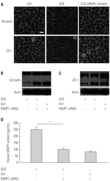
GCE leads to disruption of the cellular tight junction We treated cells with GCE for 24 h and stained them with anti- occludin or anti-ZO-1 antibody, since these proteins are impor- tant indicators for the disruption of the cellular tight junction:
Occludin plays a crucial role in the maintenance of tight junc- tions and junction remodeling, while ZO-1 is involved in the establishment of the belt-like tight junctions. In addition, these proteins are known to closely interact, although the char- acteristics and functional significance of the interaction are poorly understood.
16As shown in Fig. 2A, control cells (untreat- ed) showed strong immunofluorescent staining, with clear in- tercellular borders, while the GCE-stimulated cells displayed irregular and smeared staining patterns. Consistent with tight junction disruption, GCE treatment significantly and dose- dependently decreased TEER (Fig. 2B). Meanwhile, occludin and ZO-1 mRNA expression did not significantly differ be- tween controls and GCE-treated cells (Fig. 2C and D), where- as occludin and ZO-1 protein expression decreased depend- ing on the dose of GCE treatment (Fig. 2E and F).
GCE-induced MMP1 expression leads to tight junction disruption
To determine the relationship between tight junction disrup-
tion and MMP1 release, cells were pretreated with an MMP
inhibitor, GM6001, that can inactivate MMP1, 2, 3, 7, 8, 9, 12,
14, and 26 for 1 h prior to the 24-h treatment with GCE. Con-
focal microscopy analysis showed that GCE-treated cells had
discontinuous tight junctions, together with the altered local-
ization of occludin and ZO-1, whereas GM6001 prevented the
cellular junctions from being disrupted (Fig. 3A). Further-
more, we determine that MMP1 siRNA transfected cells main-
tained occludin and ZO-1 protein expression even after GCE treatment via immunoblotting analysis (Fig. 3B and C). The transfection efficiency of MMP-1 siRNA was confirmed by the measurement of MMP1 protein in the supernatant (Fig. 3D) ETs1 and sP1 regulate GCE-induced MMP-1 expression that leads to tight junction disruption
Since it was reported that MMP1 expression and dysregula- tion correlates with asthma severity relating to pulmonary ar- chitecture remodeling and inflammation,
20we investigated the mechanisms by which GCE regulates MMP1 expression.
First, we analyzed mRNA expressions of transcriptional fac- tors, ETS1, SP1, AP1, and NF-κB, known to play important roles in MMP1 regulation.
21,22As shown in Fig. 4A and B, ETS1 and SP1 mRNA expression was highest at 6 h and 1 h, respectively, after GCE treatment (2.7-fold and 1.5-fold, respectively, p<0.01, compared with GCE untreated cells as the control; n=3). How- ever, AP1 and NF-κB mRNA expression did not change (data not shown). Through EMSA analysis, we confirmed that ETS1 abundantly translocated into the nucleus at 1 h after GCE treat- ment, while SP1 did so at 2 h (Fig. 4C and D). Second, we evalu- ated whether ETS1 and SP1 can regulate GCE-induced MMP1 Fig. 1. Expression of matrix metalloproteinases (MMPs) and TIMP in human airway epithelial cells (NCI-H292) treated with German cockroach extract (GCE) for up to 48 h. (A) MMP1, 2, 7, 9, and 12 mRNA expressions at 6 h after GCE treatment. Time-dependent mRNA expression of (B) MMP1, (C) MMP9, and (D) TIMP1 was analyzed using real-time PCR. (E) MMP1 and (F) MMP9 protein expression were analyzed in the cell culture supernatants. Data are presented as the mean±SEM obtained in three independent experiments. *p<0.05,
†p<0.01,
‡p<0.001, compared with control (ctrl, media only) or cells at 0 h of media only. NS, not significant.
25
20
15
10
5
0
1200 1000 800 600 400 200 0
70 60 50 40 30 20 10 0 3.0 2.5 2.0 1.5 1.0 0.5 0.0 30 25 20 15 10 5 0
25
20
15
10
5
0
MMP1 0 h 1 h 2 h 6 h 12 h 24 h
0 h 1 h 2 h 6 h 12 h 24 h
6 h 12 h 24 h 48 h 6 h 12 h 24 h 48 h 0 h 1 h 2 h 6 h 12 h 24 h MMP2
NS
NS NS Ctrl
GCE
Ctrl GCE
Ctrl GCE Ctrl GCE GCE
GCE
NS
NS NS
NS NS
NS NS
NS
NS
NS
†
†
†
†
‡
*
†
* * *
*
†
*
*
*
*
MMP7 MMP9 MMP12
Human MMPs mRNA (fold increase) Human total MMP1 protein (pg/mL) Human total MMP9 protein (pg/mL) Human TIMP1 mRNA (fold increase) Human MMP1 mRNA (fold increase)
Human MMP9 mRNA (fold increase)
A
C
E
B
D
F
expression using siRNA transfection. The results indicated that ETS1, ETS2, and SP1 siRNA inhibit GCE-induced MMP1 expression (74.4, 75.2, and 60.7% reduction, respectively, compared with the GCE-stimulated cells; n=3) (Fig. 4E and F).
Furthermore, ETS1 and SP1 siRNA transfected cells were treated with GCE for 24 h and lysed for immunoblotting anal- ysis to investigate tight junction protein expression. It was previously confirmed that cells transiently transfected with ETS1 and SP1 siRNA in media only show no difference in oc- cludin and ZO-1 protein probed with anti-occludin and ZO-1 (Supplementary Fig. 1, only online). In the result, the occludin protein expression did not significantly change against siRNA
ETS1 and SP1 transfection after GCE treatment, while ZO-1 protein expressions in ETS1 and SP1 siRNA transfected cells were not influenced by GCE (Fig. 4G and H).
ERK through TLR2 controls GCE-induced MMP-1 expression, leading to tight junction protein decrease Furthermore, we investigated GCE-induced MAPK pathway activation using specific antibodies against phosphorylated ERK1/2, p38, and JNK (data not shown). While GCE activated the phosphorylation of ERK1/2 molecules (Fig. 5A), it did not activate p38 and JNK molecules (data not shown). Cell treat- ment with PD98059, a potent inhibitor of MAPK/ERK, signifi- Fig. 2. GCE induces tight junction and its proteins change. (A) Occludin and ZO-1 localization on GCE-stimulated cells was detected by immunofluores- cence staining with anti-occludin or ZO-1 and observed under confocal microscopy. Scale bar represents 10 µm. (B) Transepithelial electrical resistance was measured after 24 hours of GCE treatment. (C) Occludin and (D) ZO-1 mRNA expression by time-dependent GCE treatment. *p<0.05 and
†p<0.01, compared with control (ctrl, cells in media only). (E) Occludin and (F) ZO-1 protein expression by dose-dependent GCE treatment probed with anti-occlu- din or ZO-1. Control (Ctrl) is protein from cells in media only probed with anti-occludin or ZO-1. Data are presented as the mean±SEM obtained in three in- dependent experiments. Representative images are presented. GCE, German cockroach extract; ZO, zonula occludens; NS, not significant.
4
3
2
1
0 5
4
3
2
1
0
300 250 200 150 100 50
0 Ctrl GCE 10 µg/mL GCE 50 µg/mL
0.5 h 1 h 2 h 6 h 12 h 24 h 0.5 h 1 h 2 h 6 h 12 h 24 h
Ctrl 2 10 30 50 (µg/mL)
Ctrl 2 10 30 50 (µg/mL) NS
NS NS NS
Ctrl GCE Ctrl
GCE
GCE
GCE Ctrl
GCE
Occludin
ZO-1
NS
NS
NS
NS
NS
NS NS
NS
*
†
Human ZO-1 mRNA (fold increase)
Human occludin mRNA (fold increase) TEER (Ω · cm
2)
A B
D
F C
E
Ctrl GCE
Occludin
Actin
ZO-1
Actin
cantly decreased GCE-induced MMP1 expression (n=3; 65.9%
reduction, compared with that in the GCE only-treated cells;
p<0.05) (Fig. 5B). Furthermore, this treatment prevented the GCE-induced translocation of ETS1 and SP1 (Fig. 5C and D).
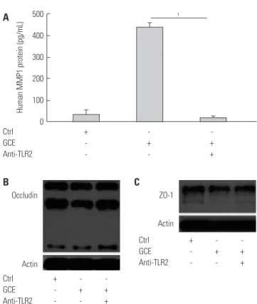
While PD98059 inhibitor did not affect GCE-induced occlu- din decrease, it did prevent GCE-induced decreases in ZO-1 levels (Fig. 5E and F). Additionally, we investigated whether TLRs, which play a key role in innate immune system re- sponses, can affect GCE-induced MMP1 release, leading to tight junction alterations. A specific anti-TLR2 antibody pre- vented GCE-mediated MMP1 expression (97% of reduction,
compared with that in the GCE only-treated cells; n=3; p<0.01) (Fig. 6A) and GCE-induced decrease of occludin protein ex- pression as well (Fig. 6B). However, ZO-1 protein expression was not affected by anti-TLR antibody pretreatment (Fig. 6C).
DIsCUssION
Cockroach allergen was shown to contain a protease that as- sociates with PAR2 and TLR2 motifs, which can modulate in- flammatory cytokine and chemokine activity, leading to direct activation of the innate immune system.
5,7Furthermore, cock- roach extract increases bronchial airway epithelial permeabil- ity, although the roles of different components have not been elucidated.
23Here, we demonstrated in vitro that GCE signifi- cantly induces MMP1 expression. MMPs are key molecules in- volved in cell proliferation and migration, tissue growth, re- modeling, and regeneration and have high proteolytic activity and broad substrate specificity.
10Recently, MMPs were identi- fied as likely to be involved in the pathogenesis of asthma, pri- marily in the asthma-associated airway remodeling.
24Although the role of MMP9 in asthma pathogenesis has been extensive- ly studied, the levels of other MMPs, including MMP-1, 2, 3, or 12, in individuals with asthma were shown to be increased as well, indicating that their levels in sputum, BAL, and exhaled breath condensates correlate with disease exacerbation.
25,26Additionally, it was reported that MMP1 is highly expressed in airway epithelial cells, inflammatory cells, and even airway smooth muscle cells in asthmatic patients.
20,27,28Many studies have indicated that various aeroallergens, such as house dust mite and pollen, exhibit proteolytic activi- ties, which may contribute to asthma pathogenesis by damag- ing the barrier function of the airway epithelium.
29,30However, only a small number of studies have investigated the effects of cockroach allergen on the epithelial barrier.
5In this study, we selected occludin and ZO-1 as the indicators of tight junction disruption, because occludin is a key component of a tight seal formed between the cells and because ZO-1 directly in- teracts with occludin to ensure proper localization of the tight junctions.
16We showed that GCE induces morphological changes and a GCE dose-dependent reduction of tight junc- tion protein levels, in combination with an increase in the epi- thelial permeability, suggesting that GCE may lead to the degra- dation of epithelial tight junction proteins. Previously, cockroach antigen treatment was shown to induce VEGF expression and decrease electrical resistance in bronchial epithelial cells, al- though the cockroach role was not investigated.
23To date, the activity of cockroach allergen has been explained by its effects on PAR2, which can lead to disease pathogenesis or exacerba- tion.
5It was also suggested that these cockroach allergen ac- tivities mediated by PAR2 may be further complicated by the involvement of TLRs, showing that cockroach allergen con- tains both TLR4 and TLR2 motifs
7and that PAR2 and TLR4 Occludin
ZO-1
Occludin
Actin
ZO-1
Actin
GCE + + -
Ctrl - - +
MMP1 siRNA - + +
GCE + + -
Ctrl - - +
MMP1 siRNA - + +
GCE + + -
Ctrl - - +
MMP1 siRNA - + + Ctrl GCE GCE+MMPs inhibitor A
B C
300 250 200 150 100 50 0
*
Human MMP1 protein (pg/mL)
D
Fig. 3. GCE-induced MMP1 expression influences tight junction disrup-
tion. (A) Representative images show occludin and ZO-1 localization in
cells pretreated with MMP inhibitor GM6001 and treated with GCE. Scale
bar represents 10 µm. (B) Occludin and (C) ZO-1 protein was detected in
cells transiently transfected with MMP1 siRNA and treated with GCE by
anti-occludin or ZO-1. Representative images of three independent ex-
periments are presented. (D) MMP1 protein expression abundance was
measured in the supernatant of cells transiently transfected with MMP1
siRNA and treated with GCE by ELISA. *p<0.05, compared with the GCE
only-treated cells. GCE, German cockroach extract; MMP, matrix metallo-
proteinase; ZO, zonula occludens; siRNA, small interfering RNA.
Ctrl + - - -
GCE - + + +
ETS1 siRNA - - + -
ETS2 siRNA - - - +
GCE + + + - - -
Ctrl - - - + + +
ETS1 siRNA - + - - + -
SP1 siRNA - - + - - +
GCE + + + - - -
Ctrl - - - + + +
ETS1 siRNA - + - - + -
SP1 siRNA - - + - - +
Ctrl + - - GCE - + +
SP1 siRNA - - +
160 140 120 100 80 60 40 20 0
250 200 150 100 50 0
* *
†Human MMP1 protein (pg/mL) Human MMP1 protein (pg/mL)
E
G H
F
Fig. 4. GCE-induced MMP1 transcriptional regulation influences tight junction disruption. (A) ETS1 and (B) SP1 mRNA was expressed in GCE-treated cells. (C and D) Nuclear extracts were collected to assess the translocation of ETS1 and SP1. (E and F) MMP1 protein was measured in transiently trans- fected cells with ETS1, ETS2, or SP1 siRNA and treated with GCE. (G and H) Cell lysates were collected to assess the expression of occludin and ZO-1 pro- tein. Data are represented as the mean±SEM obtained in three independent experiments. *p<0.05,
†p<0.01, compared with cells in media only or treated with GCE. Images are representative of three individual experiments. GCE, German cockroach extract; MMP, matrix metalloproteinase; ZO, zonula oc- cludens; siRNA, small interfering RNA; NS, not significant.
2.0
1.5
1.0
0.5
0.0 6
5 4 3 2 1
0 0 h 1 h 2 h 6 h 12 h 24 h
1 h 2 h
GCE GCE
ETS1
1 h 2 h
SP1 0 h 1 h 2 h 6 h 12 h 24 h
NS NS
*
*
Ctrl GCE Ctrl
GCE
NS NS NS
†
* NS
NS
NS
Human SP1 mRNA (fold increase)
Human ETS1 mRNA (fold increase)
B
D A
C
Occludin
Actin
ZO-1
Actin
signal cooperatively.
31,32Our data demonstrated that GCE induces MMP1 expression and tight junction disruption, which play important roles in disease-associated airway remodeling.
19Therefore, we inves- tigated whether MMP1 cleaves occludin and ZO-1, the compo- nents of tight junctions. In the result, GM6001, a potent MMP inhibitor, and MMP1 siRNA prevented the degradation of tight junction proteins, showing that tight junction disruption may be increased by GCE-induced MMP1 expression in airway epi- thelial cells. MMP1 is a collagenase that can be detected in the small airways and lung parenchyma of asthmatic patients.
33,34MMP1 expression was shown to directly correlate with the airway obstruction in BAL fluid and to be associated with the development of asthma symptoms, although the mechanisms thereof remain unclear.
20,28,35Inadequate tight junction forma- tion has been confirmed in asthmatic samples, compared with normal cultures, using anti-ZO-1 and anti-occludin antibod- ies, suggesting that respiratory viruses, air pollutants, and pro- teolytically active allergens can cause epithelial desquamation through tight junction disruption and the disruption of epithe- lial barrier function.
18,20,29,30,36Although the role of MMPs in asthma pathogenesis has been Phospho p44/42
Total p44/42
Ctrl + - -
GCE - + + PD98059 - - +
Ctrl + - -
GCE - + + Anti-TLR2 - - + A
400
300
200
100
0
500 400 300 200 100
* 0
†