INTRODUCTION
Background and Purpose
Peripheral arterial occlusive disease (PAOD) is primarily caused by atherosclerosis. The purpose of treatment is to reduce the symptoms of limb ischemia such as claudication and to salvage the limbs. Interventional procedures
Korean Guidelines for Interventional Recanalization of Lower Extremity Arteries
Young Hwan Kim, MD
1, Jae Ik Bae, MD
2, Yong Sun Jeon, MD
3, Chang Won Kim, MD
4, Hwan Jun Jae, MD
5, Kwang Bo Park, MD
6, Young Kwon Cho, MD
7*, Man Deuk Kim, MD
81Department of Radiology, Keimyung University College of Medicine, Daegu 700-712, Korea; 2Mint Radiologic Clinic, Seongnam 463-950, Korea;
3Department of Radiology, Inha University College of Medicine, Incheon 400-711, Korea; 4Department of Radiology, Pusan National University College of Medicine, Busan 602-739, Korea; 5Department of Radiology, Seoul National University College of Medicine, Seoul 110-744, Korea;
6Department of Radiology, Sungkyunkwan University College of Medicine, Seoul 135-710, Korea; 7Department of Radiology, Eulji University College of Medicine, Seoul 139-872, Korea; 8Department of Radiology, Yonsei University College of Medicine, Seoul 120-752, Korea
Peripheral arterial occlusive disease caused by atherosclerosis can present with intermittent claudication or critical limb ischemia. Proper diagnosis and management is warranted to improve symptoms and salvage limbs. With the introduction of new techniques and dedicated materials, endovascular recanalization is widely performed for the treatment of peripheral arterial occlusive disease because it is less invasive than surgery. However, there are various opinions regarding the appropriate indications and procedure methods for interventional recanalization according to operator and institution in Korea. Therefore, we intend to provide evidence based guidelines for interventional recanalization by multidisciplinary consensus. These guidelines are the result of a close collaboration between physicians from many different areas of expertise including interventional radiology, interventional cardiology, and vascular surgery. The goal of these guidelines is to ensure better treatment, to serve as a guide to the clinician, and consequently, to contribute to public health care.
Index terms: Guideline; Peripheral arterial disease; Diagnosis and management; Intervention Korean J Radiol 2015;16(4):696-722
(angioplasty or stent placement) for the recanalization of lower extremity arteries in patients with chronic stenosis or occlusion of lower extremity arteries have been widely performed in recent years as medical equipment and technology have developed.
The indications for these interventional procedures are gradually increasing with the rapid development of
pISSN 1229-6929 · eISSN 2005-8330
Received March 16, 2015; accepted after revision April 21, 2015.
This article is based on a study first reported in the Journal of the Korean Society of Radiology [Kim YH, Bae JI, Jeon YS, Kim CW, Jae HJ, Park KB, Jo YK, Kim MD. Guideline for interventional recanalization of lower extremity artery. J Korean Soc Radiol 2014 Nov;71(5):205- 230][Kim YH, Bae JI, Jeon YS, Kim CW, Jae HJ, Park KB, Cho YK, Kim MD. Erratum: Guideline for interventional recanalization of lower extremity artery. J Korean Soc Radiol 2014 Dec;71(6):329-329].
*Current affiliation: Department of Radiology, Kangdong Sacred Heart Hospital, Hallym University College of Medicine, Seoul, Korea.
Corresponding author: Young Hwan Kim, MD, Department of Radiology, Dongsan Medical Center, Keimyung University College of Medicine, 56 Dalseong-ro, Jung-gu, Daegu 700-712, Korea.
• Tel: (8253) 250-7767 • Fax: (8253) 250-7766 • E-mail: yhkim68@dsmc.or.kr
This is an Open Access article distributed under the terms of the Creative Commons Attribution Non-Commercial License (http://
creativecommons.org/licenses/by-nc/3.0) which permits unrestricted non-commercial use, distribution, and reproduction in any medium, provided the original work is properly cited.
sources described above then developed a new proposal by implementing the rule to suitably transform the guidelines of other countries for domestic circumstances in Korea.
Subject of Application and Scope
Adult men and women with limb ischemia symptoms caused by chronic stenosis or occlusion of the lower extremity arteries that occurred due to atherosclerosis or diabetes were targeted. Acute limb occlusion and chronic limb occlusion caused by other underlying diseases other than diabetes or atherosclerosis were excluded. The guidelines were limited to proposing recommendations for interventional recanalization, therefore the proposal of recommendations for medical treatment, exercise therapy, and surgical operations were excluded. Cases with lack of evidence and controversial cases were excluded from the guidelines.
Level of Evidence and Classification of Recommendation The level of evidence (LOE) and the classification of recommendations (COR) followed the criteria used in the American College of Cardiology/American Heart Association (ACC/AHA) 2005 guideline. COR was separated into three categories: Recommendation I (strong recommendation), Recommendation II (weak recommendation), and
Recommendation III (contraindication). Recommendation I was defined as conditions for which there was solid evidence for and/or general agreement that a given procedure or treatment is effective, useful, and beneficial and will not be changed by further research. Recommendation II was defined as conditions with conflicting evidence and/
or a divergence of opinion about the usefulness/efficacy of a procedure or treatment. IIa was defined as cases for which the weight of evidence/opinion was in favor of usefulness/efficacy. IIb was defined as cases for which the usefulness/efficacy was less well established by evidence/
opinion. Recommendation III was defined as conditions for which there was evidence and/or general agreement that a procedure/treatment is not useful/effective and may be harmful in some cases (Table 1). LOE was classified into three steps: A, B, and C. Evidence A was defined as data derived from multiple randomized clinical trials or meta- analysis. Evidence B was defined as data derived from a single randomized clinical trial or non-randomized studies.
Evidence C was defined as only consensus opinion of experts, case studies, or standard of care (Table 2).
equipment and procedures, and there are a variety of views about the indications for patients and procedure methods among medical institutions and operators. In the United States, the American Heart Association (AHA) presented guideline recommendations for interventional procedures in lower extremity arteries in 2005 and announced an amendment in 2011 as procedures developed. In Europe and North America, Transatlantic Intersociety Consensus (TASC), a working group formed in 2001 and centered on interventional radiology, vascular surgery, and cardiology, issued guidelines for interventional procedures and a revision in 2007. Furthermore, in 2011, new guidelines were issued with the European Heart Association as the focus. Through these efforts, by establishing the standard for procedures and raising the awareness of physicians on the front lines of medicine to the importance of the procedures, a cautious approach is encouraging. Moreover, these guidelines can be used as basic standards for social insurance wages and judgment standard for reducing medical costs.
However, even though a number of studies like this have been conducted, correct clinical guidelines for our situation in Korea have not yet been established. Thus, experts from academies (the Korean Society of Interventional Radiology [KSIR], the Korean Society for Vascular Surgery, the Korean Society of Interventional Cardiology, and the Korean Society of Radiology and Nursing) related to interventional procedures in Korea came together and agreed to develop clinical guidelines. We would like to agree on the proposal of recommendation by presenting evidence-based treatment recommendations through a multidisciplinary approach, to serve as a guide for interventional procedures by providing up-to-date and accurate information to health care providers working at primary, secondary and tertiary hospitals. Furthermore, we would like to help patients themselves choose appropriate medical services by recognizing accurate information and further contribute to public health promotion.
Guideline Development Method
Considering that the development of de novo domestic clinical guidelines is a difficult undertaking, these guidelines were developed by adapting the pre-existing guidelines of other countries. But if there were no existing guidelines, we evaluated existing documents with good quality using the systemic document review methodology.
We proposed initial recommendations based on the two
Independence of Support and Editing
This study was developed as a project for creating the clinical guidelines of the Korean Society of Radiology and the KSIR. During the entire process of guideline development, there was no influence from the Korean Society of Radiology and the KSIR and there was no external support from any other academies, institutions and interest groups. All members who participated in this clinical guideline development process have signed agreements confirming that there was no conflict of interest in connection with the studies.
Guideline Revision
We will conduct revision at intervals of 3–5 years by using an adaptation development method when new examination and treatment methods for interventional recanalization of lower extremity arteries are introduced and research results are accumulated.
Guideline Adaptation Process
Configuration and Role of the Committee
The steering committee was composed of the president and executives of the KSIR. The steering committee fixed the subject and the goal, assigned the guideline development chairman and committee members and approved the guideline development budget. The guideline development committee was made up of 20 committee members including a chairman and a secretary.
Guideline development committee members were educated about guideline development methodology and the
evaluation of guidelines based on the Appraisal of Guideline for Research and Evaluation II (AGREE II). This was
accomplished by inviting methodology experts and hosting workshops. After the workshops, we discussed the purpose of the guidelines, the range of development including writing topics, the subjects of application and user groups, development method, the determination of the LOE, COR, the selection of the consensus development method, internal and external review processes, revision processes and formation of the committee of detail associated with guideline development during the first conference of the guideline development committee. The committee of detail was composed of the guideline evaluation committee, the writing committee, and the editing committee.
Five members were involved in the guideline evaluation committee and evaluated the pre-existing guidelines based on AGREE II. Eight members were involved in the writing committee and were in charge of drawing up the guideline draft and the proposal of recommendations. The editing committee was composed of 5 members and was in charge of reviewing the recommendation levels, the LOE, and the guideline draft by performing peer review.
Evaluation and Selection of Guidelines in Other Countries In order to select a high-quality guideline for reference in the adaptation process, we searched for existing guidelines.
We recovered 70 documents by mixing search index words such as PAOD, endovascular treatment, limb ischemia and guidelines with the use of the PUBMED, OVID, SCOPUS, WEB OF SCIENCE, and COCHRANE search engine.
The guideline evaluation committee determined the inclusion and exclusion criteria for quote-worthy documents among the obtained documents. The inclusion criteria were set as evidence-based guidelines, international guidelines written in English, and recent guidelines written since Table 1. Classification of Recommendations
Class Description
I Conditions for which there is evidence for and/or general agreement that given procedure or treatment is beneficial, useful, and effective
II Conditions for which there is conflicting evidence and/or divergence of opinion about usefulness/efficacy of procedure or treatment
IIa Weight of evidence/opinion is in favor of usefulness/efficacy IIb Usefulness/efficacy is less well established by evidence/opinion
III Conditions for which there is evidence and/or general agreement that procedure/treatment is not useful/effective and is some cases may be harmful
Table 2. Levels of Evidence
Level Description
A Data derived from multiple randomized clinical trials or meta-analysis
B Data derived from single randomized clinical trial or nonrandomized studies
C Only consensus opinion of experts, case studies, or standard of care
2005. Guidelines that did not represent the organization, those written by one person and translations of single guidelines were excluded.
A total of 6 guidelines were chosen based on this inclusion criterion. 4 guideline evaluation committee members evaluated these guidelines based on AGREE II, which is the most commonly used tool internationally for the quality assessment of guidelines. AGREE II is made up of 23 sub-items within 6 assessment categories and each item is given a score on a 7-point Likert scale. All guidelines evaluation committee members received systemic education about AGREE II with methodology experts through case- by-case analysis by participating in the workshop. In comparing the results of 4 evaluation committee members, cases for which the difference between the highest record and the lowest record was more than 4 points were defined as a disagreement. If more than 5 items were disagreed upon, reevaluation was conducted after coordinating opinions about inconsistent evaluation items. Based on the re-evaluated results after opinion coordination, 5 guidelines that had standardized scores of more than 50%
in all categories and in particular, over 70% in the rigour of development category, were finally selected (1-5).
Deduction and Selection of Key Question
The guideline development committee shared roles by assigning committee members according to sub-themes of writing items for the deduction of the clinical question at the second conference. The third conference of the guideline development committee selected the final key question after reviewing whether population, intervention, comparison, and outcome (PICO), the essential structural component of the key question, was well equipped and appropriate as a clinical question. The key question was agreed upon using the nominal group technique. Consensus was defined as more than 75% of the panel selecting 1 and 2 on the 5-Likert scale (1, agree entirely; 2, agree generally; 3, agree partially; 4, do not agree generally; 5, do not agree entirely). If the consensus was less than 75%, a second round of voting was carried out after discussion and a modification of phrases. If agreement was not reached in the second round of voting, the key question was dismissed.
Ten development committee members participated in the panel. Seventeen of a total of 40 key questions that were drawn up were agreed upon by committee members and were selected as the final clinical question. The selected key questions were arranged and the <Key Question PICO
Data Extraction Form> was developed (Table 3).
Document Search
Because of the adaptation development process, the deduction of the proposal of recommendations for the clinical question mostly referred to existing selected guidelines. However, for clinical questions without existing guidelines, a library search expert was involved in order to find good quality documents by sub-themes. PubMed and Cochrane library databases were used for evidence based documents searches. The searching parameters were restricted to publications before June 30, 2013, studies done on humans only, and studies published in English.
After developing the searching formulas for each sub-theme, the search results were reviewed and evidential studies were selected for each related key question. When a more recent systemic review or meta-analysis study was found, papers previously published with a lower LOE were excluded along with the corresponding case reports. The search terms for PAOD were Peripheral Arterial Disease (MeSH), and Arterial Occlusive Disease (MeSH), and related search terms were input by connecting AND/OR.
The search formulas by sub-themes using Mesh terms in PubMed and Cochrane library were as follows:
Diagnosis of Peripheral Arterial Occlusive Disease (Peripheral Arterial Disease/diagnosis [MeSH]) AND (Arterial Occlusive Diseases/diagnosis [MeSH]) AND (Ankle Brachial Index [MeSH Major Topic] OR Ultrasonography, Doppler, Duplex [MeSH] OR Computed Tomography Angiography OR Magnetic Resonance Angiography [MeSH]
OR Angiography [MeSH])
Patient Care for Contrast Medium-Related Nephrotoxicity (Kidney Diseases/prevention and control [MeSH] OR Kidney Diseases/therapy [MeSH]) AND (Contrast induced [Title]) AND (Angiography [MeSH])
Indication of Recanalization and the Establishment of Procedure Plan
(Peripheral Arterial Disease/therapy [MeSH]) AND (Arterial Occlusive Diseases/therapy [MeSH]) AND (Endovascular Procedures/instrumentation, methods, therapy [MeSH])
Interventional Procedure Method
(Peripheral Arterial Disease [MeSH]) AND (Arterial Occlusive Diseases [MeSH]) AND (Endovascular Procedures/
Table 3. Key Question PICO Data Extraction Form Sub-ThemesClinical QuestionsP (Population)I (Intervention)C (Comparison)O (Outcome) DiagnosisScreening test for diagnosis of peripheral arterial occlusive diseaseIntermittent claudication, critical limb ischemia patients or adult with risk factor of peripheral arterial occlusive disease
Hemodynamic examinationImaging examinationSensitivity, specificity Vascular imaging examination for localization of peripheral arterial occlusive disease
Patients with peripheral arterial occlusive diseaseNoninvasive imaging examinationInvasive imaging examinationSensitivity, specificity Indications for interventional recanalization
Implementation subject of recanalization (including intervention and surgery) of lower extremity artery (divided into intermittent claudication and critical limb ischemia)
Patients with intermittent claudication or critical limb ischemiaIntervention or surgeryMedication or exercise therapyImprovement of symptom, quality of life, morbidity, mortality Which lesion can be applied for interventional recanalization of lower extremity artery compared with surgery?
Patients with intermittent claudication or critical limb ischemiaInterventionSurgeryPatency rate, quality of life, amputation free survival rate, morbidity, mortality Patients care before interventional procedure
Laboratory test evaluating contrast medium related renal injury and preprocedural care for preventing renal injury Patients with intermittent claudication or critical limb ischemia who have plan for interventional recanalization Laboratory test or care for preventing contrast medium related renal injury
ObservationIncidence of contrast induced nephropathy, morbidity, mortality Is drug therapy needed before and after procedure?Patients with intermittent claudication or critical limb ischemiaDrug therapyObservationMorbidity, mortality Establishment of procedure planSelection criteria of target artery for treatment Patients with intermittent claudication or critical limb ischemiaInterventionConservative treatmentImprovement of symptom, quality of life, morbidity, mortality Treatment plan of multiple lesionsPatients with multiple stenotic or occlusive lesions in lower extremity artery
InterventionConservative treatmentImprovement of symptom, quality of life, morbidity, mortality Aorto-iliac artery interventional procedure
In which case is primary stent placement needed for interventional racanalization of aortoiliac artery?
Patients with stenotic or occlusive lesions in aortoiliac arteryPrimary stent placementBalloon angioplastyPatency rate, complication When is bailout stent placement needed during interventional recanalization of aortoiliac artery Patients with stenotic or occlusive lesions in aortoiliac arteryBailout stent placementBalloon angioplastyPatency rate, complication Is kissing stent needed for treatment of lesions involving aortoiliac bifurcation?Patients with stenotic or occlusive lesions in aortoiliac bifurcationKissing stent placementStent placement, balloon angioplastyPatency rate, complication
instrumentation, methods [MeSH]) AND (Iliac Artery [MeSH]
OR Popliteal Artery [MeSH] OR Femoral Artery [MeSH] OR Infrapoplliteal Artery) AND (Angioplasty [MeSH] OR Stents [MeSH] OR Drug-Eluting Balloon [All Fields] OR Drug-Eluting Stents [MeSH] OR Stent Graft [All Fields] OR Atherectomy [MeSH] OR Cryoplasty [All Fields])
Deduction of Recommendations
Writing committee members who were assigned to specific sub-themes drew up the proposal of recommendations for clinical questions. A single draft of the proposal of recommendations was deduced by collecting common information and removing unnecessary information after analyzing the recommendations extracted from the selected guidelines. If there was no data for reference in existing guidelines of clinical questions, a new proposal of recommendations was developed based on the results of evaluating the quality of documents through the documents search and review process. Thus a total of 63 proposals of recommendations were drawn up. We created a recommendation data extraction form, which the panel used as a reference during the Delphi consensus process.
At the 4th guideline development committee member conference, a preliminary examination of the draft was conducted under the agreement of 11 development
committee members who participated in the conference. In this process, vague phrases and sentences were checked and the draft of the proposal of recommendations was modified prior to the Delphi consensus process. Fourteen of the recommendations were deleted because no final agreement was reached due to opinions that information remained ambiguous and difficult to understand despite modification of phrases or sentences or the recommendations
were unpractical in Korea, etc. The remaining 49 recommendations were selected by Delphi consensus survey and a <Recommendation Data Extraction Form> was created (Table 4). The nominal group consensus was used for the agreement method and the criteria of adoption and rejection was the same as the selection of key questions.
Agreement Method and Panel Selection for Recommendation Adoption
For formal mutual agreement of the final adoption of recommendations, a modified Delphi technique was applied. By sending an official document for cooperation explaining the objectives and necessity of a guideline for interventional recanalization of the lower extremity Table 3. Key Question PICO Data Extraction Form (Continued) Sub-ThemesClinical QuestionsP (Population)I (Intervention)C (Comparison)O (Outcome) Femoro-popliteal artery interventional procedure
Is primary stent placement needed for short segment lesion of femoropopliteal artery? Patients with short segment stenotic or occlusive lesions in femoropopliteal arteryPrimary stent placementBalloon angioplastyPatency rate, limb salvage rate, complication When is bailout stent placement needed during interventional recanalization of femoropopliteal artery?
Patients with stenotic or occlusive lesions in femoropopliteal arteryBailout stent placementBalloon angioplastyPatency rate, limb salvage rate, complication Is subintimal angioplasty needed for long segment occlusive lesion of femoropopliteal artery?
Patients with long segment occlusive lesions in femoropopliteal arterySubintimal balloon angioplastyStent placementPatency rate, limb salvage rate, complication Are new procedures more effective than plain old balloon angioplasty or stent placement for interventional recanalization of femoropopliteal artery?
Patients with stenotic or occlusive lesions in femoropopliteal arteryPlain old balloon angioplasty or stent placement Drug eluting balloon angioplasty, drug eluting stent, cutting balloon angioplasty, atherectomy, laser therapy, stent graft
Patency rate, limb salvage rate, complication Infrapopliteal artery interventional procedure
How would guide wire passage during interventional recanalization of infrapopliteal artery?
Patients with stenotic or occlusive lesions in infrapopliteal arteryIntraluminal guide wire passageSubintimal guide wire passageSuccess rate, patency rate, complication In which case is stent placement needed for interventional racanalization of infrapopliteal artery?
Patients with stenotic or occlusive lesions in infrapopliteal arteryStent placementBalloon angioplastyPatency rate, limb salvage rate, complication
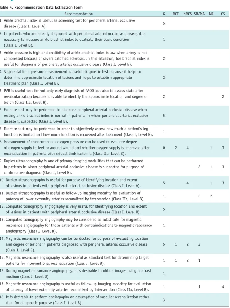
Table 4. Recommendation Data Extraction Form
Recommendation G RCT NRCS SR/MA NR CS
1. Ankle brachial index is useful as screening test for peripheral arterial occlusive
disease (Class I, Level A). 5
2. In patients who are already diagnosed with peripheral arterial occlusive disease, it is necessary to measure ankle brachial index to evaluate their basic condition
(Class I, Level B).
1
3. Ankle pressure is high and credibility of ankle brachial index is low when artery is not compressed because of severe calcified sclerosis. In this situation, toe brachial index is useful for diagnosis of peripheral arterial occlusive disease (Class I, Level B).
2
4. Segmental limb pressure measurement is useful diagnostic test because it helps to determine approximate location of lesions and helps to establish appropriate treatment plan (Class I, Level B).
2
5. PVR is useful test for not only early diagnosis of PAOD but also to assess state after revascularization because it is able to identify the approximate location and degree of lesion (Class IIa, Level B).
2 2
6. Exercise test may be performed to diagnose peripheral arterial occlusive disease when resting ankle brachial index is normal in patients in whom peripheral arterial occlusive disease is suspected (Class I, Level B).
5
7. Exercise test may be performed in order to objectively assess how much a patient’s leg function is limited and how much function is recovered after treatment (Class I, Level B). 1 8. Measurement of transcutaneous oxygen pressure can be used to evaluate degree
of oxygen supply to feet or around wound and whether oxygen supply is improved after recanalization in patients with critical limb ischemia (Class IIa, Level B).
0 2 4 1 3
9. Duplex ultrasonography is one of primary imaging modalities that can be performed in patients in whom peripheral arterial occlusive disease is suspected for purpose of confirmative diagnosis (Class I, Level B).
1 2 1 3
10. Duplex ultrasonography is useful for purpose of identifying location and extent
of lesions in patients with peripheral arterial occlusive disease (Class I, Level A). 5 4 1 3 11. Duplex ultrasonography is useful as follow-up imaging modality for evaluation of
patency of lower extremity arteries recanalized by intervention (Class IIa, Level B). 1 2 2 12. Computed tomography angiography is very useful for identifying location and extent
of lesions in patients with peripheral arterial occlusive disease (Class I, Level B). 5 13. Computed tomography angiography may be considered as substitute for magnetic
resonance angiography for those patients with contraindications to magnetic resonance angiography (Class I, Level B).
1
14. Magnetic resonance angiography can be conducted for purpose of evaluating location and degree of lesions in patients diagnosed with peripheral arterial occlusive disease (Class I, Level B).
5 1 2 3
15. Magnetic resonance angiography is also useful as standard test for determining target
patients for interventional recanalization (Class I, Level B). 1 1 2 1
16. During magnetic resonance angiography, it is desirable to obtain images using contrast
medium (Class I, Level B). 1
17. Magnetic resonance angiography is useful as follow-up imaging modality for evaluation
of patency of lower extremity arteries recanalized by intervention (Class IIa, Level B). 1 1 4 18. It is desirable to perform angiography on assumption of vascular recanalization rather
than for diagnostic purpose (Class I, Level B). 3
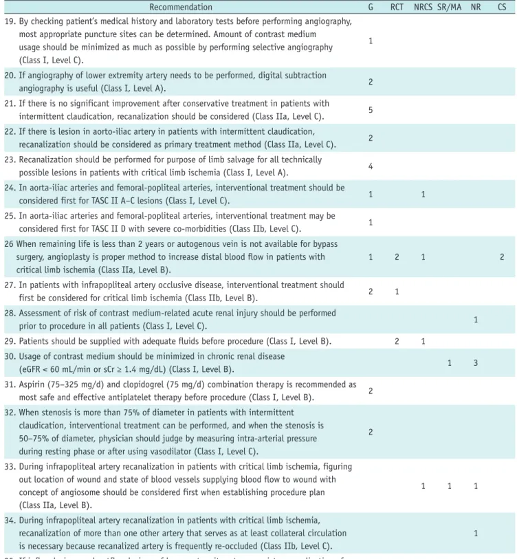
Table 4. Recommendation Data Extraction Form (Continued)
Recommendation G RCT NRCS SR/MA NR CS
19. By checking patient’s medical history and laboratory tests before performing angiography, most appropriate puncture sites can be determined. Amount of contrast medium
usage should be minimized as much as possible by performing selective angiography (Class I, Level C).
1
20. If angiography of lower extremity artery needs to be performed, digital subtraction
angiography is useful (Class I, Level A). 2
21. If there is no significant improvement after conservative treatment in patients with
intermittent claudication, recanalization should be considered (Class IIa, Level C). 5 22. If there is lesion in aorto-iliac artery in patients with intermittent claudication,
recanalization should be considered as primary treatment method (Class IIa, Level C). 2 23. Recanalization should be performed for purpose of limb salvage for all technically
possible lesions in patients with critical limb ischemia (Class I, Level A). 4 24. In aorta-iliac arteries and femoral-popliteal arteries, interventional treatment should be
considered first for TASC II A–C lesions (Class I, Level C). 1 1
25. In aorta-iliac arteries and femoral-popliteal arteries, interventional treatment may be considered first for TASC II D with severe co-morbidities (Class IIb, Level C). 1 26 When remaining life is less than 2 years or autogenous vein is not available for bypass
surgery, angioplasty is proper method to increase distal blood flow in patients with critical limb ischemia (Class IIa, Level B).
1 2 1 2
27. In patients with infrapopliteal artery occlusive disease, interventional treatment should
first be considered for critical limb ischemia (Class IIb, Level B). 2 1
28. Assessment of risk of contrast medium-related acute renal injury should be performed
prior to procedure in all patients (Class I, Level C). 1
29. Patients should be supplied with adequate fluids before procedure (Class I, Level B). 2 1 30. Usage of contrast medium should be minimized in chronic renal disease
(eGFR < 60 mL/min or sCr ≥ 1.4 mg/dL) (Class I, Level B). 1 3
31. Aspirin (75–325 mg/d) and clopidogrel (75 mg/d) combination therapy is recommended as
most safe and effective antiplatelet therapy before procedure (Class I, Level B). 2 32. When stenosis is more than 75% of diameter in patients with intermittent
claudication, interventional treatment can be performed, and when the stenosis is 50–75% of diameter, physician should judge by measuring intra-arterial pressure during resting phase or after using vasodilator (Class I, Level C).
2
33. During infrapopliteal artery recanalization in patients with critical limb ischemia, figuring out location of wound and state of blood vessels supplying blood flow to wound with concept of angiosome should be considered first when establishing procedure plan (Class IIa, Level B).
1 1 1
34. During infrapopliteal artery recanalization in patients with critical limb ischemia, recanalization of more than one other artery that serves as at least collateral circulation is necessary because recanalized artery is frequently re-occluded (Class IIb, Level C).
1
35. If inflow lesions and outflow lesions of lower extremity artery coexist, recanalization of
inflow lesions should be performed first (Class I, Level C). 1
36. When symptoms continue even after recanalization of inflow lesions in patients with both inflow and outflow lesions, recanalization of outflow lesions should be performed (Class I, Level B).
1
37. If it is not clear whether hemodynamically significant inflow disease exists, intra-arterial pressure of each suprainguinal lesion should be measured before and after
vasodilator infusion (Class I, Level C).
1
arteries to associated academies, namely the Korean Society of Interventional Cardiology and the Korean Society for Vascular Surgery, panels with representatives and expertise from the associated academies were assigned evenly. The panel was composed of 36 members (the KSIR, 15; the Korean Society of Interventional Cardiology, 9; and the Korean Society for Vascular Surgery, 12). Members of the guideline development committee, who were involved in the deduction of clinical questions and recommendations during the process of developing the guidelines, were excluded. To assist the panel, the recommendation data extraction form and related references were provided
by e-mail. The vote was performed anonymously. After sending the disclosure sheet on conflict of interest to all panel members, the members signed the document confirming that they did not receive any support from interest groups related to the recommendation. The degree of consensus was quantitatively analyzed using a 9-Likert scale (1, strongly disagree; 9, entirely agree). In the response scale, 7–9 points were considered to agree with the recommendations. When more than 70% of the panel agreed, the proposal of recommendations was considered to have reached consensus. For recommendations without consensus, a second round of voting was conducted after Table 4. Recommendation Data Extraction Form (Continued)
Recommendation G RCT NRCS SR/MA NR CS
38. If short-term and long-term outcome is similar and there is no difference in co-morbidities in patients with critical limb ischemia accompanied by ipsilateral
femoro-popliteal artery lesion and infrapopliteal artery lesion, angioplasty is recommended first (Class IIa, Level C).
2
39. Primary stent placement is first considered in long segment stenosis or complete occlusion
of common and external iliac artery (Class I, Level A). 2 1 1
40. Bailout stent placement is performed in cases with more than 5 mm Hg pressure difference crossing lesion, more than 30% residual stenosis, or flow-limiting intimal dissection after balloon angioplasty (Class IIa, Level C).
2
41. Kissing stent can be considered first in cases where degree of risk for aorto-bifemoral bypass surgery is significant in stenotic or occlusive lesions involving aortic bifurcation and bilateral common iliac artery (Class IIb, Level C).
8
42. Primary stent placement is not recommended for short segments of femoral-popliteal
artery (Class III, Level A). 1 7 1 1
43. Bailout stent placement is recommended when there is residual stenosis of more than
30% or flow-limiting dissection after balloon angioplasty (Class IIa, Level C). 1 44. Subintimal balloon angioplasty can be performed to improve limb salvage rate in patients
who have limitations in surgical treatment and who also have occlusive lesions longer than 10 cm in femoro-popliteal artery (Class IIb, Level C).
2 1
45. Efficacy of drug-eluting stent, atherectomy, cutting balloon angioplasty, and laser therapy in interventional treatment of femoro-popliteal artery has not been established yet (Class IIb, Level A).
1 9 1
46. Angioplasty with drug eluting balloons had good patency rate compared to plain old balloon angioplasty, but clear clinical effect has not been proved yet with respect to cost and risk of bailout stent placement (Class IIb, Level A).
3 1
47. During infrapopliteal angioplasty, guide wire passage into true lumen of lesion is
primarily attempted for stenotic lesions (Class IIb, Level A). 1 3
48. During infrapopliteal angioplasty, intraluminal guide wire passage at proximal part of lesion is primarily attempted for calcified complete occlusion lesions, and if fails, tandem subintimal guide wire passage is attempted (Class IIb, Level B).
1 6
49. Primary stent placement in infrapopliteal arteries is not desirable, but it can be considered
as bailout method after balloon angioplasty (Class IIa, Level A). 6 3 4 1 5
CS = case series study, G = guideline, NR = non-systemic narrative review, NRCS = non-randomized controlled study, RCT = randomized controlled study, SR/MA = systemic review/meta-analysis
providing all information to panelists through an online discussion session on matters that required clarification.
If an agreement was not reached, the recommendations were rejected. Twenty five out of 36 panel members participated in the vote (the KSIR, 12; the Korean Society of Interventional Cardiology, 4; the Korean Society for Vascular Surgery, 9). During the first round of voting, one recommendation did not meet the agreement condition.
The agreement condition for a different recommendation was barely met, but with many disputes. After providing all information, re-voting was conducted, but the two recommendations did not reach agreement.
Guideline Drafting
Based on the adopted recommendations, the writing committee members assigned to specific sub-themes wrote a guideline draft.
Review and Guideline Determination Process
The editing committee evaluated the written guideline draft based on the selected recommendations. The guidelines were internally evaluated through an expert advisory conference to discuss the expertise of relevant academies associated with the guideline including problems in the recommendations, background and description of the evidence, etc. After the guidelines were determined by internal evaluation, the final guidelines were established through a public hearing with the relevant field experts and stakeholders.
CONTENTS
Diagnosis of Peripheral Arterial Occlusive Disease Peripheral arterial occlusive disease should be diagnosed from a combination of the patient’s symptoms, physical, hemodynamic, and vascular imaging examination.
Symptoms are intermittent claudication and critical limb ischemia (CLI). Diseases that need to be distinguished from intermittent claudication are chronic compartment syndrome, venous claudication, nerve root compression, Baker’s cyst, spinal stenosis, arthritis of foot and ankle etc. CLI should be differentiated from diabetic neuropathy, complex regional pain syndrome, nerve root compression, night cramp, and Buerger’s disease. Physical examination includes blood pressure in both arms, cardiac sound, color and temperature of foot, the presence of leg muscle atrophy, loss of leg hair, palpation of femoral artery, radial
artery, ulnar artery, brachial artery, carotid artery, popliteal artery, dorsalis pedis artery and posterior tibial artery.
Hemodynamic Examination Ankle Brachial Index
The ankle brachial index (ABI) is the ratio of the ankle artery blood pressure to the patient’s arm blood pressure.
ABI is the simplest and most non-invasive examination among the tests available for the diagnosis of PAOD because blood pressure is easily measured at the same time in both arms and ankles using a cuff. Therefore, ABI can be used as a screening test for PAOD.
Resting ABI should be measured in all patients with intermittent claudication or incurable foot wounds, all patients aged 50–69 years with diabetes or smoking history, and all persons over the age of 70 (1-6).
The possibility of wound healing and limb salvage as well as patient survival can be predicted to some extent through ABI. ABI is useful as an evaluation index of the therapeutic effect of interventional recanalization of lower extremity arteries. ABI is also measured to assess basic status in patients who are already diagnosed with PAOD.
The method of ABI measurement is as follows. Systolic blood pressure is measured using a cuff enclosing each brachial artery, dorsalis pedis artery, and posterior tibial artery in both the right and left side with the patient lying down. The same cuff should be wrapped around all four arteries and a cuff with a 10–12 cm width is usually used (1). A hand held Doppler is recommended to detect blood flow, but plethysmography or a stethoscope, and automatic blood pressure measurement devices can also be used.
Higher brachial pressure in either arm and higher ankle pressure in the dorsalis pedis artery and the posterior tibial artery are considered variables for ABI (1, 3). Under normal conditions, ankle blood pressure is 10–15 mm Hg higher than that of the brachial artery, so ABI should be more than 1.00. If the resting ABI is less than 0.9, PAOD can be diagnosed with 95% sensitivity and 100% specificity (7).
Ankle blood pressure is higher than normal when the blood vessel is not compressed because of severe calcified sclerosis of the arteries caused by diabetes, old age or dialysis. As a result, additional tests should be required for patients with diabetes, old age, and those receiving dialysis, because it is difficult to diagnose PAOD or
determine its severity in these patients using only ABI. ABI greater than 1.4 can be attributed to the severe calcified
ankle artery and additional tests are required. Typical tests that can be added are toe brachial index (TBI), pulse volume recording (PVR), transcutaneous oxygen pressure, and vascular imaging examination (1, 3). Toe pressure is usually measured by placing the cuff around the big toe or the second toe and attaching a blood volume measurement instrument to the tip of the toe. TBI is obtained by comparing with brachial pressure, similar to the ABI. The normal range of TBI is more than 0.7 and under 0.7 may indicate PAOD (1, 3).
Recommendation
1. Ankle brachial index is useful as a screening test for peripheral arterial occlusive disease (Class I, Level A).
2. In patients who are already diagnosed with peripheral arterial occlusive disease, it is necessary to measure the ankle brachial index to evaluate their basic condition (Class I, Level B).
3. The ankle pressure is high and the credibility of the ankle brachial index is low when the artery is not compressed because of severe calcified sclerosis.
In this situation, toe brachial index is useful for diagnosis of peripheral arterial occlusive disease (Class I, Level B).
Segmental Limb Pressure
Segmental limb pressure (SLP) can be used to measure arterial blood pressure divided into several segments from thigh to ankle. Systolic blood pressure is measured by wrapping a cuff around four areas (upper thigh, lower thigh, upper leg, and ankle) then inflating and deflating each cuff in a manner similar to when measuring ankle blood pressure. In some cases, measurements may be performed with only three areas (thigh, upper leg, ankle).
Measuring SLP is very helpful for establishing a treatment plan because the approximate location of lesions can be estimated. A significant difference in blood pressure between the brachial and upper thigh indicates lesions in the aorta and iliac artery, a difference between the upper thigh and lower thigh indicates lesions in the superficial femoral artery, a difference between the lower thigh and upper leg indicates lesions in the distal superficial femoral artery or popliteal artery, and a difference between the upper leg and ankle indicates stenosis of the infrapopliteal artery (1, 3).
Recommendation
1. Segmental limb pressure measurement is a useful diagnostic test because it helps to determine the approximate location of lesions and helps to establish an appropriate treatment plan (Class I, Level B).
Pulse Volume Recording
Pulsatile arterial inflow to the limbs causes periodic changes in blood volume in the leg. PVR is used to measure this change. Plethysmography is a device that records volume changes in organs or limbs. According to the measurement method, it is classified into: pneumoplethysmography, a recording wave formed by converting the volume change of air injected into the pneumatic cuff into pressure, photoplethysmography, which detects color changes in skin that occur with the cardiac beat using infrared sensors on the skin, and strain gauge plethysmography, displayed as a wave form by converting the volume change of the limb concurrent with the cardiac beat into electrical resistance using a thin rubber tube filled with mercury or indium- gallium. Among these, pneumoplethysmography is most commonly used.
Using plethysmography, not only can SLP be measured but PVR can also be obtained. In general, it senses volume changes and produces a wave form when the pressure is increased to 65 mm Hg by the injection of a suitable amount of air measured by the pneumatic cuff. Change in pressure according to volume changes in the measured part is calculated by a formula. It is similar to the change in arterial blood pressure, and can estimate the degree of arterial occlusion through the change in the wave form and amplitude.
Normal PVR is similar to the artery waveform and consists of rapid systolic up-stroke, rapid down-stroke, and prominent dicrotic notch. If the degree of arterial occlusion is severe, the waveform is weakened, the slope becomes flat, the width becomes wide and the dicrotic notch disappears. PVR can determine the approximate location of the lesion because it is measured on each segment of the limbs and can also determine the degree of stenosis through waveform analysis. Thus, it is a useful test not only for early diagnosis of PAOD but also to assess the state after revascularization (7). It is also a useful test in the case of severe calcification because change in volume according to blood flow is not associated with the presence of calcification on the vascular wall (3).
Recommendation
1. Pulse volume recording is a useful test for not only early diagnosis of PAOD but also to assess the state after revascularization because it is able to identify the approximate location and degree of the lesion (Class IIa, Level B).
Exercise Test
The exercise test is useful for identifying decreases in arterial pressure caused by stenotic lesions that are not apparent in the resting phase due to collateral flow by increasing blood flow demand in the leg muscles with exercise. The exercise test is helpful for diagnosis when resting ABI is normal in patients in whom PAOD is suspected. It is also helpful for evaluating how much the leg function of a patient with PAOD is limited and how much function is recovered after treatment. Furthermore, it enables differentiation of claudication caused by PAOD from other causes, and can also provide personalized data for exercise treatment that may be needed in the future (3).
An exercise test should be carried out by a protocol defined under the strict surveillance of the patient’s condition, and patients should have knowledge about potential symptoms that may occur during the test.
The treadmill is employed universally as an exercise load method. The patient is tested by walking until pain occurs (up to 5 minutes) at the speed of 3.2 km/hr (2 mile/hr) and with a 10–12% slope. If treadmill exercise is not possible, climbing stairs or walking down the aisle can be used.
If the patient cannot move, other methods that involve repeating active pedal plantar flexion or inducing reactive hyperemia by blocking blood flow in the thigh using a cuff for 3–5 minutes until the pressure exceeds systolic arterial pressure and then letting it flow (1).
In the resting phase, initial ABI is measured and ABI is measured again after the patient exercises. A decrease in ABI of 15–20% after exercise would be indicative of PAOD (1).
Recommendation
1. The exercise test may be performed to diagnose peripheral arterial occlusive disease when resting ankle brachial index is normal in patients in whom peripheral arterial occlusive disease is suspected (Class I, Level B).
2. The exercise test may be performed in order to objectively assess how much a patient’s leg function
is limited and how much function is recovered after treatment (Class I, Level B).
Transcutaneous Oxygen Pressure
Transcutaneous oxygen pressure is a test for directly measuring the diffusion of oxygen in the blood stream passing through the skin by attaching a probe to the skin.
Ultimately, it is a test that reflects microcirculation in the skin rather than a measurement of blood pressure in the foot. Therefore, it can evaluate whether blood flow and oxygen are properly supplied to the wound site in CLI or not. It can also assess whether blood flow to the wound site is sufficiently restored for wound healing after recanalization (8). In cases where amputation is inevitable, it is also measured in order to predict wound healing on the amputated limb (9).
Recommendation
1. The measurement of transcutaneous oxygen pressure can be used to evaluate the degree of oxygen supply to the feet or around a wound and whether the oxygen supply is improved after recanalization in patients with critical limb ischemia (Class IIa, Level B).
Vascular Imaging Examination Duplex Ultrasonography
Duplex ultrasonography (DUS) is a fast and non- invasive diagnostic modality that can be directly applied in outpatient clinics or at the bedside. It has several advantages including direct visualization of the arterial wall using gray scale imaging, observation of blood flow using color Doppler and power Doppler, measurement of blood flow, and assessment of the degree of stenosis through Doppler waveform analysis. The sensitivity and specificity of DUS are 90% and 95%, respectively for diagnosis of stenosis with a diameter of more than 50%. Thus, DUS can be used as a primary imaging test for definite diagnosis for patients in whom PAOD is suspected (10, 11). Furthermore, DUS is also useful for evaluating the anatomical location and degree of stenosis (12). On the basis of DUS information, interventional recanalization of lower extremity arteries or bypass surgery can be performed. However, the results can be different depending on the examiner’s skill and experience, and it is difficult to evaluate the iliac artery, which is located in a deep place and hidden by the intestine. There is also a limitation for evaluating vessels