Natriuretic peptides (NPs) have been found to be useful markers in differentiating acute dyspneic patients presenting to the emergency department (ED) and emerg- ed as potent prognostic markers for patients with congestive heart failure (CHF).
The best-established and widely used clinical application of BNP and NT- proBNP testing is for the emergent diagnosis of CHF in patients presenting with acute dyspnea. BNP and NT-proBNP, as the European Society of Cardiology recommended, are helpful in the diagnosis of HF and providing prognostic potential; as well at a low-normal concentration in untreated patients makes HF unlikely as the cause of symptoms.1The Food and Drug Administration (FDA) approved a cutoff value of BNP for the diagnosis of CHF is 100 pg/mL. In NT- proBNP, the optimal cutoff values for confirmatory (rule in) decision limits for CHF are 450, 900 and 1,800 pg/mL for ages less than 50 years, between 50 to 75 years, and older than 75 years of age. The exclusionary (rule out) decision limit of NT-proBNP for cardiogenic acute dyspnea in all ages is less than 300 pg/mL.2
Interpretation and Use of Natriuretic Peptides in Non-Congestive Heart Failure Settings
Shih-Hung Tsai,
1Yen-Yue Lin,
1Shi-Jye Chu,
1Ching-Wang Hsu,
1and Shu-Meng Cheng
21Department of Emergency Medicine, 2Division of Cardiology, Department of Internal Medicine, Tri-Service General Hospital, National Defense Medical Center, Taipei, Taiwan.
Natriuretic peptides (NPs) have been found to be useful markers in differentiating acute dyspneic patients presenting to the emergency department (ED) and emerged as potent prognostic markers for patients with congestive heart failure (CHF). The best-established and widely used clinical application of BNP and NT-proBNP testing is for the emergent diagnosis of CHF in patients presenting with acute dyspnea. Nevertheless, elevated NPs levels can be found in many circumstances involving left ventricular (LV) dysfunction or hypertrophy; right ventricular (RV) dysfunction secondary to pulmonary diseases; cardiac inflammatory or infectious diseases;
endocrinology diseases and high output status without decreased LV ejection fraction. Even in the absence of significant clinical evidence of volume overload or LV dysfunction, markedly elevated NP levels can be found in patients with multiple comorbidities with a certain degree of prognostic value. Potential clinical applications of NPs are expanded accompanied by emerging reports regarding screening the presence of secondary cardiac dysfunction;
monitoring the therapeutic responses, risk stratifications and providing prognostic values in many settings.
Clinicians need to have expanded knowledge regarding the interpretation of elevated NPs levels and potential clinical applications of NPs. Clinicians should recognize that currently the only reasonable application for routine practice is limited to differentiation of acute dyspnea, rule-out-diagnostic-tests, monitoring of therapeutic responses and prognosis of acute or decompensated CHF. The rationales as well the potential applications of NPs in these settings are discussed in this review article.
Key Words: Natriuretic peptides, acute coronary syndrome, cardiac dysrhythmia, pulmonary embolism, pulmonary hypertension, hyperthyroidism, cirrhosis of liver, renal failure, sepsis, stroke, carbon monoxide intoxication
Received: March 10, 2009 Revised: May 21, 2009 Accepted: May 28, 2009
Corresponding author: Dr. Shih-Hung Tsai, Department of Emergency Medicine, Tri-Service General Hospital, National Defense Medical Center, No. 325, Cheng-Kung Road, Sec. 2, Neihu 114, Taipei, Taiwan.
Tel: 886-2-87923311-12731, Fax: 886-2-27955682 E-mail: [email protected]
∙The authors have no financial conflicts of interest.
© Copyright:
Yonsei University College of Medicine 2010
INTRODUCTION
However, with the expanding clinical uses of NPs, many circumstances that directly or indirectly influence the heart functions have been found to associate with elevated NPs levels. Emerging reports have also suggested that the determination of NPs might be helpful in screening the presence of secondary cardiac dysfunction, monitoring the therapeutic responses, and providing prognostic values in many settings. Thus clinicians need to have expanded knowledge regarding the interpretation of elevated NPs levels and potential clinical applications of NPs.
In this review, we focus our discussion in current inves- tigations on the interpretation and uses of elevated NPs levels in various non-CHF settings. The rationales as well the potential applications of NPs in these clinical settings are discussed.
Since 1956, Kisch found secretory granules in the guinea pig atrium; the heart is recognized not only as the pump of the circulatory system but also an endocrine organ.3,4The secretory granule was determined to be atrial natriuretic peptide (ANP). Brain natriuretic peptide was identified from the porcine brain tissue initially and was found primarily synthesized from the ventricle. The name was subsequently changed to B-type natriuretic peptide (BNP). C-type nat- riuretic peptide (CNP) is produced by vascular endothelial
cells and the kidney and is structurally similar to ANP and BNP.
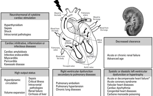
BNP can be produced in both atria and ventricles, and is upregulated in failing ventricular myocardium. In response to increased myocardial stretch and wall stress, ventricular myocytes secret the pro-hormone pre-proBNP, which is then cleaved into biologically active BNP and the inactive byproduct N-terminal-proBNP (NT-proBNP). The biolo- gical actions of NPs are mediated through membrane- bound natriuretic peptide receptors (NPR) that are linked to a cyclic guanosine monophosphate-dependent signaling cascade, including NPR-A, which preferentially binds ANP and BNP, and NPR-B, which preferentially binds CNP. Elevated BNP levels have been demonstrated to res- ponsed to increased angiotensin II and sympathetic tones.5 Elevated NPs levels can be also found in many circum- stances involving LV dysfunction or hypertrophy; right ventricular (RV) dysfunction secondary to pulmonary diseases; cardiac inflammatory or infectious diseases; and endocrinology diseases and high output status without decreased left ventricular ejection fraction (EF), e.g., sepsis, renal failure, cirrhosis of liver, or intracranial pathologies.
Even in the absence of significant clinical evidence of volume overload or LV dysfunction, markedly elevated NP levels can be found in patients with multiple comorbi- dities with certain degree of prognostic value.6The causes and mechanisms of elevated NPs levels are summarized in Fig. 1. The potential clinical applications in the non-HF settings are summarized in Table 1.
PATHOPHYSIOLOGICAL CONSIDERATIONS OF NPs
Fig. 1. The causes and mechanisms of elevated natriuretic peptides levels. *The main reason of elevated natriuretic peptides and the best- established use of these testing. Circumstances in the example list may share more than one mechanism.
Cardiac amyloidosis Infective endocarditis Myocarditis Pericarditis Kawasaki disease
Cardiac inhiltrative, inflammation or infectious diseases Hyperthyroidism
Sepsis Shock
Intracranial pathologies Neurohormonal of cytokine
cardiac stimulation
Hyperdynamic circulation
Volume expansion Sepsis Critical illness Intracranial
pathologies Renal failure Cirrhosis of liver High-output status
Acute or chronic renal failure Advanced age
Decreased clearance
Acute or decompensate heart failure*
Acute coronary syndrome Valvular heart diseases Cardiac dysrhythmia Congenital heart diseases Carbone monoxide poisoning
Systolic or diastolic left ventricular dysfunction or hypertrophy
Pulmonary embolism Pulmonary hypertension Chronic lung diseases
Right ventricular dysfunction secondary to pulmonary diseases
NPs induce vasodilatation, increase diuresis, and inhibit renin and aldosterone production. The function of CNP seems to be the regulation of regional blood flow. The net effects are decreasing cardiac preload and afterload. BNP is eliminated by binding to the NPR-C or degradation by neutral endopeptidase on endothelial cells, smooth muscle cells, cardiac myocytes, renal epithelium, and fibroblasts.
NT-proBNP is cleared mainly by the kidney.7BNP has a relatively shorter half-life of about 20 minutes; the half-life of NT-proBNP is about 60-90 minutes and would be expected to be longer in the setting of renal dysfunction.
Obese patients (especially those who have body mass index greater than 30) tend to have lower BNP levels than others. Neural endopeptidases that can be secreted by adipose tissue may be related to increased BNP clearance in obese patients.8
During the past few years, several fully automated, rapid assays for the determinations of BNP and NT-proBNP have become commercially available, including both high- throughput automated plateforms and point-of care tests.
Most immunoassays currently used in the clinical setting, however, do not determine precise molecular forms of these NPs, such as different cleavage points and post-translatio-
nal modifications, which may vary depending on the pathophysiological state of CHF. In a recent study using quantitative mass spectral methodology, the authors report- ed the absence of circulating 32-amino acid BNP in ad- vanced heart failure, suggesting the existence of altered forms of BNP that contribute to the high levels measured by conventional point-of-care tests.9Plastic tubes containing ethylenedinitrolotetraacetic acid (EDTA) are desirable for BNP determination and refrigeration is required if the interval between blood collection and analysis is over 4 hours; whereas NT-proBNP can be measured in both serum or plasma, collected in glass or plastic tubes, and has no significant loss of immunoreactivity after 48 hours at room temperature. Although these existing BNP assays correlate closely, BNP assays are not currently analytical equivalent due to lack of assay standardization.10A multicenter colla- borative proficiency testing study conducted in 90 Italian laboratories had demonstrates that there are significant differences in analytical characteristics and measured values among the most popular commercial methods for BNP and NT-proBNP. Thus, clinicians should be very careful when comparing results obtained by laboratories that use different methods.11
Table 1. Potential Clinical Applications of Natriuretic Peptides in Selected Diseases Diseases severity /
Diseases Screening* risk stratification / Prognosis
monitoring of therapy
Heart failure� + + +
Acute coronary syndrome + + +
Cardiac procedures + + +
Pulmonary embolism + + +
Pulmonary hypertension + + +
Chronic lung diseases + + -
Valvular heart diseases + + +
Cardiac dysrhythmia + + N / A
Cardiac inflammatory or infectious diseases + + + / -
Cardiogenic syncope + N / A N / A
Sleep apnea + + N / A
Hypertension + + N / A
Sepsis + + +
Renal failure + + +
Cirrhosis of liver + + +
Hyperthyroidism + + N / A
Intracranial pathologies + + +
Epilepsy / Seizures + - -
Carbone monoxide poisoning + N / A N / A
N/A, not available.
*Screening for the presence of cardiac dysfunction.
�The best-established clinical application of these natriuretic peptides testing.
Increased BNP levels correlate grossly with the severity of left ventricular (LV) dysfunction, both clinically and hemodynamically. It has been suggested that NT-proBNP levels also correlate and provide important prognostic information beyond echocardiographic parameters of car- diac structure and function.12 In 72 patients with various degrees of LV systolic dysfunction, a head-to-head com- parison for performance of BNP and NT-proBNP to detect LV systolic dysfunction was performed. Similar to BNP, NT-proBNP is a promising marker in identifying LV systolic dysfunction. Although both assays are reliable and have good analytical performance, their diagnostic cut-off value is dynamic and population-dependent. The slightly wider detection range and the more stable structure of NT- proBNP compared to the BNP assay suggest that NT- proBNP could play an additional role in the evaluation of patients with LV systolic dysfunction.13
Acute coronary syndrome (ACS)
In a prospective observational study of 100 patients pre- senting with chest pain, Brown, et al. had reported that mean BNP levels in patients with cardiac chest pain was significantly greater than that for patients with non-cardiac chest pain. Comparing to cardiac troponin (cTn) T, BNP was less specific for myocardial injury, but more sensitive for the detection of acute cardiac chest pain. Combining BNP and cTn T significantly increased the sensitivity from 55.6% to 95.6% in detecting cardiac chest pain and was also a satisfactory rule out test (negative predictive value, NPV of 96%) for excluding chest pain that had a cardiac origin.14Anemia (Hb < 12 g/dL for females; < 13 g/dL for males) was also associated with elevated NPs levels in patients with known or suspected coronary artery disease.15 NT-proBNP is also a sensitive marker of myocardial ische- mia that rises much higher in the earlier phase compared with conventional markers of myocardial damage, especi- ally in non-ST elevation -MI patients.16
In patients with ACS, BNP is a powerful indicator of acute HF.17In a prospective study involving 74 patients with ST elevation MI, increased BNP levels are associated with progressive ventricular dilatation and development of clinical heart failure and powerful markers of LV systolic dysfunction as well poor prognosis after MI.18In a study following 276 patients presented to the emergency depart- ment with chest pain for 90 days, baseline elevated BNP and NT-proBNP levels were predictive of adverse events, i.e., ED re-visiting for chest pain, cardiac readmission, and death at 30 and 90 days.19Besides, BNP levels were signi- ficantly higher in patients with multivessels than those
with only single-vessel diseases.20Multivariate logistic reg- ression analysis incorporating age, gender, history of hypertension, diabetes, LVEF, cTn I, and therapeutic regimens indicated that BNP was an independent predictor of cardiac death in ACS patients.21In an outpatient follow- up study, ACS patients who had newly elevated BNP levels at 4 months were at increased risk of death or newly developed HF. Patients with elevated BNP levels at study entry and with BNP levels lower than 80 pg/mL at 4 months tended to have only modestly increased risk compared to patients with BNP levels lower than 80 pg/mL at both visits. Patients with BNP levels of more than 840 pg/mL and increased cTn levels are at a particularly high risk for mortality.22 In addition to Thrombolysis In Myocardial Infarction (TIMI) risk score, multi-marker risk approach based on cTnT, C-reactive protein (CRP), and NT-proBNP was associated with adverse events at 6 months.23Changes in BNP levels over time were associated with long-term clinical outcomes.24
Percutaneous cardiac intervention, cardiac surgery, heart transplantation, and the use of extracorporeal membrane oxygenation
BNP levels obtained at post-PCI 24th hour were inde- pendent predictors of major adverse cardiac events (MACE), including cardiac death, MI, hospitalization with angina or repeat revascularization during 12 months of follow-up after elective successful PCI.25In 26 patients that received the implantation of a paclitaxel-eluting stent, symptomatic re-stenosis after implantation of a paclitaxel-eluting stent could be predicted by the combination of a basal plasma BNP level of more than 50 pg/mL and a positive or incon- clusive conventional exercise test. Thus, determining basal BNP levels could improve both the detection and exclu- sion of asymptomatic re-stenosis.26In 135 patients under- going various cardiac procedures, serum NT-proBNP is a good predictor for complications after cardiac surgery, is as good as the European system for cardiac operative risk evaluation, and better than EF alone.27In 85 patients under- going first-time elective CABG, preoperative BNP levels could be used to predict the development of postoperative new-onset atrial fibrillation (AFib), a need for inotropic support, and a requirement for intra-aortic balloon pump.28 In 149 that patients underwent cardiac surgery, BNP levels measured at the beginning of the rehabilitation program had been found to be independent predictors of late AFib after cardiac surgery.29
Despite the absence of a ventricular stretch or intracar- diac pressure derangement, elevated BNP levels were found after cardiac transplantation; thus it had been impli- cated as a marker for allograft rejection, adverse prognosis, and coronary graft vasculopathy in these patients.30BNP
CARDIAC DISEASES
levels have high sensitivity and NPV for acute rejection in pediatric heart transplant patients. In 86 that patients underwent a total of 560 endomyocardial biopsies in patients > 1 year post-transplant, a BNP level of < 100 pg/mL correlated with a < 1% chance of aortic regurgitation (AR) and may obviate the need for biopsies in some cases.31 In an observational study that included 71 heart transplant patients, the increase between the two serial determinations of BNP levels at the end of the first year post-transplant indicated a subgroup of patients with a poorer outcome.32
In 19 children supported with a pulsatile ventricular assist device, extremely high levels of NPs reflect the sev- erity of myocardial failure before extracorporeal membrane oxygenation implantation. During mechanical support, the decline of NPs appears to be an additional tool in the pre- selection of potential weaning candidates.33
RV dysfunction secondary to pulmonary diseases
Pulmonary embolism
NPs levels are elevated in most patients with acute PE complicated with RV overload or dysfunction.34In 93 consecutive outpatients diagnosed with acute PE by means of helical computed tomography (CT), plasma levels of NT-proBNP greater than 500 ng/L were independently associated with central PE (in the main trunk or right or left main branches of the pulmonary artery) and perhaps as a predictor of death.35 The presence of RV dysfunction should be strongly considered in patients with plasma BNP levels of more than 90 pg/mL.36 BNP levels significantly associated with the severity of pulmonary hypertension and increased ventilation-perfusion mismatch on scintigraphy also corresponded to incremental increases in the levels of ANP and BNP. The combination of NT-proBNP or cTn tests to echocardiography could aid risk stratification in patients with acute PE.37
An aggressive therapy such as thrombolytic therapy may be beneficial in patients with acute massive or non-mas- sive PE with RV dysfunction. Thrombolysis significantly reduced systolic pulmonary pressure, and declined BNP and cTn I levels were indicators of the efficacy of pharma- cological treatment in patients with acute PE.38After pulmonary thromboendarterectomy, plasma BNP levels markedly decreased in association with a reduction of total pulmonary resistance in survivors. The changes of plasma BNP levels were closely correlated with the total pulmo- nary resistance. Sustained elevated plasma BNP levels indicated the presence of residual pulmonary hypertension after operation. In hemodynamically stable PE, BNP/cTnI measurement could be helpful on admission, especially for ruling out circulatory failure and in-hospital death.39
Pulmonary hypertension
In patients with RV pressure overload due to primary pulmonary hypertension (PPH) and thromboembolism, plasma BNP levels correlated with mean pulmonary artery pressure, right atrial pressure, RV end-diastolic pressure, and total pulmonary resistance.40In 49 dyspneic patients (25 with chronic obstructive pulmonary disease (COPD), 8 with interstitial pneumonia, 16 with sequelae of tuberculo- sis), hypoxemia was more severe in patients with higher BNP levels than those with lower BNP levels.41In a study involving 61 patients with various forms of chronic pulmo- nary hypertension, plasma NT-proBNP levels can be used to determine the clinical severity and are independently associated with long-term mortality.42
Chronic lung diseases and respiratory failure
Elevated BNP levels have been found in hypoxemic pa- tients with COPD, particularly in patients with cor pulmo- nale when compared with patients with COPD alone. BNP levels increased in proportion to the severity of RV dysfunc- tion and could be a useful indicator of RV dysfunction.43 Adding mild diuretics to the standard treatment for an acute exacerbation (AE) of COPD may rapidly reduce plasma BNP levels in COPD patients with AE who have elevated BNP levels without any clinical evidence of cor pulmonale.44In 208 patients presenting to the ED with an AE of COPD, BNP levels were significantly elevated during AE compared to recovery. In these patients, BNP levels independently predict the need for intensive care unit (ICU). However, BNP levels failed to predict short- and long-term mortality in these patients.45
Cardiovascular dysfunction is responsible for a majority of weaning failure. In 52 patients recovering from acute respiratory failure, the extubation failure group had signi- ficantly greater increases in BNP levels at the end of spontaneous breathing trial (SBT) than the extubation success groups. Thus, measuring the percentage change in the BNP levels during a SBT may help to improve the predictive value of SBT on weaning outcomes.46
Valvular heart disease
Mitral valve
Natriuretic peptides levels increase with increasing severity of mitral regurgitation (MR) and had been shown to be a marker of poor outcome in patients with organic MR.47
NT-proBNP levels were significantly greater in patients with severe mitral stenosis (MS) than in moderate MS.
NT-proBNP levels were also significantly higher in patients with mitral valve replacement than in controls. There were positive correlations between BNP level with the degree of MS, pulmonary artery pressure, and a negative correlation
with mitral valve area.48 Serum NT-proBNP levels cor- related well with echocardiographic findings and functio- nal class in patients with MS.49
Aortic valve
In Patients < 70 years old with mild to moderate aortic stenosis (AS) with a peak transaortic gradient > 20 mm Hg, NT-proBNP levels increased even in asymptomatic patients with AS and correlated with echocardiographic parameters related to the severity of AS and degree of diastolic dysfunction.50Preoperative NT-proBNP correlated significantly with diastolic and systolic LV wall stress assessed by echocardiography. Independent to preopera- tive LV mass index, higher pre-operative levels of NT- proBNP predicted a greater magnitude of total LV mass regression at follow-up.51
NPs could be used as an additional noninvasive method in monitoring disease progression and have potential prog- nostic usefulness in adjunct to echocardiography. In high- risk patients, BNP has been also considered to be useful for monitoring the progression of VHD.52Detection of a cardiac murmur during routine medical examinations of hospitalized patients is associated with increased risk of death within a year. Determining NT-proBNP gives signi- ficant additional prognostic information of a murmur and could obviate the need for echocardiography in selected patients with a murmur and normal NT-proBNP for whom surgery is not feasible.53
Cardiac dysrhythmia
In 60 patients with persistent AFib evaluated before cardioversion and 1, 30 and 180 days after cardioversion, a medium-low elevation of NT-proBNP indicated only an acute response to the distension of the atrial tissue induced by the arrhythmia; in contrast, markedly elevated values might be caused by ventricular dysfunction or hypertro- phy. AFib may cause enduringly elevated ANP and BNP level. Changes in left atria (LA) volume correlated well with the changes in ANP and BNP levels following car- dioversion; and atrial volume seems to be an important determinant of ANP and BNP in AFib.54 In 43 patients undergoing persistent/permanent AFib ablation, sinus rhythm (SR) following persistent/permanent AFib ablation is associated with a dramatic decrease in NPs with signi- ficantly improved cardiac functions, even after relatively extensive atrial ablation.55Plasma NT-proBNP levels obtained before electrical cardioversion did neither predict cardioversion success nor relapse of AFib in patients without HF. However, maintained SR during follow-up was associated with a significant reduction in NT-proBNP levels.56Significant reduction in BNP levels after pul- monary vein isolation was associated with therapeutic
responses to antiarrhythmic drug therapy in patients with recurrent AFib after the procedure.57 In a study including 20 open-heart surgery patients with concomitant AFib, NPs levels tended to decrease in patients with stable SR at one year compared to patients in AFib.58In 105 patients presented to the ED with a recent-onset AFib and supra- ventricular tachycardia, SR was restored in a low per- centage of patients with NT-proBNP of greater than 4,500 pg/mL, while the vast majority of those with values < 1,500 pg/mL was normalized even by means of antiarrhythmic drugs alone. The evaluation of NT-proBNP could be used as an alternative to echocardiography to rapidly select patients in whom cardioversion should be attempted in the ED.59
High degree atrioventricular block can induce elevated plasma BNP levels and the atrioventricular dyssynchrony further induce elevated BNP levels. In 34 patients with preserved LV systolic function on permanent RV apical pacing, RV apical pacing is associated with LV dyssyn- chrony and subsequent accelerated BNP secretion.60In a study involving 43 children and adolescents with high- grade second degree or complete atrioventricular conduc- tion block, plasma BNP levels were significantly higher in patients without pacemaker than in those with permanent pacemaker. Additionally, patients with dual-chamber pac- ing had significantly lower BNP values compared to those with single chamber ventricular pacing.61
Miscellaneous uses in cardiac diseases
Cardiac inflammatory and infectious diseases
Increased release of BNP into the circulation may be a general feature of cardiac inflammation or injury. Inflam- matory process can contribute specific cytokines leading to the disregulation of cardiac ANP and BNP production ob- served during myocardial inflammation and this process might be angiotensin receptor 1-dependent.62In 14 patients with suspected acute perimyocarditis, BNP level are incre- ased in some patients with acute perimyocarditis. BNP elevation is probably associated with hemodynamical stress caused by transient contractility abnormalities.63 In 45 patients with confirmed infective endocarditis, admission NT-proBNP levels of more than 1,500 pg/mL had signifi- cantly lower event-free survival than others. The combina- tion of admission NT-proBNP and cTnI levels appeared to have even greater value for risk stratification in these patients.64In a prospective study involving 45 patients with acute and convalescent rheumatic fever, BNP levels were higher compared to those of healthy subjects and could be used as a complementary tool in the treatment and pro- gnosis of acute rheumatic fever.65In 13 pediatric patients with Kawasaki disease, NT-proBNP levels increased in the
acute phase and decreased significantly in the convalescent phase.66
Restrictive cardiomyopathy and constrictive pericarditis Restrictive cardiomyopathy (RCM) and constrictive peri- carditis (CP) are difficult to distinguish clinically and pose a diagnostic challenge. In 11 patients suspected of having either CP or RCM suffered New York Heart Association (NYHA) stage III-IV CHF with similar elevation in intra- cardiac pressures, BNP levels were markedly elevated in 5 patients with RCM than 6 patients with CP.67In addition, patients with idiopathic CP had significantly lower levels of BNP than those with RCM and those with CP secon- dary to radiation and cardiac surgery.68
Cardiogenic syncope
In 61 patients admitted to a university hospital due to syn- cope, NT-proBNP at a cut-off of 164 pg/mL identified patients with cardiac syncope and patients requiring inter- ventional therapy with high sensitivity and NPV. Assess- ment of NT-proBNP might be helpful in differentiating cardiac from non-cardiac syncope and could effectively reduce the use of Holter echocardiography, telemetry mon- itoring, stress tests, echocardiography, coronary angio- graphies, and electrophysiological examinations in synco- pal patients.69
Sleep apnea and morbid obesity
Cardiac structural and functional abnormalities were ob- served with increased severity of sleep apnea. In 235 patients evaluated in a sleep clinic for obstructive sleep apnea, elevated BNP levels seemed to reflect an increased likelihood of LV hypertrophy in patients with severe sleep apnea.70 In a cross-sectional study involving 110 morbid obesity, healthy young female patients, NT-proBNP levels were independently predicted by sleep-disordered breath- ing and insulin resistance.71
Hypertension
In 154 untreated hypertensive patients without heart failure or AFib, logarithmic BNP was positively correlated with age, pulse pressure, cardiac hypertrophy, and diastolic dys- function, and negatively correlated with hemoglobin, renin levels, and E/A ratio. Thus BNP might be useful in initial assessment of the severity of essential hypertension and may be also valuable for risk stratification.72
Aortic dissection
Both acute aortic dissection (AAD) and chronic aneury- sms of the ascending aorta were associated with long- standing hypertension, which is often accompanied by LV diastolic dysfunction and AR. In 18 patients with AAD,
both patients with AAD and chronic uncomplicated aneurysms had significant higher BNP levels than normal controls.73
Congenital heart diseases
Although clinical and echocardiographic signs define hemo- dynamic significance of patent ductus arteriosus (PDA), they are not predictive of the need for PDA intervention in the first days of life. In a prospective blinded study involving 67 preterm infants on the second day of life, plasma BNP levels positively correlated with ductal size and LA/Aorta- ratio significantly.74 Plasma NT-proBNP levels on day 3 are a good marker for hemodynamic significant PDA in preterm infants. Serial measurements of NT-proBNP may be useful in assessing the clinical course of PDA.75
Cardiac amyloidosis
In a study evaluating heart involvement in 152 patients with amyloidosis, NT-proBNP is a sensitive marker of cardiac dysfunction and a strong predictor of outcome in cardiac amyloidosis and had been shown to decline with treatment.76
Infectious diseases and sepsis
Ventricular dysfunction with reduced EF and biventricular dilatation is present in most patients with severe sepsis and septic shock. Plasma ANP, NT-proBNP, and BNP were significantly elevated in septic shock patients than healthy control subjects. In a study involving 54 patients without heart diseases, BNP levels could be elevated in the acute phase of community-acquired microbial infections, parti- cularly in patients with diabetes mellitus (DM) and lower respiratory tract infection, even in the absence of severe sepsis or septic shock.77 In a study involving 50 patients with septic shock, elevated BNP levels were significantly associated with myocardial dysfunction and severity of global tissue hypoxia. BNP levels were positively correlated with CRP levels in septic patients without clinical or echo- cardiographic evidence of volume overload or systolic dysfunction.78In patients with community-acquired pneu- monia (CAP), BNP level was an independent predictor of treatment failure and death. In a study evaluating 302 consecutive patients presenting to the ED with CAP, when used in conjunction with the pneumonia severity index (PSI), BNP levels significantly improved the risk prediction when compared with the PSI alone.79Patients with septic
CAUSES OF ELEVATED NPs LEVELS DUE TO HYPERDYNAMIC STATUS, OTHER SECONDARY CARDIAC DYSFUNCTIONS,
OR DECREASED CLEARANCE
shock had significantly higher BNP levels on admission compared to patients with early sepsis and healthy subjects.
BNP levels for patients with septic shock were positively correlated with Sequential Organ Failure Assessment scores and prognosticated survival.80 NT-proBNP is an independent prognostic marker of mortality in severe sepsis and both NT-proBNP obtained on admission and 72-hours after admission are significantly higher in nonsurvivors than survivors.81
Renal failure
In a study involving 164 hemodialysis patients, elevated BNP levels were associated with volume overload, LV hypertrophy, cardiovascular disease and DM, and may be a useful parameter for assessing the risk of cardiac death in these patients.82Renal function clearly influences the diagnostic performance of NT-proBNP; Goei, et al. had showed that NT-proBNP had more favorable discrimina- tive value in patients with glomerular filtration rate (GFR) more than 90 mL/min/1.73 m2and lost its prognostic value in patients with GFR less than 30 mL/min/1.73 m2.83Unlike NT-proBNP, BNP levels are relatively independent of GFR. BNP may therefore be the more appropriate bio- marker to screen for cardiac dysfunction in patients with renal failure.84BNP levels could be a valuable tool for risk stratification of hemodialysis patients by confining echo- cardiographic studies to only patients with BNP levels above the established cutoff values.85
On the basis of the German Diabetes and Dialysis Study which evaluated atorvastatin in 1255 type 2 DM hemo- dialysis patients with a median follow-up of 4 years, patients with baseline NT-proBNP of more than 9,252 pg/mL exhibited a more than four-fold risk of stroke and a more than two-fold risk of sudden death, cardiovascular events, and mortality compared with patients with baseline NT-proBNP less than 1,433 pg/mL. Doubling of NT- proBNP increased the risk of death by 46%.86
Cirrhosis of liver
In patients with cirrhosis of the liver, elevated proBNP and BNP levels reflect increased cardiac ventricular generation of NPs and thus indicates the presence of cardiac dys- function. Hyperdynamic systemic circulation could also be contributed to elevated NPs levels in patients with cirrhosis of liver. In a study evaluating 52 non-alcoholic cirrhotic patients, BNP levels were significantly higher in cirrhotic patients complicated with ascites, spontaneous bacterial peritonitis and hepatic encephalopathy history, but no significant differences were observed for the presence of esophageal varices and bleeding history. BNP levels signi- ficantly correlated with Child score, interventricular septal thickness, and LV posterior wall thickness.87In 51 cirrhotic
patients during a hemodynamic investigation, circulating proBNP and BNP levels were related to the severity of liver decompensation (Child score, serum albumin, coa- gulation factors, and hepatic venous pressure gradient) and to markers of cardiac dysfunction (QT interval, heart rate, plasma volume).88
Critical illness and shock
Assessing cardiac filling pressure and cardiac performance are important in patients with either noncardiogenic or cardiogenic shock. Low NT-proBNP values may be useful in excluding the need for pulmonary artery catheter place- ment in patients with ICU shock and identified those at lower risk for death.89 In a prospective, blinded cohort study that incorporated 54 patients admitted to the ICUs with respiratory distress, bilateral pulmonary edema and a central venous catheter suggesting either high-pressure (cardiogenic) or low-pressure, acute lung injury acute respiratory distress syndrome (ALI/ARDS) pulmonary edema, BNP at an accepted cutoff of 100 pg/mL had a high specificity for the diagnosis of ALI/ARDS (95.2%), but the sensitivity was poor (27.3%). BNP levels drawn within 48 hours of admission to the ICU did not correlate with invasive hemodynamic measurements and did not track predictably with changes in volume status on consecutive daily measurements.90 Patients with significant blood loss (decreased hemoglobin level more than 3 g/dL) had signi- ficant lower BNP levels than those without blood loss.
Thus, BNP levels below normal are indicative of intravas- cular volume loss in traumatically injured patients.91In a study evaluating 40 patients who had pre- and post transfu- sion NT-proBNP determinations, NT-proBNP could also be used to evaluate the development of transfusion asso- ciated circulatory overload with a sensitivity of 93.8% and specificity of 83.8%. Elevated post transfusion NT-proBNP is the only independent variable for this diagnosis.92 NT- proBNP on admission is an independent prognostic marker of outcome in an unselected cohort of critically ill patients.
Measurement of NT-proBNP might facilitate triage of critical ED and ICU patients.93
Hyperthyroidism
Serum NT-proBNP levels are affected by thyroid functions and seem to be a direct stimulatory effect of thyroid hor- mones.94 A comparison between 67 patients with clinical hyperthyroidism and normal subjects, elevated BNP levels were mainly found in hyperthyroid patients who had clinical and echocardiographic evidence of LV dysfunction (increased LA diameter and decreased LVEF) but not in those with normal LV function and normal subjects.95 Multiple linear regression analysis demonstrated that free T4 and free T3 were independently associated with a high
serum NT-proBNP, whereas cardiac output and resting pulse rate were not. In 21 patients with hyperthyroidism, both NT-proBNP and BNP levels were higher than hypo- thyroid patients and normal controls; and treatment of thyroid dysfunctions could result in normalization of NT- proBNP levels in both hypothyroid and hyperthyroid groups.96
Intracranial pathologies
Acute ischemic stroke
A stress response consisting of elevated levels of cortisol and catecholamines is common after acute ischemic stroke (AIS). Activation of the hypothalamus-pituitary-adrenal axis in patients with AIS is associated with elevated levels of NPs.97NT-proBNP levels were elevated in patients with AIS. In a study involving 30 patients with an AIS, the activated neurohumoral responses occurring in patients with AIS reflect either a counterbalancing vasodilating response to the cerebral ischemia or direct myocardial dysfunction.98 The log NT-proBNP correlated positively with cTnI and heart rate, and negatively with LVEF; and patients with signs of marked myocardial ischemia or insular cortex involvement had particularly higher NT- proBNP levels. BNP levels in AIS patients without AFib were correlated with MAP on admission or the degree of reduction in MAP at day 1. While in AIS patients with AFib, BNP levels showed a negative correlation with MAP on admission.99 Even in patients without AFib, the presence of at least one LA or appendage abnormality (atrial dilatation, low flow velocity, spontaneous echocon- trast, or thrombus) had the strongest association with an elevated BNP, suggesting that BNP level could be a poten- tial marker for the presence of LA sources of emboli.100In a study involving 707 patients presenting to the ED with AIS, BNP of more than 76 pg/mL was an independent pre- dictor of cardioembolic stroke. Even among patients with transient symptoms, a high BNP level identified cardioem- bolic etiology.101Elevated NPs and cortisol levels predict long-term mortality after AIS, suggesting that this profound neurohumoral disturbance is prognostically unfavorable.102
Subarachnoid hemorrhage
Cardiovascular abnormalities are independent predictors of in-patient mortality after a subarachnoid hemorrhage (SAH).
Patients with SAH showed an increased urine output and urinary excretion of sodium as well as higher BNP levels than the controls.103,104In a study involving 50 patients with traumatic SAH, early after SAH, elevated BNP levels are associated with myocardial necrosis, pulmonary edema, and LV dysfunction. BNP levels may be elevated in patients with head injuries without echocardiographic evidence of
HF.105 In the absence of evidence for activation of NPs within the brain, prompt and consistent increase in both ANP and BNP strongly supports the view that the heart is the source of increased NPs release after acute SAH.106In a study involving 30 patients with severe isolated head in- jury, BNP levels are elevated shortly after head injury and progressively elevated through days 7-8 after the event in patients with diffused SAH as compared to patients with mild or no SAH; and in patients with elevated intracranial pressure (ICP) as compared to patients without elevated ICP as well as a poorer outcome.107In a study involving 14 patients suffering from severe traumatic brain injury, NT- proBNP levels in cerebral spinal fluid and serum were significantly elevated in patients exhibiting an ICP of more than 15 mmHg.108In a prospective cohort study incorpo- rating 300 patients with aneurysmal SAH, an initial BNP level greater than 600 pg/mL was markedly associated with death.109
Epilepsy
In a study incorporating 65 postictal children and 31 chil- dren with epilepsy after a seizure-free period for at least 2 months serving as controls, NT-proBNP levels were signi- ficantly higher 4 hours after a seizure attack compared to 24-48 hours after a seizure attack. Serum NT-proBNP levels were higher in children with tonic-clonic seizures and febrile convulsions than those with partial motor seizures, syncope, or normal subjects.110Obi, et al. had reported a patient who had episodic elevation of ANP and BNP during epileptic activity caused by a left frontotem- poral lesion. ANP and BNP were assumed to release from the heart by the epileptic stimulation on the anterior ventral region of the third ventricle.111
Carbon monoxide poisoning
The level of NT-proBNP and carbon monoxide (CO) Hb were increased in patients with CO poisoning. In a study involving 15 patient with CO poisoning, there was a posi- tive correlation between the levels of COHb and NT- proBNP. Thus, determining plasma NT-proBNP levels may contribute to the early diagnosis of cardiotoxicity in patients with CO poisoning.112
Elevated NPs levels have shown predictive value in various diseases that have direct or indirect influences on the heart functions in many non-HF circumstances, even in the absence of depressed cardiac function. It should be noted that NPs should never be interpreted without thorough clinical messages. NPs are very sensitive for primary or
CONCLUSION
secondary cardiac dysfunction, but the specificity is low.
Potential clinical applications of NPs are expanded, accom- panied by emerging reports regarding to screening for the presence of secondary cardiac dysfunction, monitoring the therapeutic responses, risk stratifications, or providing prognostic values in many settings. Clinical decision on timely and costly cardiac examinations or more risky and invasive procedures rather than deferred, conservative strategies might be facilitated by appropriate use and inter- pretation of NPs. However, clinicians should recognize that currently the only reasonable application for routine practice is limited to differentiation of acute dyspnea, rule- out-diagnostic-tests (high NPV and low positive predictive values), monitoring of therapeutic responses, and prognosis of acute or decompensated CHF.
1. Swedberg K, Cleland J, Dargie H, Drexler H, Follath F, Komajda M, et al. Guidelines for the diagnosis and treatment of chronic heart failure: executive summary (update 2005): The Task Force for the Diagnosis and Treatment of Chronic Heart Failure of the European Society of Cardiology. Eur Heart J 2005;26:1115-40.
2. Januzzi JL, van Kimmenade R, Lainchbury J, Bayes-Genis A, Ordonez-Llanos J, Santalo-Bel M, et al. NT-proBNP testing for diagnosis and short-term prognosis in acute destabilized heart failure: an international pooled analysis of 1256 patients: the International Collaborative of NT-proBNP Study. Eur Heart J 2006;27:330-7.
3. Kisch B. Electron microscopy of the atrium of the heart. I.
Guinea pig. Exp Med Surg 1956;14:99-112.
4. De Bold AJ. Heart atria granularity effects of changes in water- electrolyte balance. Proc Soc Exp Biol Med 1979;161:508-11.
5. Iwanaga Y, Nishi I, Furuichi S, Noguchi T, Sase K, Kihara Y, et al. B-type natriuretic peptide strongly reflects diastolic wall stress in patients with chronic heart failure: comparison between systolic and diastolic heart failure. J Am Coll Cardiol 2006;47:
742-8.
6. Burjonroppa SC, Tong AT, Xiao LC, Johnson MM, Yusuf SW, Lenihan DJ. Cancer patients with markedly elevated B-type natriuretic peptide may not have volume overload. Am J Clin Oncol 2007;30:287-93.
7. Schrier RW, Abraham WT. Hormones and hemodynamics in heart failure. N Engl J Med 1999;341:577-85.
8. Young JB, Correia NG, Francis GS, Maisel A, Michota F.Testing for B-type natriuretic peptide in the diagnosis and assessment of heart failure: what are the nuances? Cleve Clin J Med 2004;71 Suppl 5:S1-17.
9. Hawkridge AM, Heublein DM, Bergen HR 3rd, Cataliotti A, Burnett JC Jr, Muddiman DC. Quantitative mass spectral evidence for the absence of circulating brain natriuretic peptide (BNP-32) in severe human heart failure. Proc Natl Acad Sci U S A 2005;102:17442-7.
10. Omland T. Advances in congestive heart failure management in the intensive care unit: B-type natriuretic peptides in evaluation of acute heart failure. Crit Care Med 2008;36:S17-27.
11. Prontera C, Zaninotto M, Giovannini S, Zucchelli GC, Pilo A, Sciacovelli L, et al. Proficiency testing project for brain natriuretic peptide (BNP) and the N-terminal part of the propeptide of BNP (NT-proBNP) immunoassays: the CardioOrmocheck study. Clin Chem Lab Med 2009. In press.
12. Chen AA, Wood MJ, Krauser DG, Baggish AL, Tung R, Anwaruddin S, et al. NT-proBNP levels, echocardiographic findings, and outcomes in breathless patients: results from the ProBNP Investigation of Dyspnoea in the Emergency Department (PRIDE) echocardiographic substudy. Eur Heart J 2006;27:839-45.
13. Vanderheyden M, Bartunek, Claeys G, Manoharan G, Beckers JF, Ide L. Head to head comparison of N-terminal pro-B-type natriuretic peptide and B-type natriuretic peptide in patients with/without left ventricular systolic dysfunction. Clin Biochem 2006;39:640-5.
14. Brown A, George J, Murphy MJ, Struthers A. Could BNP screen- ing of acute chest pain cases lead to safe earlier discharge of patients with non-cardiac causes? A pilot study. QJM 2007;100:
755-61.
15. Fukuta H, Ohte N, Mukai S, Saeki T, Kobayashi K, Kimura G.
Anemia is an independent predictor for elevated plasma levels of natriuretic peptides in patients undergoing cardiac catheterization for coronary artery disease. Circ J 2008;72:212-7.
16. Ogawa A, Seino Y, Yamashita T, Ogata K, Takano T. Difference in elevation of N-terminal pro-BNP and conventional cardiac markers between patients with ST elevation vs non-ST elevation acute coronary syndrome. Circ J 2006;70:1372-8.
17. Sinclair H, Paterson M, Walker S, Beckett G, Fox KA. Predicting outcome in patients with acute coronary syndrome: evaluation of B-type natriuretic peptide and the global registry of acute coronary events (GRACE) risk score. Scott Med J 2007;52:8-13.
18. Günes Y, Okçün B, Kavlak E, Erbas C, Karcier S. Value of brain natriuretic peptide after acute myocardial infarction. Anadolu Kardiyol Derg 2008;8:182-7.
19. Kwan G, Isakson SR, Beede J, Clopton P, Maisel AS, Fitzgerald RL. Short-term serial sampling of natriuretic peptides in patients presenting with chest pain. J Am Coll Cardiol 2007;49:1186-92.
20. Palazzuoli A, Rizzello V, Calabrò A, Gallotta M, Martini G, Quatrini I, et al. Osteoprotegerin and B-type natriuretic peptide in non-ST elevation acute coronary syndromes: relation to coronary artery narrowing and plaques number. Clin Chim Acta 2008;391:
74-9.
21. Sun T, Wang L, Zhang Y. Prognostic value of B-type natriuretic peptide in patients with acute coronary syndromes. Arch Med Res 2006;37:502-5.
22. Fonarow GC, Peacock WF, Horwich TB, Phillips CO, Givertz MM, Lopatin M, et al. Usefulness of B-Type natriuretic peptide and cardiac troponin levels to predict in-hospital mortality from ADHERE. Am J Cardiol 2008;101:231-7.
23. Tello-Montoliu A, Marín F, Roldán V, Mainar L, López MT, Sogorb F, et al. A multimarker risk stratification approach to non- ST elevation acute coronary syndrome: implications of troponin T, CRP, NT pro-BNP and fibrin D-dimer levels. J Intern Med 2007;262:651-8.
24. Morrow DA, de Lemos JA, Blazing MA, Sabatine MS, Murphy SA, Jarolim P, et al. Prognostic value of serial B-type natriuretic peptide testing during follow-up of patients with unstable coro- nary artery disease. JAMA 2005;294:2866-71.
25. Yildirir A, Acikel S, Ertan C, Aydinalp A, Ozin B, Muderrisoglu H. Value of peri-procedural B-type natriuretic peptide levels in
REFERENCES
predicting cardiac events after elective percutaneous coronary intervention. Acta Cardiol 2008;63:47-52.
26. Lo Mauro R, Arcoleo F, Lo Giudice A, Picone A, Schirosa M, Raspanti G, et al. B-type natriuretic peptide as a marker of asym- ptomatic re-stenosis after coronary implantation of a paclitaxel- eluting stent. J Cardiovasc Med (Hagerstown) 2007;8:1020-3.
27. Elíasdóttir SB, Klemenzson G, Torfason B, Valsson F. Brain natriuretic peptide is a good predictor for outcome in cardiac surgery. Acta Anaesthesiol Scand 2008;52:182-7.
28. Turk T, Ata Y, Ay D, Ozkan H, Vural H, Yavuz S, et al. Plasma brain natriuretic peptide after isolated on-pump coronary artery bypass grafting: prediction of postoperative adverse outcomes.
Heart Surg Forum 2008;11:E84-9.
29. Sarzi Braga S, Vaninetti R, Pedretti RF. Plasma B-type natriuretic peptide predicts atrial fibrillation during rehabilitation after cardiac surgery. Eur J Cardiovasc Prev Rehabil 2008;15:460-6.
30. Shaw SM, Fildes J, Yonan N, Williams SG. Does brain natriu- retic peptide interact with the immune system after cardiac trans- plantation? Transplantation 2007;84:1377-81.
31. Rossano JW, Denfield SW, Kim JJ, Price JF, Jefferies JL, Decker JA, et al. B-type Natriuretic peptide is a sensitive screening test for acute rejection in pediatric heart transplant patients. J Heart Lung Transplant 2008;27:649-54.
32. Martínez-Dolz L, Almenar L, Hervás I, Moro J, Agüero J, Sánchez-Lázaro I, et al. Prognostic relationship between two serial determinations of B-type natriuretic peptide and medium- long-term events in heart transplantation. J Heart Lung Transplant 2008;27:735-40.
33. Heise G, Lemmer J, Weng Y, Hübler M, Alexi-Meskishvili V, Böttcher W, et al. Biomarker responses during mid-term mecha- nical cardiac support in children. J Heart Lung Transplant 2008;
27:150-7.
34. Pruszczyk P. N-terminal pro-brain natriuretic peptide as an indicator of right ventricular dysfunction. J Card Fail 2005;11:
S65-9.
35. Alonso-Martínez JL, Urbieta-Echezarreta M, Anniccherico- Sánchez FJ, Abínzano-Guillén ML, Garcia-Sanchotena JL. N- terminal pro-B-type natriuretic peptide predicts the burden of pulmonary embolism. Am J Med Sci 2009;337:88-92.
36. Yardan T, Altintop L, Baydin A, Yilmaz O, Guven H. B-type natriuretic peptide as an indicator of right ventricular dysfunction in acute pulmonary embolism. Int J Clin Pract 2008;62:1177-82.
37. Binder L, Pieske B, Olschewski M, Geibel A, Klostermann B, Reiner C, et al. N-terminal pro-brain natriuretic peptide or tro- ponin testing followed by echocardiography for risk stratification of acute pulmonary embolism. Circulation 2005;112:1573-9.
38. Enea I, Ceparano G, Mazzarella G, Di Sarno R, Cangiano G, Busino CA. [Biohumoral markers and right ventricular dysfunction in acute pulmonary embolism: the answer to thrombolytic therapy.] Ital Heart J Suppl 2004;5:29-35.
39. Logeart D, Lecuyer L, Thabut G, Tabet JY, Tartière JM, Chavelas C, et al. Biomarker-based strategy for screening right ventricular dysfunction in patients with non-massive pulmonary embolism.
Intensive Care Med 2007;33:286-92.
40. Yap LB, Mukerjee D, Timms PM, Ashrafian H, Coghlan JG.
Natriuretic peptides, respiratory disease, and the right heart. Chest 2004;126:1330-6.
41. Yano S, Kobayashi K, Kato K, Ikeda T. [The study of pulmonary hypertension and plasma BNP values in respiratory diseases.]
Nihon Kokyuki Gakkai Zasshi 2006;44:99-103.
42. Andreassen AK, Wergeland R, Simonsen S, Geiran O, Guevara C, Ueland T. N-terminal pro-B-type natriuretic peptide as an indicator of disease severity in a heterogeneous group of patients with chronic precapillary pulmonary hypertension. Am J Cardiol 2006;98:525-9.
43. Yap LB, Mukerjee D, Timms PM, Ashrafian H, Coghlan JG.
Natriuretic peptides, respiratory disease, and the right heart. Chest 2004;126:1330-6.
44. Kanat F, Vatansev H, Teke T. Diuretics, plasma brain natriuretic peptide and chronic obstructive pulmonary disease. Neth J Med 2007;65:296-300.
45. Stolz D, Breidthardt T, Christ-Crain M, Bingisser R, Miedinger D, Leuppi J, et al. Use of B-type natriuretic peptide in the risk stratification of acute exacerbations of COPD. Chest 2008;133:
1088-94.
46. Chien JY, Lin MS, Huang YC, Chien YF, Yu CJ, Yang PC.
Changes in B-type natriuretic peptide improve weaning outcome predicted by spontaneous breathing trial. Crit Care Med 2008;
36:1421-6.
47. Moe GW, Howlett J, Januzzi JL, Zowall H; Canadian Multi- center Improved Management of Patients With Congestive Heart Failure (IMPROVE-CHF) Study Investigators. N-terminal pro- B-type natriuretic peptide testing improves the management of patients with suspected acute heart failure: primary results of the Canadian prospective randomized multicenter IMPROVE-CHF study. Circulation 2007;115:3103-10.
48. Iltumur K, Karabulut A, Yokus B, Yavuzkir M, Taskesen T, Toprak N. N-terminal proBNP plasma levels correlate with severity of mitral stenosis. J Heart Valve Dis 2005;14:735-41.
49. Arat-Ozkan A, Kaya A, Yigit Z, Balci H, Okçün B, Yazicioglu N, et al. Serum N-terminal pro-BNP levels correlate with symp- toms and echocardiographic findings in patients with mitral stenosis. Echocardiography 2005;22:473-8.
50. Cemri M, Arslan U, Kocaman SA, Cengel A. Relationship between N-terminal pro-B type natriuretic peptide and extensive echocardiographic parameters in mild to moderate aortic stenosis.
J Postgrad Med 2008;54:12-6.
51. Della Corte A, Salerno G, Chiosi E, Iarussi D, Santarpino G, Miraglia M, et al. Preoperative, postoperative and 1-year follow-up N-terminal pro-B-type natriuretic peptide levels in severe chronic aortic regurgitation: correlations with echocardiographic findings.
Interact Cardiovasc Thorac Surg 2008;7:419-24.
52. Moura LM, Rocha-Gonçalves F, Zamorano JL, Barros I, Bettencourt P, Rajamannan N. New cardiovascular biomarkers:
clinical implications in patients with valvular heart disease. Expert Rev Cardiovasc Ther 2008;6:945-54.
53. Iversen K, Nielsen OW, Kirk V, Bay M, Hassager C, Boesgaard S, et al. Heart murmur and N-terminal pro-brain natriuretic peptide as predictors of death in 2977 consecutive hospitalized patients.
Am J Med Sci 2008;335:444-50.
54. Therkelsen SK, Groenning BA, Kjaer A, Svendsen JH, Boje Jensen G. ANP and BNP in atrial fibrillation before and after cardioversion--and their relationship to cardiac volume and function. Int J Cardiol 2008;127:396-9.
55. Sacher F, Corcuff JB, Schraub P, Le Bouffos V, Georges A, Jones SO, et al. Chronic atrial fibrillation ablation impact on endocrine and mechanical cardiac functions. Eur Heart J 2008;29:
1290-5.
56. Tveit A, Seljeflot I, Grundvold I, Abdelnoor M, Arnesen H, Smith P. Candesartan, NT-proBNP and recurrence of atrial fib-
rillation after electrical cardioversion. Int J Cardiol 2009;131:
234-9.
57. Yamada T, Murakami Y, Okada T, Yoshida N, Toyama J, Yoshida Y, et al. Plasma brain natriuretic peptide level after hybrid therapy with pulmonary vein isolation and antiarrhythmic drugs for atrial fibrillation. Int Heart J 2008;49:143-51.
58. Ahlsson A, Linde P, Rask P, Englund A. Atrial function after epicardial microwave ablation in patients with atrial fibrillation.
Scand Cardiovasc J 2008;42:192-201.
59. Magioncalda A, Sartini M, Costaguta C, Castellaneta M, Sche- none E, Tognoni E, et al. [The utility of assaying the N-terminal of brain natriuretic peptide precursor (NT pro-BNP) to predict the clinical outcome in patients with supraventricular tachyarrhy- thmias observed and treated in the emergency room.] Recenti Prog Med 2008;99:141-5.
60. Kawanishi Y, Ito T, Suwa M, Terasaki F, Futai R, Kitaura Y.
Effect of left ventricular dyssynchrony on plasma B-type natriuretic peptide levels in patients with long-term right ventricular apical pacing. Int Heart J 2008;49:165-73.
61. Koch A, Zink S, Dittrich S. Plasma levels of B-type natriuretic peptide in children and adolescents with high degree atrioven- tricular block. Int J Cardiol 2009;134:429-30.
62. Ogawa T, Veinot JP, Kuroski de Bold ML, Georgalis T, de Bold AJ. Angiotensin II receptor antagonism reverts the selective car- diac BNP upregulation and secretion observed in myocarditis.
Am J Physiol Heart Circ Physiol 2008;294:H2596-603.
63. Grabowski M, Karpin´ski G, J Filipiak K, Rdzanek A, Pietrasik A, Wretowski D, et al. Diagnostic value of BNP in suspected perim- yocarditis--a preliminary report. Kardiol Pol 2004;61:451-8.
64. Kahveci G, Bayrak F, Mutlu B, Bitigen A, Karaahmet T, Sonmez K, et al. Prognostic value of N-terminal pro-B-type natriuretic peptide in patients with active infective endocarditis. Am J Cardiol 2007;99:1429-33.
65. Bayram E, Kocatürk H, Yücel O, Atalay C, Colak MC, Atesal S.
The role of adrenomedullin and brain natriuretic peptide levels in acute rheumatic fever in adults. Anadolu Kardiyol Derg 2008;
8:188-91.
66. Zhang QY, Du JB, Chen YH, Li WZ. [Change in plasma N- terminal pro-brain natriuretic peptide in children with Kawasaki disease and its value in clinical practice.] Zhonghua Er Ke Za Zhi 2006;44:886-90.
67. Leya FS, Arab D, Joyal D, Shioura KM, Lewis BE, Steen LH, et al. The efficacy of brain natriuretic peptide levels in differen- tiating constrictive pericarditis from restrictive cardiomyopathy. J Am Coll Cardiol 2005;45:1900-2.
68. Babuin L, Alegria JR, Oh JK, Nishimura RA, Jaffe AS. Brain natriuretic peptide levels in constrictive pericarditis and restrictive cardiomyopathy. J Am Coll Cardiol 2006;47:1489-91.
69. Pfister R, Diedrichs H, Larbig R, Erdmann E, Schneider CA. NT- pro-BNP for differential diagnosis in patients with syncope. Int J Cardiol 2009;133:51-4.
70. Usui Y, Tomiyama H, Hashimoto H, Takata Y, Inoue Y, Asano K, et al. Plasma B-type natriuretic peptide level is associated with left ventricular hypertrophy among obstructive sleep apnoea patients. J Hypertens 2008;26:117-23.
71. Ybarra J, Planas F, Pou JM. Aminoterminal pro-brain natriuretic peptide (NT-proBNP) and sleep-disordered breathing in morbidly obese females: a cross-sectional study. Diab Vasc Dis Res 2008;
5:19-24.
72. Seki S, Tsurusaki T, Kasai T, Taniguchi I, Mochizuki S, Yoshi-
mura M. Clinical significance of B-type natriuretic Peptide in the assessment of untreated hypertension. Circ J 2008;72:770-7.
73. Sbarouni E, Georgiadou P, Marathias A, Geroulanos S, Krema- stinos DT. D-dimer and BNP levels in acute aortic dissection. Int J Cardiol 2007;122:170-2.
74. Czernik C, Lemmer J, Metze B, Koehne PS, Mueller C, Obladen M. B-type natriuretic peptide to predict ductus intervention in infants < 28 weeks. Pediatr Res 2008;64:286-90.
75. Farombi-Oghuvbu I, Matthews T, Mayne PD, Guerin H, Corcoran JD. N-terminal pro-B-type natriuretic peptide: a measure of significant patent ductus arteriosus. Arch Dis Child Fetal Neona- tal Ed 2008;93:F257-60.
76. Palladini G, Campana C, Klersy C, Balduini A, Vadacca G, Perfetti V, et al. Serum N-terminal pro-brain natriuretic peptide is a sensitive marker of myocardial dysfunction in AL amyloidosis.
Circulation 2003;107:2440-5.
77. Nikolaou NI, Goritsas C, Dede M, Paissios NP, Papavasileiou M, Rombola AT, et al. Brain natriuretic peptide increases in septic patients without severe sepsis or shock. Eur J Intern Med 2007;
18:535-41.
78. Nakamura T, Suzuki T, Kawagoe Y, Koide H. Polymyxin B- immobilized fiber hemoperfusion attenuates increased plasma atrial natriuretic peptide and brain natriuretic Peptide levels in patients with septic shock. ASAIO J 2008;54:210-3.
79. Christ-Crain M, Breidthardt T, Stolz D, Zobrist K, Bingisser R, Miedinger D, et al. Use of B-type natriuretic peptide in the risk stratification of community-acquired pneumonia. J Intern Med 2008;264:166-76.
80. Kandil E, Burack J, Sawas A, Bibawy H, Schwartzman A, Zenilman ME, et al. B-type natriuretic peptide: a biomarker for the diagnosis and risk stratification of patients with septic shock.
Arch Surg 2008;143:242-6.
81. Varpula M, Pulkki K, Karlsson S, Ruokonen E, Pettilä V;
FINNSEPSIS Study Group. Predictive value of N-terminal pro- brain natriuretic peptide in severe sepsis and septic shock. Crit Care Med 2007;35:1277-83.
82. Naganuma T, Sugimura K, Wada S, Yasumoto R, Sugimura T, Masuda C, et al. The prognostic role of brain natriuretic peptides in hemodialysis patients. Am J Nephrol 2002;22:437-44.
83. Goei D, Schouten O, Boersma E, Welten GM, Dunkelgrun M, Lindemans J, et al. Influence of renal function on the usefulness of N-terminal pro-B-type natriuretic peptide as a prognostic cardiac risk marker in patients undergoing noncardiac vascular surgery. Am J Cardiol 2008;101:122-6.
84. Tagore R, Ling LH, Yang H, Daw HY, Chan YH, Sethi SK.
Natriuretic peptides in chronic kidney disease. Clin J Am Soc Nephrol 2008;3:1644-51.
85. Biasioli S, Zamperetti M, Borin D, Guidi G, De Fanti E, Schiavon R. Significance of plasma B-type natriuretic peptide in hemodia- lysis patients: blood sample timing and comorbidity burden.
ASAIO J 2007;53:587-91.
86. Winkler K, Wanner C, Drechsler C, Lilienthal J, März W, Krane V; for the German Diabetes and Dialysis Study Investigators.
Change in N-terminal-pro-B-type-natriuretic-peptide and the risk of sudden death, stroke, myocardial infarction, and all-cause mortality in diabetic dialysis patients. Eur Heart J 2008 Jul 9.
[Epubahead of print].
87. Yildiz R, Yildirim B, Karincaoglu M, Harputluoglu M, Hilmioglu F. Brain natriuretic peptide and severity of disease in non- alcoholic cirrhotic patients. J Gastroenterol Hepatol 2005;20: