INTRODUCTION CASEREPORT AbdominalActinomycosisAssociatedwithaSigmoidColonPerforationinaPatientwithaVentriculoperitonealShunt
4
0
0
전체 글
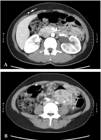
(2) Eun-Young Jung, et al.. A Fig. 2. Sigmoid colon is thickened and perforated about 1 cm in diameter.. B Fig. 1. (A) Abdominal computed tomography (CT) shows left hydroureter and hydronephrosis. VP shunt is shown (white arrow). (B) The approximately 6 × 7 cm multilobulated, poorly-margined mass in the lower left quadrant encases left ureter (white arrow).. trating lesion-like mass involving the small bowel, mesentery, and sigmoid colon. A sigmoid colon perforation, approximately 1 cm in diameter, was observed (Fig. 2). Lysis of adhesions, pus drainage, and sigmoid colon segmentectomy were performed. Pathology revealed an intense chronic inflammatory reaction with sulfur granules (Fig. 3). After 12 weeks of penicillin use, the patient's clinical manifestations and radiologic findings were improved. She was discharged without sequelae. At a follow-up visit 6 months later, there were no signs of recurrence.. DISCUSSION The ventriculoperitoneal shunt is the most widely used procedure in the treatment of hydrocephalus, but complications resulting from the Yonsei Med J Vol. 47, No. 4, 2006. Fig. 3. Histological examination of the resected tissue shows the sulfur granules characteristic of actinomycosis (H&E, × 400).. shunts have been reported in 24-47% of patients. These include: mechanical malfunction, infection, cerebrospinal fluid collection, shunt migration, 7,8 and bowel perforation. Spontaneous bowel perforation is a rare complication of the VP shunt, occurring in only 0.1% of patients, and it is asymptomatic in 42% of patients.9,10 This is the first report of actinomycosis related with a bowel perforation by a VP shunt. Furthermore, there have been no reports of hydroureter and hydronephrosis with complicated actinomycosis for a VP shunt patient. Actinomycosis is an indolent and slowly progressive infection, which releases characteristic sulfur granules. In 1846, Bradshaw published the first description of a patient with abdominal actinomycosis. Harz named this bacteria Actinomyces.
(3) Abdominal Actinomycosis Associated with Sigmoid Colon Perforation. several years later.11 Human actinomycosis is caused by Actinomycoces israelii, a gram-positive microaerophilic organism that is normally found in the mouth, the gastrointestinal tract, and the female genital tract. Disease occurs following a disruption in the mucosal barrier.12,13 All tissues and organs can be infected, but four main clinical types of infection can be distinguished, depending on the primary site of infection: cervicofacial, thoracic, abdominopelvic, and disseminated disease.14 Abdominal actinomycosis is a rare disease that is associated with appendicitis, diverticulitis, previous bowel surgery, foreign bodies, bowel perforation, and IUDassociated pelvic disease.15 The diagnosis is seldom made preoperatively due to its relative infrequency and the lack of reliable or consistent clinical manifestations. The diagnosis is usually made during exploratory laparotomy by staining and culture of Actinomyces israelii, or by histological demonstration of sulfur granules in pus, or in the surgically-resected specimen.2,3 Treatment consists of surgical resection of the infected lesion and long-term antibiotic therapy. Although therapy should be individualized, typical treatment includes intravenous administration of 18 to 24 million units of Penicillin for 2 to 6 weeks, followed by oral therapy with Penicillin or Amoxicillin for 6 to 12 months. Antibiotic agents whose success has been reported anecdotally are: tetracycline, erythromycin, clindamycin, lincomycin, and rifampin.16 Recently, abdominal actinomycosis infections in IUD users have been frequently reported. However, VP shunt-associated abdominal actinomycosis has not yet been reported. In this case, it is assumed that the spontaneous sigmoid colon perforation occurred due to continuous friction at the sigmoid colon wall after the peritoneal tube was implanted. Abdominal actinomycosis, secondary to bowel perforation, might have then occurred. This abdominal actinomycosis was complicated with hydroureter and hydronephrosis by obstruction of the ureter. In previously reported cases of bowel perforation by a VP shunt, most patients presented with the peritoneal tip of the shunt in the intestine. Moreover, the perforation was accompanied by severe complications such as intracranial sepsis,. ventriculitis, meningoencephalitis, subdural abscess, and pneumocephalus. Consequently, the VP shunt must be removed. However, in this case, the VP shunt was not removed because cerebrospinal fluid examination in the operating room did not suggest any evidence of intracranial infection, and the VP shunt was functioning properly. In conclusion, a patient with a VP shunt for several years may develop a spontaneous bowel perforation due to continuous friction on the peritoneal tip. This could then be further complicated with an abdominal abscess and actinomycosis. Abdominal actinomycosis associated with bowel perforation should be considered if patients with a VP shunt present with chronic abdominal pain and an abdominal mass.. REFERENCES 1. Chung JM, Choi YH, Kim DH, Lee CH, Kim SK, Kwon HM, et al. A case of abdominal actinomycosis. Korean J Gastroenterol 1998;31:558-61. 2. Weese WC, Smith IM. A study of 57 cases of actinomycosis over a 36 year period: a diagnostic ‘failure’ with good prognosis after treatment. Arch Intern Med 1975;135:1562-8. 3. Choi SH, Park SM, Kim YH, Kim IC. A case of abdominal actinomycosis. Korean J Gastroenterol 1992; 24:1463-7. 4. Kim MK, Sun BH. Clinical analysis of actinomycosis: 66 cases of Korean experience. J Korean Surg Soc 1997;52: 702-10. 5. Asuncion CM, Cinti DC, Hawkins HB. Abdominal manifestation of actinomycosis in IUD users. J Clin Gastroenterol 1984;6:343-8. 6. Laurent T, de Grandi P, Schnyder P. Abdominal actinomycosis associated with intrauterine device: CT features. Eur Radiol 1996;6:670-3. 7. Christoph CL, Poole CA, Kochan PS. Operative gastric perforation: a rare complication of ventriculoperitoneal shunt. Pediatr Radiol 1995;25 (Suppl 1):S173-4. 8. Choi JU, Kim DS, Kim SH. Endoscopic surgery for obstructive hydrocephalus. Yonsei Med J 1999;40:600-7. 9. Digray NC, Thappa DR, Arora M, Mengi Y, Goswamy HL. Silent bowel perforation and transanal prolapse of a ventriculoperitoneal shunt. Pediatr Surg Int 2000;16: 94-5. 10. Sathyanarayana S, Wylen EL, Baskaya MK, Nanda A. Spontaneous bowel perforation after ventriculoperitoneal shunt surgery: case report and a review of 45 cases. Surg Neurol 2000;54:388-96. 11. Harz CO. Actinomycosis bovis: Zinuer mould in that Yonsei Med J Vol. 47, No. 4, 2006.
(4) Eun-Young Jung, et al.. fabrics of the cattle. In: Broud AI, editor. Infectious disease and medical microbiology. 2nd ed. Philadelphia: W.B. Saunders Co.; 1986. p.391. 12. Hong KB, Choi SR, Park EH, Keum DJ, Kim KJ, Lee SR, et al. A case of pelvic actinomycosis invading the rectum. Korean J Gastroenterol 2002;40:343-7. 13. Robbins TS, Scott SA: Actinomycosis: the disease and its treatment. Drug Intell Clin Pharm 1981;15:99-102. 14. Son SH, Kim BS, Huh KC, Park SK. Abdominal acti-. Yonsei Med J Vol. 47, No. 4, 2006. nomycosis initially diagnosed as a colorectal cancer or periappendiceal abscess. Korean J Gastrointest Endosc 2000;21:713-22. 15. Kim H, Lee WK, Kim SH, Park SM, Kim YN, Choi SJ, et al. A case of abdominal actinomycosis. Korean J Gastrointest Endosc 2000;20:307-11. 16. Park CH, Yune OJ, Lee HY, Kim YK, Ro HK, Lee BH. A case of abdomino-pelvic actinomycosis. Korean J Intern Med 1989;36:272-6..
(5)
수치
관련 문서
The G-kSP placement algorithm proposed here aims to work around the complexity of the formulated ILP. Here, we assume that a network supports a constant number of network
This maturing market has been typified by a very rapid sales growth, an increased preference for and availability of quality products, continued entry of
The economies of the world will remain highly interdependent through trade, investment and fi nancial system linkages, driving the need for stronger global policy
The “Asset Allocation” portfolio assumes the following weights: 25% in the S&P 500, 10% in the Russell 2000, 15% in the MSCI EAFE, 5% in the MSCI EME, 25% in the
1 John Owen, Justification by Faith Alone, in The Works of John Owen, ed. John Bolt, trans. Scott Clark, "Do This and Live: Christ's Active Obedience as the
Finally the reason for the cosmetic consumption tendency dependent on the containers used was proposed.. According to a statistical report, a quarter of
A 27-year-old man with spondyloarthropathy, oblique coronal fat-saturated T2-weighted (A) and oblique coronal postcontrast fat-saturated T1-weighted (B) images show
A Study on the Meteorological Characteristics Associated with Heavy Rainfall Case of July 9, 2009 Using KLAPS Reanalysis Data..
