INTRODUCTION
Epithelial ovarian cancer (OCa) affects nearly 25000 women in North America every year and is the fifth most common malig- nancy in women with a five-year mortality of over 70%.1 Be-
cause the symptoms are often not observed until the cancer has spread extensively, less than 25% of women are diagnosed at early stage of the disease. Combined surgery and cytotoxic therapy produce favorable clinical responses in 50% to 80% of patients; however, the majority of patients relapse. Therefore, it is crucial to search for new biologically targeted treatment mo- dalities.2,3
The Müllerian duct, which is formed from the coelomic epi- thelium, develops into the Fallopian tubes, uterus, cervix, prox- imal vagina, and the surface epithelium of the ovaries in fe- males.4 Müllerian inhibiting substance (MIS), also referred to as anti-Müllerian hormone or AMH, is a glycoprotein composed of two identical 535 amino acid residues subunits with a com- bined molecular weight of 140 kDa. MIS belongs to the trans- forming growth factor-β (TGF-β) family along with bone mor- phogenetic protein, activin, and inhibin.5 Several studies have
Anti-Proliferative and Apoptotic Activities of Müllerian Inhibiting Substance Combined with Calcitriol in Ovarian Cancer Cell Lines
Yeon Soo Jung1,2, Hee Jung Kim2, Seok Kyo Seo2,3, Young Sik Choi2,3, Eun Ji Nam2,3, Sunghoon Kim2,3, Sang Wun Kim2,3, Hyuck Dong Han1, Jae Wook Kim2,3, and Young Tae Kim2,3
1Department of Obstetrics and Gynecology, Wonju Severance Christian Hospital, Yonsei University Wonju College of Medicine, Wonju;
2Institute of Women’s Life Medical Science, Yonsei University College of Medicine, Seoul;
3Department of Obstetrics and Gynecology, Severance Hospital, Yonsei University College of Medicine, Seoul, Korea.
Purpose: This study aimed to investigate whether Müllerian inhibiting substance (MIS) in combination with calcitriol modulates proliferation and apoptosis of human ovarian cancer (OCa) cell lines (SKOV3, OVCAR3, and OVCA433) and identify the signaling pathway by which MIS mediates apoptosis.
Materials and Methods: OCa cell lines were treated with MIS in the absence or presence of calcitriol. Cell viability and prolifera- tion were evaluated using the Cell Counting Kit-8 assay and apoptosis was evaluated by DNA fragmentation assay. Western blot and enzyme-linked immunosorbent assay were used to determine the signaling pathway.
Results: The cells showed specific staining for the MIS type II receptor. Treatment of OCa cells with MIS and calcitriol led to dose- and time-dependent inhibition of cell growth and survival. The combination treatment significantly suppressed cell growth, down- regulated the expression of B-cell lymphoma 2 (Bcl-2), and up-regulated the expressions of Bcl-2 associated X protein, caspase-3, and caspase-9 through the extracellular signal-regulated kinase signaling pathway.
Conclusion: These results, coupled with a much-needed decrease in the toxic side effects of currently employed therapeutic agents, provide a strong rationale for testing the therapeutic potential of MIS, alone or in combination with calcitriol, in the treat- ment of OCa.
Key Words: Ovarian cancer, Müllerian inhibiting substance, calcitriol, antiproliferation, apoptosis Yonsei Med J 2016 Jan;57(1):33-40
http://dx.doi.org/10.3349/ymj.2016.57.1.33 pISSN: 0513-5796 · eISSN: 1976-2437
Received: May 18, 2015 Revised: July 28, 2015 Accepted: August 6, 2015
Corresponding author: Dr. Young Tae Kim, Department of Obstetrics and Gyne- cology, Severance Hospital, Yonsei University College of Medicine, 50-1 Yonsei-ro, Seodaemun-gu, Seoul 03722, Korea.
Tel: 82-2-2228-2230, Fax: 82-2-313-8357, E-mail: [email protected]
•The authors have no financial conflicts of interest.
© Copyright: Yonsei University College of Medicine 2016
This is an Open Access article distributed under the terms of the Creative Com- mons Attribution Non-Commercial License (http://creativecommons.org/ licenses/
by-nc/3.0) which permits unrestricted non-commercial use, distribution, and repro- duction in any medium, provided the original work is properly cited.
suggested that MIS inhibits the growth of cell lines and tissues of MIS receptor-expressing gynecological malignancies such as breast, endometrial, cervical, and ovarian cancer.6-10
Calcitriol (1, 25-dihydroxycholecalciferol), the hormonally active form of Vitamin D (Vit D), has long been known as an important regulator of calcium homeostasis and bone metab- olism through its activity in the intestines, bones, kidneys, and the parathyroid glands.11 Emerging evidence indicates that calcitriol may be implicated in the regulation of other impor- tant biological processes such as insulin secretion, immune response, pro-differentiation, anti-proliferation, pro-apopto- sis, anti-angiogenesis, inhibition of invasion and metastasis, and antiinflammation.12 It has recently been suggested that MIS constitutes a novel target regulated by calcitriol in prostate cells, and that induction of MIS expression may play an impor- tant role in the anti-cancer activity of calcitriol.13,14
Growing evidence suggesting that MIS and calcitriol can act synergistically to inhibit the growth of tumor cells prompted us to examine the effects of MIS in combination with calcitriol on OCa cell lines. Therefore, the purpose of this study was to inves- tigate whether MIS in combination with calcitriol modulates the proliferation and apoptosis of human OCa cell lines (SKOV3, OVCAR3, and OVCA433) and to identify the signaling pathway by which MIS and calcitriol mediate proliferation and apoptosis.
MATERIALS AND METHODS
Cell culturing
Human epithelial OCa cell lines (SKOV3, OVCAR3, and OVCA 433) were cultured in RPMI-1640 medium (Gibco-BRL, Gaith- ersburg, MD, USA) supplemented with 10% fetal bovine serum (FBS), 100 U/mL penicillin, and 100 μg/mL streptomycin. The cells were incubated in a humidified atmosphere containing 5% CO2 at 37°C.
MIS type II receptor (MISRII) detection
MIS type II receptor (MISRII) expression was examined in SKOV3, OVCAR3, and OVCA433 cell lines by western blot anal- ysis, performed with rabbit polyclonal antihuman MISRII anti- serum (Abcam, Cambridge, MA, USA).
Cell viability and proliferation assay
Cell viability and proliferation were measured using the Cell Counting Kit-8 (CCK-8) assay (Dojindo Laboratories, Kuma- moto, Japan). Cells were seeded in a 96-well flat-bottomed plate (2×10³ cells in 100 μL per well), incubated overnight to al- low for cell attachment and recovery, and then exposed to MIS (USCN Life Science Inc., Wuhan, Hubei, China) and calcitriol (Sigma, St. Louis, MO, USA) dissolved in methanol for 24, 48, 72, and 96 h. Next, CCK-8 solution (10 μL) was added to each well, and the cells were incubated for an additional two hours.
Absorbance at 450 nm was measured with a microplate reader.
Treatment of cells with inhibitors
Cells were seeded at a density of 5×10³ cells/100-mm dish. Fol- lowing the 48-h incubation, they were washed with a serum- free medium and then transferred into media without FBS at least 16 h prior to the start of the experiments. Cells were pre- treated with 20 μM each of the following inhibitors: SB203580 (Sigma), a p38 mitogen-activated protein kinase (p38 MAPK) inhibitor; PD98059 (Sigma), an extracellular signal-regulated kinase (ERK) inhibitor; LY294002, a phosphoinositide 3-kinase (PI3K) inhibitor; and SP600125 (Sigma), a c-Jun amino-termi- nal kinase (JNK) inhibitor.
Western blot analysis
Following treatment, cells were collected and centrifuged and whole-cell lysates were prepared using a lysis buffer. Total pro- tein concentration was determined using a DC protein assay kit (Bio-Rad, Hercules, CA, USA). Thirty micrograms of pro- tein was directly separated by 10% SDS-polyacrylamide gel electrophoresis (PAGE) and transferred to polyvinylidene diflu- oride (PVDF, Bio-Rad Laboratories). Blocked membranes were
Fig. 1. Effects of MIS and calcitriol on the viability of ovarian cancer (OCa) cells. (A) Cells were treated with a range of MIS (35.5–284 nM) to determine the IC50 for each cell line. (B) Cells were treated with a range of calcitriol concentrations (1–100 µM) to determine the IC50 for each cell line. Values are presented as a percentage of the control and were calculated using the following equation: [(mean absorbance of treated cells)/(mean absorbance of control cells)]×100. Data are expressed as mean±SD from three independent experiments. For all subsequent experiments, MIS and calcitriol concentrations were held constant at or near their IC50, 71 nM and 50 μM, respectively. MIS, Müllerian inhibiting substance.
120 100 80 60 40 20 0
120 100 80 60 40 20
0 nM 35.5 nM 71 nM 142 nM 284 nM 0 0 μM 1 μM 5 μM 10 μM 50 μM 100 μM
MIS Calcitriol
Cell viability (%) Cell viability (%)
SKOV3 OVCAR3 OVCA433
SKOV3 OVCAR3 OVCA433
A B
5.00
4.00
3.00
2.00
1.00
0.00
Day 0 Day 1 Day 2 Day 3 Day 4 SKOV3
Relative cell growth
Control MIS Calcitriol MIS+calcitriol
Day 0 Day 1 Day 2 Day 3 Day 4 5.000
4.000
3.000
2.000
1.000
0.000
OVCAR3
Relative cell growth
Control MIS Calcitriol MIS+calcitriol
Fig. 2. Viability and proliferation assays conducted with ovarian cancer (OCa) cell lines treated with MIS and calcitriol (relative cell growth as a function of time). MIS and calcitriol concentrations were held constant at or near their IC50; 71 nM MIS and 50 μM calcitriol. Cells were treated with vehicles (control), 71 nM MIS (MIS), 50 μM calcitriol (calcitriol), or both agents (MIS+calcitriol). Combined use of calcitriol and MIS reduces cell proliferation of human OCa cell lines (SKOV3, OVCAR3, and OVCA433). Cell growth was monitored for 96 h post treatment. For the combination treatment, cells were pretreated with calcitriol (50 μM) for 4 h prior to the addition of MIS (71 nM). Cell proliferation was measured using the Cell Counting Kit-8 solution as described in the Ma- terials. Results are representative of three experiments. Data in the bar graph represent mean±SD. *p<0.05 vs. control, †p<0.05 vs. MIS. MIS, Müllerian inhibiting substance.
5.000
4.000
3.000
2.000
1.000
0.000
OVCAR3
Relative cell growth
Control MIS Calcitriol MIS+calcitriol
**
*,†
Day 0 Day 1 Day 2 Day 3 Day 4
5.00
4.00
3.00
2.00
1.00
0.00
OVCA433
Relative cell growth
Control MIS Calcitriol MIS+calcitriol
**
*,†
Day 0 Day 1 Day 2 Day 3 Day 4 5.00
4.00
3.00
2.00
1.00
0.00
OVCA433
Relative cell growth
Control MIS Calcitriol MIS+calcitriol
Day 0 Day 1 Day 2 Day 3 Day 4
5.00
4.00
3.00
2.00
1.00
0.00
SKOV3
Relative cell growth
Control MIS Calcitriol MIS+calcitriol
**
*,†
Day 0 Day 1 Day 2 Day 3 Day 4
then incubated with primary antibodies overnight at 4°C. B- cell lymphoma 2 (Bcl-2), Bcl-2 associated X protein (BAX), ERK, phospho-ERK, JNK, and phosphor-JNK purchased from Cell signaling Technology (Beverly, MA, USA); and caspase-3 and caspase-9 from Biovision (Milpitas, CA, USA) were used
at a 1:1000 dilution. Subsequent to washing, the membranes were incubated with a horseradish peroxidase-conjugated an- ti-rabbit or anti-mouse secondary antibody (Santa Cruz, Bio- technology, Santa Cruz, CA, USA). The blots were re-probed with anti-β-actin antibody as a loading control. Protein bands
Fig. 3. Calcitriol enhances MIS-induced apoptosis in OCa cell lines. Cells (1×10⁵/well) were pretreated with or without calcitriol (50 μM) for 4 h prior to the addition of MIS (71 nM). Apoptosis was measured as cellular DNA fragmentation determined by ELISA. Results are representative of three exper- iments. Significant inhibition relative to the control (MIS 0 nM) is indicated by an asterisk and the cross indicates a significant difference relative to the MIS treatment (MIS 71 nM). Data are presented as mean±SD. *p<0.05 vs. control, †p<0.05 vs. MIS. MIS, Müllerian inhibiting substance; ELISA, enzyme-linked immunosorbent assay; OD, optical density.
DNA fragment (OD 450 nm)
0.800 0.600 0.400 0.200 0.000
SKOV3
Control MIS Calcitriol MIS+
calcitriol
*
*,†
DNA fragment (OD 450 nm)
1.500 1.000 0.500 0.000
OVCAR3
* *
*,†
DNA fragment (OD 450 nm)
0.800 0.600 0.400 0.200 0.000
OVCA433
* *
*,†
Control MIS Calcitriol MIS+ Control MIS Calcitriol calcitriol
MIS+
calcitriol
Fig. 4. MIS and calcitriol alter the expression of regulatory proteins in SKOV3. Cells were treated with vehicles (control), 71 nM MIS (MIS), 50 μM cal- citriol (calcitriol), or both (MIS+calcitriol). For the combined treatment, cells were incubated in the presence or absence of calcitriol (50 μM) for 4 h followed by stimulation with MIS (71 nM) for 48 h. (A) Western blot analysis of caspase-9 and caspase-3. (B) Western blot analysis of Bcl-2 and BAX.
Band intensities were quantitated and data are presented as mean±SD. *p<0.05 vs. control, †p<0.05 vs. MIS. MIS, Müllerian inhibiting substance; Bcl- 2, B-cell lymphoma 2; BAX, Bcl-2 associated X protein.
MIS Calcitriol
Caspase-9 (47, 37, 35 Kda) Cleaved
– + – +
– – + +
β-actin
MIS Calcitriol
Bcl-2
– + – +
– – + +
β-actin
MIS Calcitriol
Caspase-3 (35, 19, 17 Kda) Cleaved
– + – +
– – + +
β-actin A
Level of Bcl-2 (% of control)
120 100 80 60 40 20 0
* *
*,†
MIS Calcitriol BAX
– + – +
– – + +
β-actin
Level of BAX (% of control)
250
200
150
100
50
0
*,†
Control MIS Calcitriol MIS+ Control MIS Calcitriol
calcitriol
MIS+
calcitriol B
were visualized using an enhanced chemiluminescence sys- tem (ECLTM; Amersham, Little Chalfont, UK) and band inten- sities were quantified using the Luminescent image analyzer (LAS 4000 mini; Fujifilm, Uppsala, Sweden). All experiments were independently repeated three times.
DNA fragmentation assay
The DNA fragmentation assay was performed using enzyme- linked immunosorbent assay (ELISA) with a DNA fragmenta- tion Kit (Roche Applied Science, Indianapolis, IN, USA). Cells were seeded at a density of 1×10⁵ cells per well in 96-well plates.
Subsequent to 24 h growth, the medium was changed to a se- rum-free one, and cells were grown for an additional 24 h. In order to label the DNA, the medium was replaced with 10%
FBS-Dulbecco’s modified Eagle medium, 10 μM 5-bromo-2’- deoxyuridine was added to each well, and cells were incubat- ed for 24 h. Cells were treated with calcitriol for 4 h and then incubated with MIS for an additional 96 h. Cells were then lysed in 200 μL of incubation buffer, and soluble DNA fragments were quantified using the Cellular DNA fragmentation ELISA kit according to the manufacturer’s instructions. All experi- ments were performed in triplicate.
Statistical analysis
Data were analyzed by Student’s t-test or ANOVA for each of the repeated experiments. For all analyses, statistically signifi- cant differences were designated as p<0.05. Results are shown as mean±SD.
RESULTS
MISRII is expressed in ovarian cancer cell lines Before evaluating the response of OCa cells to MIS, we first de- termined whether the cells expressed MISRII. Thus, expression of MISRII was examined in SKOV3, OVCAR3, and OVCA433 cell lines by western blot analysis, using a rabbit polyclonal an- tihuman MISRII antiserum. All cell lines showed specific stain- ing for MISRII, although expression in OVCAR3 was higher than in SKOV3 and OVCA433 (data not shown).
Growth inhibitory concentrations for MIS and calcitriol OCa cells were treated with a range of MIS (35.5–284 nM) and calcitriol concentrations (1–100 μM) to determine the IC50 for each cell line. For all subsequent experiments, MIS and cal- citriol concentrations were held constant at or near their IC50, 71 nM and 50 μM, respectively (Fig. 1).
Cooperative effects of MIS and calcitriol on suppres- sion of ovarian cancer cell proliferation
SKOV3, OVCAR3, and OVCA433 cells were grown in plates for 48 h, and then treated with 71 nM MIS and 50 μM calcitriol.
After 24, 48, 72, and 96 h of treatment, the cells were incubated with CCK-8 solution and absorbance was read at 450 nm. As shown in Fig. 2, cell proliferation decreased significantly fol- lowing 72 h of exposure to MIS combined with calcitriol.
Calcitriol enhanced MIS-induced apoptosis in ovarian cancer cell lines
The degree of apoptosis was analyzed by measuring the level of cellular DNA fragmentation using ELISA. A significant in-
Time p-ERK ERK
0 10 min 30 min 1 h 2 h 4 h 24 h
Contol 10 min 30 min 1 h 2 h 4 h 24 h
Level of phospho-ERK (% of control)
900 800 700 600 500 400 300 200 100 0
DNA fragmentation (OD 450 nm)
1.8 1.6 1.4 1.2 1.0 0.8 0.6 0.4 0.2 0.0
ERK
*
† †
MIS PD98059
– + – +
– – + +
p-ERK ERK
Control MIS Cal MIS+cal
Level of phospho-ERK (% of control)
1000 800 600 400 200 0 MIS Calcitriol
– + – +
– – + +
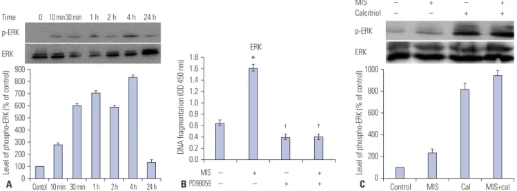
Fig. 5. Calcitriol increases MIS-induced ERK phosphorylation in SKOV3 cells. (A) Western blot analysis of ERK and phosphorylated ERK levels in SKOV3 cells treated with MIS. Bands were detected with anti-ERK and anti phospho-ERK antibodies. (B) SKOV3 cells were treated with 71 nM MIS, 20 μM PD98059, or both for 2 h and the degree of apoptosis was analyzed by ELISA measuring the level of cellular DNA fragmentation. Significant increase relative to controls (MIS 0 nM) is indicated by asterisks and crosses indicate a significant difference (p<0.05) compared with MIS treatment (MIS 71 nM). (C) SKOV3 cells were incubated with or without calcitriol (50 μM) for 4 h prior to the addition of MIS (71 nM). Subsequent to 48 h of incubation, cells were harvested and ERK activation was analyzed by western blot analysis using anti-phospho-ERK antibody. *p<0.05 vs. control, †p<0.05 vs. MIS. MIS, Müllerian inhibiting substance; ERK, extracellular signal-regulated kinase; ELISA, enzyme-linked immunosorbent assay; OD, optical density.
A B C
crease in DNA fragmentation was apparent in cells treated with MIS (71 nM) combined with calcitriol (50 μM), compared with the control. In addition, the results for the combination treatment were significantly different from MIS alone treat- ment in all cell lines (Fig. 3).
MIS and calcitriol alter the expression of regulatory proteins in SKOV3
To demonstrate that MIS and calcitriol-reduced cell prolifera- tion was due to apoptotic activity, the levels of Bcl-2, BAX, cas- pase-3, and caspase-9 were examined. SKOV3 cells were pre- treated with or without calcitriol (50 μM) for 4 h prior to the addition of MIS (71 nM). Following 48 h of incubation, cells were analyzed by western blot analysis with anti-caspase-9 an- tibody and anti-caspase-3 antibody; the expression of the apoptosis-related proteins Bcl-2 and BAX were also evaluated.
Treatment of SKOV3 with MIS plus calcitriol induced apopto- sis, as evidenced by an increase in the levels of BAX, caspase-3, and caspase-9 and a decrease in the levels of Bcl-2 (Fig. 4).
The effects of MIS plus calcitriol on the phosphorylation of ERK in SKOV3
Potential molecular mechanisms underlying the synergistic effect of calcitriol and MIS were assessed by western blot anal- ysis following treatment with chemical inhibitors; p38 MAPK activation was inhibited with 20 μM SB203580, PI3K was in- hibited with 20 μM LY294002, ERK was inhibited with 20 μM PD98059, and JNK signaling was inhibited with 20 μM SP600125. Although MIS did not activate the p38 MAPK, PI3K, or JNK pathway (data not shown), Fig. 5A indicates that the ERK pathway is activated by MIS. SKOV3 cells were then treat- ed with 71 nM MIS, 20 μM PD98059, or both for 2 h and then the degree of apoptosis was analyzed by measuring the level of cellular DNA fragmentation using ELISA. A significant in- crease in fragmentation was observed for the MIS treatment vs. the control (p<0.05) and MIS vs. PD98059 (p<0.05) (Fig.
5B). Next, cells were incubated with or without calcitriol (50 μM) for 4 h prior to the addition of MIS (71 nM). Following 48 h of incubation, cells were harvested and ERK activation was analyzed by western blotting using an anti-phospho-ERK an- tibody. The combination treatment resulted in the most in- tense specific band compared to control, MIS alone, and cal- citriol alone (Fig. 5C).
DISCUSSION
The clinical use of cytotoxic drugs has had a significant impact on neoplastic diseases. However, their therapeutic effectiveness is limited because of their narrow therapeutic index and the onset of chemoresistance. Therefore, many efforts are currently being directed at finding new therapeutic options that may overcome these problems.
Our present study was aimed at further elucidating the mo- lecular mechanisms underlying the anti-proliferative and can- cer preventive effects of MIS and calcitriol in order to develop strategies to improve OCa treatment. The results suggested that calcitriol enhanced the antitumor activity of MIS in OCa cells by down-regulating the expression of Bcl-2 and up-regu- lating the expression of BAX, caspase-3, and caspase-9 through the ERK signaling pathways.
Initial in vitro studies using human OCa cell lines or tissues and several follow-up studies have revealed that MIS inhibits the growth of human cancer cells including breast, cervical, en- dometrial, prostate cancer, and ocular melanoma.6,8,9,15-17 In ad- dition, a recent study indicates that MIS also plays a role in cell cycle arrest and apoptosis of endometriosis.18
The prophylactic and therapeutic activities of Vit D against the most common types of cancer have been extensively inves- tigated both in vitro and in vivo.19-21 The most striking results have been obtained from studies on breast cancer, prostate cancer, and colorectal cancer.19,22 Experimental observations suggest that the chemopreventive effects of Vit D are due main- ly to its ability to modulate important biological functions such as cell proliferation, cell differentiation, growth factor gene ex- pression, signal transduction, and apoptosis.23,24 Interestingly, recent studies have shown that calcitriol may also affect OCa cell proliferation by decreasing human telomerase reverse tran- scriptase mRNA through a small non-coding RNA.25
Vit D and TGF-β have similar effects on cell growth and dif- ferentiation. Experimental stress studies indicate that Vit D may increase the expression levels of TGF-β and its receptors or TGF-β secretion in certain cell types.26-28 The Feldman research group has demonstrated that MIS, a TGF-β family member, constitutes a novel target gene regulated by calcitriol in pros- tate cells.14 Exposing prostate cancer cells to calcitriol for 24 h resulted in a considerable increase in the expression of MIS mRNA. In addition, HeLa cells transfected with an MIS pro- moter-luciferase construct and a Vit D receptor expression vector demonstrated a significant (two- to four-fold) induction of MIS promoter-luciferase following treatment with calcitriol, suggesting that the MIS promoter is responsive to calcitriol.13,14
Determining the utility of MIS as an anticancer drug would most likely involve administering MIS to patients as an adju- vant in combination with other drugs. Therefore, elucidating the anti-proliferation and apoptosis signaling mechanisms downstream of MIS is necessary before combining MIS with commonly used cytotoxic drugs. Moreover, it is important to test for synergy or additivity between MIS and other drugs to ensure that they do not counteract with each other. Since little is known as for the signaling pathways by which MIS mediates proliferation inhibition and apoptosis in OCa cell lines, we in- vestigated several potential molecular mechanisms using chemical inhibitors of the ERK, p38 MAPK, PI3K, and JNK signaling pathways. Our results demonstrated that MIS is not dependent on the p38 MAPK, PI3K, or JNK pathways, but that
the ERK pathway is activated by MIS. Consistent with our findings, Renlund, et al.29 reported that MIS does not activate the JNK pathway. In addition, they identified the JNK inhibi- tor, SP600125, as an activator of the MIS signal transduction pathway.
Numerous case studies have demonstrated that serum MIS levels can be increased >1000-fold above the normal range without any significant adverse reactions; therefore, the thera- peutic administration of MIS to cancer patients may be well tolerated.10 However, purified recombinant MIS is difficult and expensive to obtain, and the clinical use of calcitriol is lim- ited, because of the adverse effects of hypercalcemia. Thus, sev- eral important issues remain to be resolved prior to clinical use, including indication, appropriate doses, blood concentration, adverse effects, resistance, drug interactions, and effectiveness in vivo.
Despite these issues, our findings indicate that treatment with MIS in combination with calcitriol may be an effective clinical strategy for treating ovarian cancer, since combination of the two agents enhances the anti-proliferative and apoptot- ic effects of each agent alone. These results, coupled with the need for a decrease in the toxic side effects of currently em- ployed therapeutic agents, provide a strong rationale for test- ing the therapeutic potential of MIS, alone or in combination with calcitriol, in the treatment of OCa. Future studies should address the exact biological functions of MIS and of the extent of the MIS-stimulated anti-proliferative and apoptotic activi- ties of calcitriol.
REFERENCES
1. Jemal A, Murray T, Samuels A, Ghafoor A, Ward E, Thun MJ. Can- cer statistics, 2003. CA Cancer J Clin 2003;53:5-26.
2. Berkenblit A, Cannistra SA. Advances in the management of epi- thelial ovarian cancer. J Reprod Med 2005;50:426-38.
3. Salom E, Almeida Z, Mirhashemi R. Management of recurrent ovarian cancer: evidence-based decisions. Curr Opin Oncol 2002;
14:519-27.
4. MacLaughlin DT, Teixeira J, Donahoe PK. Perspective: reproduc- tive tract development--new discoveries and future directions.
Endocrinology 2001;142:2167-72.
5. Coughlin JP, Donahoe PK, Budzik GP, MacLaughlin DT. Müllerian inhibiting substance blocks autophosphorylation of the EGF re- ceptor by inhibiting tyrosine kinase. Mol Cell Endocrinol 1987;
49:75-86.
6. Gupta V, Carey JL, Kawakubo H, Muzikansky A, Green JE, Dona- hoe PK, et al. Mullerian inhibiting substance suppresses tumor growth in the C3(1)T antigen transgenic mouse mammary carci- noma model. Proc Natl Acad Sci U S A 2005;102:3219-24.
7. Segev DL, Ha TU, Tran TT, Kenneally M, Harkin P, Jung M, et al.
Mullerian inhibiting substance inhibits breast cancer cell growth through an NFkappa B-mediated pathway. J Biol Chem 2000;275:
28371-9.
8. Renaud EJ, MacLaughlin DT, Oliva E, Rueda BR, Donahoe PK.
Endometrial cancer is a receptor-mediated target for Mullerian Inhibiting Substance. Proc Natl Acad Sci U S A 2005;102:111-6.
9. Barbie TU, Barbie DA, MacLaughlin DT, Maheswaran S, Donahoe
PK. Mullerian Inhibiting Substance inhibits cervical cancer cell growth via a pathway involving p130 and p107. Proc Natl Acad Sci U S A 2003;100:15601-6.
10. MacLaughlin DT, Donahoe PK. Müllerian inhibiting substance/
anti-Müllerian hormone: a potential therapeutic agent for human ovarian and other cancers. Future Oncol 2010;6:391-405.
11. Ponsonby AL, Lucas RM, Lewis S, Halliday J. Vitamin D status during pregnancy and aspects of offspring health. Nutrients 2010;
2:389-407.
12. Giammanco M, Di Majo D, La Guardia M, Aiello S, Crescimannno M, Flandina C, et al. Vitamin D in cancer chemoprevention. Pharm Biol 2015;53:1399-434.
13. Malloy PJ, Peng L, Wang J, Feldman D. Interaction of the vitamin D receptor with a vitamin D response element in the Mullerian- inhibiting substance (MIS) promoter: regulation of MIS expres- sion by calcitriol in prostate cancer cells. Endocrinology 2009;150:
1580-7.
14. Krishnan AV, Moreno J, Nonn L, Malloy P, Swami S, Peng L, et al.
Novel pathways that contribute to the anti-proliferative and che- mopreventive activities of calcitriol in prostate cancer. J Steroid Biochem Mol Biol 2007;103:694-702.
15. Hoshiya Y, Gupta V, Segev DL, Hoshiya M, Carey JL, Sasur LM, et al.
Mullerian Inhibiting Substance induces NFkB signaling in breast and prostate cancer cells. Mol Cell Endocrinol 2003;211:43-9.
16. Parry RL, Chin TW, Epstein J, Hudson PL, Powell DM, Donahoe PK. Recombinant human mullerian inhibiting substance inhibits human ocular melanoma cell lines in vitro and in vivo. Cancer Res 1992;52:1182-6.
17. Kim HS, Sung YJ, Paik S. Cancer Cell Line Panels Empower Ge- nomics-Based Discovery of Precision Cancer Medicine. Yonsei Med J 2015;56:1186-98.
18. Namkung J, Song JY, Jo HH, Kim MR, Lew YO, Donahoe PK, et al.
Mullerian inhibiting substance induces apoptosis of human en- dometrial stromal cells in endometriosis. J Clin Endocrinol Metab 2012;97:3224-30.
19. Leyssens C, Verlinden L, Verstuyf A. Antineoplastic effects of 1,25(OH)2D3 and its analogs in breast, prostate and colorectal cancer. Endocr Relat Cancer 2013;20:R31-47.
20. Pereira F, Larriba MJ, Muñoz A. Vitamin D and colon cancer. En- docr Relat Cancer 2012;19:R51-71.
21. Swami S, Krishnan AV, Feldman D. Vitamin D metabolism and action in the prostate: implications for health and disease. Mol Cell Endocrinol 2011;347:61-9.
22. Krishnan AV, Feldman D. Molecular pathways mediating the anti- inflammatory effects of calcitriol: implications for prostate cancer chemoprevention and treatment. Endocr Relat Cancer 2010;17:
R19-38.
23. Gocek E, Studzinski GP. Vitamin D and differentiation in cancer.
Crit Rev Clin Lab Sci 2009;46:190-209.
24. Haussler MR, Whitfield GK, Kaneko I, Haussler CA, Hsieh D, Hsieh JC, et al. Molecular mechanisms of vitamin D action. Calcif Tissue Int 2013;92:77-98.
25. Kasiappan R, Shen Z, Tse AK, Jinwal U, Tang J, Lungchukiet P, et al. 1,25-Dihydroxyvitamin D3 suppresses telomerase expression and human cancer growth through microRNA-498. J Biol Chem 2012;287:41297-309.
26. Daniel C, Schroder O, Zahn N, Gaschott T, Steinhilber D, Stein JM. The TGFbeta/Smad 3-signaling pathway is involved in butyr- ate-mediated vitamin D receptor (VDR)-expression. J Cell Bio- chem 2007;102:1420-31.
27. Tu H, Flanders WD, Ahearn TU, Daniel CR, Gonzalez-Feliciano AG, Long Q, et al. Effects of calcium and vitamin D3 on transforming growth factors in rectal mucosa of sporadic colorectal adenoma pa-
tients: a randomized controlled trial. Mol Carcinog 2015;54:270-80.
28. Bizzarri M, Cucina A, Valente MG, Tagliaferri F, Borrelli V, Stipa F, et al. Melatonin and vitamin D3 increase TGF-beta1 release and induce growth inhibition in breast cancer cell cultures. J Surg Res 2003;110:332-7.
29. Renlund N, Pieretti-Vanmarcke R, O’Neill FH, Zhang L, Donahoe PK, Teixeira J. c-Jun N-terminal kinase inhibitor II (SP600125) ac- tivates Mullerian inhibiting substance type II receptor-mediated signal transduction. Endocrinology 2008;149:108-15.