• 검색 결과가 없습니다.

Suppressive Effects of Fucoxanthin on Degranulation in IgE-antigen complex-stimulated RBL-2H3 Cells

N/A
N/A
Protected

Academic year: 2021

Share "Suppressive Effects of Fucoxanthin on Degranulation in IgE-antigen complex-stimulated RBL-2H3 Cells"

Copied!
7
0
0

로드 중.... (전체 텍스트 보기)

전체 글

(1)

Suppressive Effects of Fucoxanthin on Degranulation in IgE-antigen complex-stimulated RBL-2H3 Cells

Seung Namkoong

1

, Haemi Joo

2

, Seon-A Jang

2

, Ye-Jin Kim

3

, Taeseong Kim

3

and Eun-Hwa Sohn

3*

1

Department of Physical Therapy, Kangwon National University, Samcheok 245-710, Korea

2

Radiation Health Research Institute, Korea Hydro & Nuclear Power Co., Ltd, Seoul 132-703, Korea

3

Department of Herbal Medicine Resource, Kangwon National University, Samcheok 245-710, Korea

Abstract - The marine carotenoid fucoxanthin can be found in marine brown seaweeds, macroalgae, diatoms, and microalgae, and has remarkable biological properties. Numerous studies have shown that fucoxanthin has considerable potential and promising applications in human health, but the underlying mechanisms involved in its anti-allergic activity are not fully understood. We here investigated the mechanisms by anti-allergic activity of fucoxanthin fraction from Eisenia bicyclis in immunoglobulin E-antigen complex (IgE/DNP-BSA)-stimulated RBL-2H3 mast cells. This study we found that the fucoxanthin inhibits the release of β-hexosaminidase and suppressed not only transcriptional activation of NF-κB, but also phosphorylation of ERK and JNK in IgE/DNP-BSA-treated RBL-2H3 cells. Fucoxanthin may be useful for preventing allergic diseases, including asthma and atopic dermatitis.

Key words - Allergy, β-Hexosaminidase, MAPK, NF-κB

*Corresponding author. E-mail : ehson@kangwon.ac.kr

Introduction

Mast cells and basophils play important roles initiating and perpetuating the inflammatory responses that mediate allergic reactions by secreting abundant levels of proinflammatory mediators such as histamine and several cytokines, including interleukin (IL)-4, IL-5, IL-13, and tumor necrosis factor (TNF)-α (Matsuda et al., 2008). Because mast cells play essential roles in provoking the pathogenesis of allergic reactions via the degranulation process, measuring the degree of degranulation reflects the level of mast cell activation. β -Hexosaminidase released by these cells during this process has been reported to be a suitable marker for determining the degree of degranulation (Naal et al., 2004). The rat basophilic leukemia cell line RBL-2H3, which expresses FcεRI, is widely used to study the molecular basis of mast cell activation (Huang et al., 2008). This cell line has also been used to develop anti-allergy drugs that reduce allergic symptoms, including steroids that have anti-histamine actions and anti- inflammatory drugs that inhibit cytokine production (Matsuda et al., 2008; Huang et al., 2008). However, the effectiveness

of such drugs is limited by their side effects. These problems have led to increasing interest in traditional herbal medicines that have been used to treat allergic diseases. As a result, more and more studies examining the efficacy of natural extracts and compounds isolated from natural extracts to prevent and treat allergic disorders are being performed.

Marine environment has been the source of diverse life forms that produce different biologically active compounds.

Metabolites from marine plants have been reported to have outstanding biological activities and profound applications in nutraceuticals, cosmeceuticals, and pharmaceuticals. Eisenia bicyclis is a species of the kelp, a member of the brown algae group of seaweeds, best known for its use in Asia. E. bicyclis is high in calcium, iodine, iron, magnesium, and vitamin A as well as being a good dietary source for many other minerals.

They also contain the storage polysaccharide laminarin, alginate,

fucoxanthin and fucoidan with immunological activities (Joe

et al., 2006). It has been also known that brown algae extracts

closely linked to its protective properties against free radical

attack and have anti-oxidant, immunomodulatory effects (Hu

et al., 2010; Kang et al., 2004; Namkoong et al., 2011) and

numerous studies have shown that fucoxanthin has considerable

potential and promising applications in human health (Peng

(2)

Fig. 1. Structure of fucoxanthin.

et al., 2011).

Although E. bicyclis and its component, fucoxanthin have these biological activities, no reported study has evaluated the anti-allergic effect of E. bicyclis or fucoxanthin and the molecular mechanisms involved. Therefore, we examined the fucoxanthin (Fig. 1) isolated from E. bicyclis on the anti- allergic effects by checking the release of β-hexosaminidase induced by IgE/DNP-BSA in RBL-2H3 mast cells and the molecular mechanisms such as MAPKs-NF-κB activities, which are activated during allergic inflammation.

Materials and Methods

Preparation of fucoxanthin

The fronds of Eisenia bicyclis were harvested from the coast of Ulleung Island, East Sea, South Korea in June 2009.

Fresh E. bicyclis was washed three times with tap water to remove salt and impurities, then dried at room temperature for 3 days and stored at -20℃. The dried samples were homo- genized, using a grinder, before extraction. The fucoxanthin fraction was prepared by solvent partition method from the acetone extract of fresh E. bicyclis. Fucoxanthin fraction was used for CPC (centrifugal partition chromatography) using a two-phase solvent system of n-hexane-ethyl acetate-ethanol- water (5:5:7:3, v/v/v/v). The flow rate of mobile phase was 2 ml/min with descending mode while rotating at 1000 rpm.

The eluate was monitored at 410 nm. The content and struc- ture of fucoxanthin in the CPC fraction were confirmed with HPLC, UV, APCI/MS and NMR spectra. A preparative CPC yielded 20 mg of fucoxanthin (87% recovery from fucoxanthin fraction) in a two-step separation from 516 mg of fucoxanthin fraction containing 4.59% fucoxanthin. The purity of the isolated fucoxanthin was about 81% in the first CPC step and over 95% in the second CPC step based on the calibration curve. This fucoxanthin was kindly provided by Dr. Um (Natural Product Research Center, KIST Gangneung Institute, Korea)

(Kim et al., 2011).

Cell culture

The RBL-2H3 rat mast cell line, HaCaT human keratinocyte cell line, NIH3T3 mouse fibroblast cell line were obtained from the American Type Culture Collection (Rockville, MD) and grown in minimum essential medium (MEM) with 15%

fetal bovine serum (FBS), 2 mM L-glutamine, 100 U/ml penicillin, and 100 μg/ml streptomycin at 37℃ in a humidified incubator with a 5% CO

2

/ 95% air atmosphere.

Reagents

The anti-dinitrophenyl (DNP)-IgE and 4-nitrophenyl N- acetyl-β-D-glucosaminide were from Sigma-Aldrich, DNP- bovine serum albumin (BSA) was from Biosearch Technologies, minimum essential medium was from Invitrogen, fetal bovine serum (FBS) was from WelGENE, the protein assay kit was from BioRad Laboratories, anti-pERK, anti-ERK, anti-pJNK, anti-JNK, and anti-p-p38 were from Cell Signaling Technology, anti-p65 and anti-p38 were from Santa Cruz Biotechnology, anti-β-actin was from Sigma-Aldrich, anti-α-tubulin was from Abfrontier, the ECL chemiluminescence system was from GE Healthcare, and the polyvinylidene difluoride (PVDF) membrane was from Millipore.

XTT assay for cell survivability

Cell survivability was examined using the XTT assay kit, according to the manufacturer’s instructions. The spleen was removed aseptically and dissociated into a single cell suspen- sion in culture medium. Cells (5×10

5

cells/well) were in- cubated with various fucoxanthin concentrations (0.1, 1, and 10 μg/ml). After incubating the cells for 20 h, a mixture of 25 μl of phenazine methosulfate (PMS; electron-coupling reagent) and 25 μl of XTT [2,3-bis(2-methoxy-4-nitro-5- sulfophenyl)-2H-tetrazolium-5-carboxanilide] was added to each well. The cells were further incubated for 4 h to allow XTT formazan production. The absorbance was determined with a microplate reader at a test wavelength of 450 nm and a reference wavelength of 690 nm.

Nitrite determination

Cells were incubated in either medium alone or medium

(3)

supplemented for 24 h and the accumulation of nitrite in culture supernatants was measured using the assay system described by Ding et al. (1988). One hundred micro litter aliquots of culture supernatants were mixed with an equal volume of Griess reagent (mixture at 1:1 of naphthyleth- ylenediamine dihydrochloride and 1% sulphanilamide in 5%

H

3

PO

4

) and incubated at room temperature for 10 min. Nitrite concentration was calculated from a NaNO

2

standard curve.

β-Hexosaminidase release assay

Degranulation of RBL-2H3 cells was evaluated by measuring the activity of the granule-stored enzyme-β-hexosaminidase secreted in the extracellular medium. Cells were cultured in 24-well plates (2×10

5

cells/well) overnight. The cells were sensitized with anti-DNP-IgE (100 ng/ml) for 16 h at 37℃.

After washing the cells with TGCM buffer (136 mM NaCl, 2.68 mM KCl, 0.36 mM NaH

2

PO

2

·H

2

O, 1 mM CaCl

2

, 0.5 mM MgCl

2

, 11.9 mM NaHCO

3

, 5 mM dextrose, 1 g/L gelatin, pH 7.4), they were pretreated with fucoxanthin (0.1, 1, and 10 μ g/ml) for 30 min and then treated with DNP-BSA (1 μg/ml) for 30 min at 37℃. Aliquots of the cellular supernatant (15 μl) were transferred to 96well plates and incubated with 60 μl of substrate (1 mM p-nitrophenyl-N-acetyl-β-D-glucosaminide in citrate 0.05 M, pH 4.5) for 60 min at 37℃. The cells were lysed with 0.1% Triton X-100 before removing the super- natant to measure the total β-hexosaminidase activity. The reaction was stopped by adding 150 μl of Na

2

CO

3

NaHCO

3

buffer 0.1 M, pH 10. The absorbance at 405 nm was measured with a microplate reader (Themo Labsystems). The results were presented as the percentage of total β-hexosaminidase content of the cells determined by cell lysis with 0.1% Triton X-100.

% Degranulation = OD

supernatant

/ (OD

supernatant

+ OD

triton x-100

) x100

Western blotting

Total cell lysates were plated at 3×10

7

cells/ml and treated with fucoxanthin (0.1, 1, and 10 μg/ml) and DNP-BSA (0.01 μg/ml) for 15 min and then harvested and lysed in a lysis buffer containing 20 mM Tris, pH 7.6, 150 mM NaCl, and 1%

Triton X-100 with a protease inhibitor cocktail. Protein con-

tents were measured using a protein assay kit (Bio-Rad).

Samples were diluted with 1× lysis buffer containing 1% β -mercaptoethanol. Equal amounts of cellular protein (50 μg) were resolved by 10% SDS-PAGE and transferred onto nitro- cellulose membranes. After blocking, membranes were in- cubated with the target antibody and then with horseradish peroxidase-conjugated secondary antibody to IgG. Immuno- reactive proteins were visualized using the ECL Western blot detection system. The protein level was compared to a loading control, such as β-actin or non-phosphorylated protein.

Subcellular fractionation

Cytosolic and nuclear extracts were prepared. In brief, RBL-2H3 cells (5×10

6

cells/ml) were plated into 100 mm dishes and treated with fucoxanthin (0.1, 1, and 10 μg/ml) and IgE/DNP-BSA (0.01 μg/ml) for 4 h. The harvested cells were resuspended in 0.2 ml of buffer A (10 mM HEPES at pH 7.5, 1.5 mM MgCl

2

, 10 mM KCl, 1 mM DDT, 0.1% NP-40, 0.2 mM PMSF). The cells were lysed on ice for 15 min, and centrifuged (5,000 g, 5 min, 4℃). The supernatant was collected as cytosolic extracts. The nucleic pellet was washed with buffer A lacking NP-40, and resuspended in 25 μl of buffer C (20 mM HEPES, pH 7.5, 25% glycerol, 0.42 M NaCl, 0.2 mM EDTA, 1.5 mM MgCl

2

, 1 mM DDT, 0.2 mM PMSF). After incubation on ice for 30 min, nuclear debris was spin down (13,000 g, 10 min, 4℃). The supernatant was collected as nuclear extracts. The protein concentration was measured using a protein assay kit (Bio-Rad).

Statistical analyses

Each experiment was repeated three or four times, and the results of a representative experiment are shown. The results are expressed as the means±SEM and were compared using Student’s t-test. A statistical probability of p < 0.05 was con- sidered significant (# p < 0.05, ## p < 0.01, * p < 0.05, and **

p < 0.01).

Results and Discussion

Traditional medicines isolated from natural products often

have positive effects in the prevention and healing of various

immune disorders, such as allergy and atopic inflammation.

(4)

Fig. 2. The effects of fucoxanthin for cell viability and antigen- induced β-hexosaminidase in RBL-2H3 cells. (A) The cells were treated with various concentration of fucoxanthin for 24 h. Cell viability was assessed using XTT assay. Absorbance was measure data at 450 nm and 650 nm. (B) The cells were sensitized by overnight incubation with 1 μg/ml of DNP- specific IgE in medium. These DNP-IgE-sensitized RBL-2H3 cells were pre-incubated with various concentration of fuco- xanthin (FX) for 30 min and then incubated with antigen (IgE/DNP-BSA) for 15 min in order to measure the release of β-hexosaminidase. Each bar shows the means ± SEM of four independent experiments.

##

P<0.01: significantly different from control group, **P<0.01, *P<0.05: significantly different from IgE/DNP-BSA alone. KF: ketotifen fumarate at 100 μM.

In this study, the fucoxanthin (95% purity level) fraction of Eisenia bicyclis showed potential anti-allergic effects by decreasing β-hexosaminidase release in mast cells.

Mast cells are primary effector cells involved in the allergic or immediate hypersensitivity responses (Theoharides and Kalogeromitros, 2006). The antigen crosslinking of the IgE- FcεRI complexes through the aggregation of IgE and FcεRI on mast cells results in the release of β-hexosaminidase, which is a marker of mast cell degranulation. The release of β -hexosaminidase and histamines also causes the production of proinflammatory cytokines, such as IL-4, IL-6, and TNF- α, which can potentiate inflammatory immune responses through the subsequent induction of other atopic inflammatory mediators. Thus, the modulation of this initial process is con- sidered a rational approach for regulating the early phase of allergic responses (Gilfillan and Tkaczyk, 2006; Theoharides and Kalogeromitros, 2006). Rat mast cell line RBL-2H3 cells were used to determine the effect of fucoxanthin on the secre- tion of β-hexosaminidase. Initially, we measured the cytotoxicity of fucoxanthin on RBL-2H3 cells using the XTT assay.

Fucoxanthin at concentrations ranging from 0.1-10 μg/ml did not significantly affect the cytotoxicity in 24 h (Fig. 2A).

Thus, we treated DNP-IgE-sensitized RBL-2H3 cells with fucoxanthin ranging from 0.1-10 μg/ml in subsequent experi- ments. Fucoxanthin significantly suppressed the DNP-BSA induced β-hexosaminidase secretion in IgE-sensitized RBL- 2H3 cells at 1 and 10 μg/ml and the effects are dose-dependent (Fig. 2B). Ketotifen fumarate, an anti-allergic drug, also decreased the β-hexosaminidase secretion. The results showed that fucoxanthin significantly inhibited antigen-induced mast cell degranulation.

Atopic dermatitis is characterized by allergic skin inflam- mation. Pathological changes in atopic skin are observed as epidermal thickening and marked infiltration of inflammatory cells (Soter, 1989). Here, we evaluated the effects of fucoxanthin on the allergic skin diseases using the kinetic proliferation and the regulation of NO production in human keratinocytes (HaCaT) and mouse fibroblast (NIH3T3) cells (Park et al., 2010), but fucoxanthin has no significant effects (Fig. 3 and 4) in both of cells.

We subsequently evaluated the related mechanisms of fucoxanthin on degranulation including NF-κB activation

and the phosphorylation of MAPKs. We detected the inhibitory effects of fucoxanthin on IgE/DNP-BSA-induced nuclear translocation of NF-κB (p65). Increased expression of NF-κ B (p65) was observed in the nucleus after treatment with fucoxanthin in IgE/DNP-BSA-stimulated RBL-2H3 cells for 4 h (Fig. 5). The relative intensity of NF-κB (p65) tran- slocation in the nucleus was increased significantly in IgE/

DNP-BSA-stimulated RBL-2H3 cells compared with the no

(5)

Fig. 3. The effects of fucoxanthin on proliferation in keratinocytes (HaCaT) and fibroblast (NIH3T3) cells. (A) HaCaT and (B) NIH3T3 cells were treated with fucoxantin (FX) for 22, 44, 66, 88 hrs. The proliferations of cells were assessed by XTT assay. Cell proliferations were represented by the percentage of control. The results are mean ± SEM of quintuplicates from a representative experiment.

Fig. 4. The effects of the fucoxantin (FX) on the production of NO in (A) HaCaT keratinocytes and (B) NIH3T3 fibroblast cells.

These results are means ± SEM of quintuplicates from a representative experiment.

Fig. 5. Effects of fucoxantin on NF-κB activation in IgE/DNP- BSA-stimulated RBL-2H3 mast cells. The cells were sensitized by overnight incubation with 1 μg/ml of DNP-specific IgE in medium. This DNP-IgE-sensitized RBL-2H3 cells were pre- incubated with various concentration of fucoxanthin (FX, 0.1, 1, and 10 μg/ml) for 30 min and then incubated with antigen (IgE/DNP-BSA) for 15 min. After isolation of cytosolic (C-) and nuclear (N-) fraction, the translocation of NF-κB (p65) was assessed by Western blotting described in methods respec- tively.

antigen treatments. In contrast, the relative intensity of NF-κ B (p65) translocation in the nucleus was decreased con- siderably, to the control basal level, after the addition of 10 μg/

ml fucoxanthin. These data demonstrate that fucoxanthin attenuated NF-κB activation and might affect the degranulation of histamine in mast cells.

NF-κB is a key transcription factor that regulates the expression of genes involved in immune and inflammatory responses that require inflammatory cytokine production.

NF-κB translocation and the MAPKs pathway are regarded

as important processes in the regulation of the innate and

acquired immune responses and chronic allergic inflam-

mation (Karin and Lin, 2002; Barnes, 2008). NF-κB is also a

critical transcription factor that regulates Th2 cell differ-

entiation and Th2-dependent airway inflammation in asthma

(Yang et al., 1998). Increased NF-κB activity has been

(6)

Fig. 6. Effects of fucoxantin on phosphorylations of MAPKs in IgE/DNP-BSA-stimulated RBL-2H3 mast cells. The cells were sensitized by overnight incubation with 1 μg/ml of DNP- specific IgE in medium. This DNP-IgE-sensitized RBL-2H3 cells were pre-incubated with various concentration of fuco- xanthin (FX, 0.1, 1, and 10 μg/ml) for 30 min and then in- cubated with antigen (DNP-BSA) for 15 min. The phosphoryl- ations of p38 MAP kinase such as p38, JNK and ERK were assessed by Western blotting described in methods.

reported in asthma, an allergic disease, and the inhibition of NF-κB activity decreased asthma (Barnes, 2008). Thus, we suggest that fucoxanthin could have an anti-allergic effect based on the decrease in activated NF-κB it causes.

Conventional MAP kinases are classified into three families:

the c-Jun N-terminal kinases (JNKs), the p38 MAP kinases, and the extracellular signal-regulated kinases (ERKs). Intra- cellular signal transduction, including the phosphorylation of p38 MAPK, is subsequently followed by NF-κB translo- cation, leading to the production of cytokines and chemokines.

We also showed that fucoxanthin significantly suppressed the IgE/DNP-BSA-activated phosphorylation of ERK and JNK but not p38 in RBL-2H3 mast cells (Fig. 6). It has been reported that MAPK activation can activate transcription factors that result in the expression of IL-4, IL-5, and IL-13 in human T cells in response to antigen exposure in allergic disease (Barnes, 2008). Recently, Lee et al. (2012) reported that the glycoprotein isolated from Cudrania tricuspidata inhibits release of β-hexosaminidase via down regulation of

MAPK/NF-κB on the stage of mast cell degranulation.

We found here that fucoxanthin may exert inhibitory effects on the antigen-induced degranulation by suppressing the transcription of NF-κB and the activation of ERK-JNK but not of p38 in signaling pathways. From these results, we suggest that fucoxanthin might be useful as a therapeutic agent for treating various forms of allergic inflammation, including asthma and atopic dermatitis.

Literature Cited

Barnes, P.J. 2008. Role of GATA-3 in allergic diseases. Curr.

Mol. Med. 8:330-334.

Ding, A.H., C.F. Nathan and D.J. Stuehr. 1988. Release of reactive nitrogen intermediates and reactive oxygen inter- mediates from mouse peritoneal macrophages. Comparison of activating cytokines and evidence for independent pro- duction. J. Immunol. 141:2407-2412.

Gilfillan, A.M. and C. Tkaczyk. 2006. Integrated signaling pathways for mast-cell activation. Nat. Rev. Immunol. 6:

218-230.

Hu, T., D. Liu, Y. Chen, J. Wu and S. Wang. 2010. Antioxidant activity of sulfated polysaccharide fractions extracted from Undaria pinnitafida in vitro. Int. J. Biol. Macromol. 46:

193-198.

Huang, F., K. Yamaki, X. Tong, L. Fu, R. Zhang, Y. Cai, R.

Yanagisawa, K. Inoue, H. Takano and S. Yoshino. 2008.

Inhibition of the antigen-induced activation of RBL-2H3 cells by sinomenine. Int. Immunopharmacol. 8:502-507.

Joe, M.J., S.N. Kim, H.Y. Choi, W.S. Shin, G.M. Park, D.W.

Kang and Y.K. Kim. 2006. The inhibitory effects of eckol and dieckol from Ecklonia stolonifera on the expression of matrix metalloproteinase-1 in human dermal fibroblasts. Biol.

Pharm. Bull. 29:1735-1739.

Kang, K., H.J. Hwang, D.H. Hong, Y. Park, S.H. Kim, B.H. Lee and H.C. Shin. 2004. Antioxidant and antiinflammatory activities of ventol, a phlorotannin-rich natural agent derived from Ecklonia cava, and its effect on proteoglycan deg- radation in cartilage explant culture. Res. Commun. Mol.

Pathol. Pharmacol. 115-116:77-95.

Karin, M. and A. Lin. 2002. NF-κB at the crossroads of life and death. Nat. Immunol. 3:221-227.

Kim, S.M., Y.F. Shang and B.H. Um. 2011. A preparative

method for isolation of fucoxanthin from Eisenia bicyclis by

(7)

centrifugal partition chromatography. Phytochem. Anal. 22 (4):322-329.

Lee, J., S.J. Lee and K.T. Lim. 2012. CTB glycoprotein (75 kDa) inhibits IgE releasing, TNF-α and IL-6 expressed by bisphenol A in vivo and in vitro. Food Chem. Toxicol. 50 (6):2109-2117.

Matsuda, H., Q. Wang, K. Matsuhira, S. Nakamura, D. Yuan and M. Yoshikawa. 2008. Inhibitory effects of thunberginols A and B isolated from Hydrangeae Dulcis Folium on mRNA expression of cytokines and on activation of activator protein- 1 in RBL-2H3 cells. Phytomedicine 15:177-184.

Namkoong, S., S.C. Kang, H. Do, K.H. Jang, S.A. Jang, M.G.

Choung and E.H. Sohn. 2011. Immunomodulatory effects of supplementation with extracts from the marine brown alga Eisenia bicyclis on macrophages. J. Korean Plant Res. 24(3):

298-303.

Naal, R.M., J. Tabb, D. Holowka and B. Baird. 2004. In situ measurement of degranulation as a biosensor based on RBL- 2H3 mast cells. Biosens Bioelectron. 20(4):791-796.

Park, H.J., D.H. Jung, H. Joo, N.S. Kang, S.A. Jang, J.G. Lee and E.H. Sohn. 2010. The comparative study of anti-allergic and anti-inflammatory effects by fermented red ginseng and red ginseng. J. Korean Plant Res. 23(5): 393-485 (in Korean).

Peng, J., J.P. Yuan, C.F. Wu and J.H. Wang. 2011. Fucoxanthin, a marine carotenoid present in brown seaweeds and diatoms:

metabolism and bioactivities relevant to human health. Mar.

Drugs. 9(10):1806-1828.

Soter, N.A. 1989. Morphology of atopic eczema. Allergy 44 (Suppl 9):16-19.

Theoharides, T.C. and D. Kalogeromitros. 2006. The critical role of mast cells in allergy and inflammation. Ann. NY Acad. Sci. 1088:78-99.

Yang, L., L. Cohn, D.H. Zhang, R. Homer, A. Ray and P. Ray.

1998. Essential role of nuclear factor B in the induction of eosinophilia in allergic airway inflammation. J. Exp. Med.

188:1739-1750.

(Received 30 May 2012 ; Revised 15 June 2012 ; Accepted 25 June 2012)

수치

Fig. 1. Structure of fucoxanthin.
Fig. 2. The effects of fucoxanthin for cell viability and antigen-  induced β-hexosaminidase in RBL-2H3 cells
Fig. 4. The effects of the fucoxantin (FX) on the production of NO in (A) HaCaT keratinocytes and (B) NIH3T3 fibroblast cells
Fig. 6. Effects of fucoxantin on phosphorylations of MAPKs in  IgE/DNP-BSA-stimulated RBL-2H3 mast cells

참조

관련 문서

tricuspidata on the production of proinflammatory cytokines in TNFα+IFNγ-stimulated HaCaT cells ...15 Fig.5: The cell viability of sub-fractions from 70% EtOH

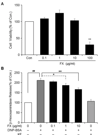
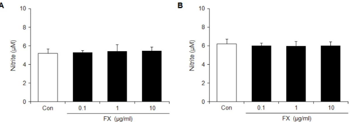
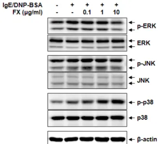
As for the research method, RBL-2H3 cells, a mast cell line, were treated with supercritical carbon dioxide extracted Chamaecyparis obtusa oil (C oil), German chamomile oil (G

 The biological effect of ionizing radiation is a consequence of the energy transfer, by ionization and excitation, to cells in the body.  Factors of radiation effects

This study supports that flavonoids present in PCE has anti-adipogenic and anti- diabetic effects by activating AMPK and normalizing the expression of

Effects of pulse frequency of low-level laser thrapy (LLLT)on bone nodule formation in rat calvarial cells.. Low-level laser therapy stimulats

 Often found connected to other molecules on the outsides of cells --- cellular recognition, cell signaling, cell

• Generation of different specialized kinds of cells from zygote (fertilized egg) or other precursor cells.. – Generate blood cells, muscle

4 &gt; Effects of Taro on COX-2 expression and iNOS expression(hot water) in human thyroid cancer cells. The cells were pretreated for 48hours with either